Phospho-Tau (S396) Recombinant Rabbit Monoclonal Antibody [SN62-09]
Catalog# ET1611-68
Phospho-Tau (S396) Recombinant Rabbit Monoclonal Antibody [SN62-09]
-
WB
-
IF-Cell
-
IF-Tissue
-
IHC-P
-
IHC-Fr
-
IP
-
Mouse
-
Rat
-
Human
-
HA750268
不含抗保成分
-
Cynomolgus monkey
-
Pig
-
unconjugated
概述
产品名称
Phospho-Tau (S396) Recombinant Rabbit Monoclonal Antibody [SN62-09]
抗体类型
Recombinant Rabbit monoclonal Antibody
免疫原
Synthetic phospho-peptide corresponding to residues surrounding Ser396 of human Tau.
种属反应性
Mouse, Rat, Human (Predicted: Cynomolgus monkey, Pig)
验证应用
WB, IF-Cell, IF-Tissue, IHC-P, IHC-Fr, IP
分子量
Predicted band size: 79 kDa
阳性对照
SHSY5Y cell lysates, N2A, PC-12, rat brain tissue, mouse brain tissue, mouse kidney tissue.
偶联
unconjugated
克隆号
SN62-09
RRID
产品特性
形态
Liquid
存放说明
Shipped at 4℃. Store at +4℃ short term (1-2 weeks). It is recommended to aliquot into single-use upon delivery. Store at -20℃ long term.
存储缓冲液
1*TBS (pH7.4), 0.05% BSA, 40% Glycerol. Preservative: 0.05% Sodium Azide.
亚型
IgG
纯化方式
Protein A affinity purified.
应用稀释度
-
WB
-
1:500-1:2,000
-
IF-Cell
-
1:100-1:500
-
IF-Tissue
-
1:100-1:500
-
IHC-P
-
1:50-1:200
-
IHC-Fr
-
1:200-1:500
-
IP
-
1-2μg/sample
靶点
功能
Tau, also known as MAPT (microtubule-associated protein tau), MAPTL, MTBT1 or TAU, is a 758 amino acid protein that localizes to the cytoplasm, as well as to the cytoskeleton and the cell membrane, and contains four Tau/MAP repeats. Expressed in neuronal tissue and existing as multiple alternatively spliced isoforms, Tau functions to promote microtubule assembly and stability and is thought to be involved in the maintenance of neuronal polarity. Tau may also link microtubules with neural plasma membrane components and, in addition to its role in microtubule stability, is also necessary for cytoskeletal plasticity. Tau is highly subject to a variety of post-translational modifications, including phosphorylation on serine and threonine residues, polyubiquitination (and subsequent proteasomal degradation) and glycation of specific Tau isoforms. Defects in the gene encoding Tau are associated with Alzheimers disease, pallido-ponto-nigral degeneration (PPND), corticobasal degeneration (CBD) and progressive supranuclear palsy (PSP).
背景文献
1. Manczak M & Reddy PH Abnormal interaction of oligomeric amyloid- with phosphorylated tau: implications to synaptic dysfunction and neuronal damage. J Alzheimers Dis 36:285-95 (2013).
2. Murakami K et al. SOD1 (copper/zinc superoxide dismutase) deficiency drives amyloid protein oligomerization and memory loss in mouse model of Alzheimer disease. J Biol Chem 286:44557-68 (2011).
组织特异性
Expressed in neurons. Isoform PNS-tau is expressed in the peripheral nervous system while the others are expressed in the central nervous system.
翻译后修饰
Phosphorylation at serine and threonine residues in S-P or T-P motifs by proline-directed protein kinases (PDPK1, CDK1, CDK5, GSK3, MAPK) (only 2-3 sites per protein in interphase, seven-fold increase in mitosis, and in the form associated with paired helical filaments (PHF-tau)), and at serine residues in K-X-G-S motifs by MAP/microtubule affinity-regulating kinase (MARK1, MARK2, MARK3 or MARK4), causing detachment from microtubules, and their disassembly. Phosphorylation decreases with age. Phosphorylation within tau/MAP's repeat domain or in flanking regions seems to reduce tau/MAP's interaction with, respectively, microtubules or plasma membrane components. Phosphorylation on Ser-610, Ser-622, Ser-641 and Ser-673 in several isoforms during mitosis. Phosphorylation at Ser-548 by GSK3B reduces ability to bind and stabilize microtubules. Phosphorylation at Ser-579 by BRSK1 and BRSK2 in neurons affects ability to bind microtubules and plays a role in neuron polarization. Phosphorylated at Ser-554, Ser-579, Ser-602, Ser-606 and Ser-669 by PHK. Phosphorylation at Ser-214 by SGK1 mediates microtubule depolymerization and neurite formation in hippocampal neurons. There is a reciprocal down-regulation of phosphorylation and O-GlcNAcylation. Phosphorylation on Ser-717 completely abolishes the O-GlcNAcylation on this site, while phosphorylation on Ser-713 and Ser-721 reduces glycosylation by a factor of 2 and 4 respectively. Phosphorylation on Ser-721 is reduced by about 41.5% by GlcNAcylation on Ser-717. Dephosphorylated at several serine and threonine residues by the serine/threonine phosphatase PPP5C.; Polyubiquitinated. Requires functional TRAF6 and may provoke SQSTM1-dependent degradation by the proteasome (By similarity). PHF-tau can be modified by three different forms of polyubiquitination. 'Lys-48'-linked polyubiquitination is the major form, 'Lys-6'-linked and 'Lys-11'-linked polyubiquitination also occur.; O-glycosylated. O-GlcNAcylation content is around 8.2%. There is reciprocal down-regulation of phosphorylation and O-GlcNAcylation. Phosphorylation on Ser-717 completely abolishes the O-GlcNAcylation on this site, while phosphorylation on Ser-713 and Ser-721 reduces O-GlcNAcylation by a factor of 2 and 4 respectively. O-GlcNAcylation on Ser-717 decreases the phosphorylation on Ser-721 by about 41.5%.; Glycation of PHF-tau, but not normal brain TAU/MAPT. Glycation is a non-enzymatic post-translational modification that involves a covalent linkage between a sugar and an amino group of a protein molecule forming ketoamine. Subsequent oxidation, fragmentation and/or cross-linking of ketoamine leads to the production of advanced glycation endproducts (AGES). Glycation may play a role in stabilizing PHF aggregation leading to tangle formation in AD.
亚细胞定位
Cell membrane, Cell projection, Cytoplasm, Cytoskeleton, Membrane, Microtubule, Secreted.
别名
AI413597 antibody
AW045860 antibody
DDPAC antibody
FLJ31424 antibody
FTDP 17 antibody
G protein beta1/gamma2 subunit interacting factor 1 antibody
MAPT antibody
MAPTL antibody
MGC134287 antibody
MGC138549 antibody
展开AI413597 antibody
AW045860 antibody
DDPAC antibody
FLJ31424 antibody
FTDP 17 antibody
G protein beta1/gamma2 subunit interacting factor 1 antibody
MAPT antibody
MAPTL antibody
MGC134287 antibody
MGC138549 antibody
MGC156663 antibody
Microtubule associated protein tau antibody
Microtubule associated protein tau isoform 4 antibody
Microtubule-associated protein tau antibody
MSTD antibody
Mtapt antibody
MTBT1 antibody
MTBT2 antibody
Neurofibrillary tangle protein antibody
Paired helical filament tau antibody
Paired helical filament-tau antibody
PHF tau antibody
PHF-tau antibody
PPND antibody
PPP1R103 antibody
Protein phosphatase 1, regulatory subunit 103 antibody
pTau antibody
RNPTAU antibody
TAU antibody
TAU_HUMAN antibody
Tauopathy and respiratory failure, included antibody
折叠图片
-

Western blot analysis of Phospho-Tau (S396) on SHSY5Y cell lysates. Proteins were transferred to a PVDF membrane and blocked with 5% BSA in PBS for 1 hour at room temperature. The primary antibody (ET1611-68, 1/500) was used in 5% BSA at room temperature for 2 hours. Goat Anti-Rabbit IgG - HRP Secondary Antibody (HA1001) at 1:5,000 dilution was used for 1 hour at room temperature.
-

☑ Cell treatment (CT)
Western blot analysis of Phospho-Tau(S396) on SHSY5Y cell lysates.
Lane 1: SHSY5Y cells, whole cell lysate, 10ug/lane
Lane 2: SHSY5Y cells treated with 2.8ug/ul lambda-PP for 30 minutes, whole cell lysates, 10ug/lane
All lanes :
Anti-Phospho-Tau(S396) antibody (ET1611-68) at 1/500 dilution. Anti-GAPDH antibody (ET1601-4) at 1/10,000 dilution. Goat Anti-Rabbit IgG H&L (HRP) (HA1001) at 1/200,000 dilution.
Predicted band size: 79 kDa
Observed band size: 70/130 kDa
Blocking and diluting buffer: 5% BSA.
Exposure time: 2 minutes 34 seconds -

ICC staining of Phospho-Tau (S396) in N2A cells (green). Formalin fixed cells were permeabilized with 0.1% Triton X-100 in TBS for 10 minutes at room temperature and blocked with 1% Blocker BSA for 15 minutes at room temperature. Cells were probed with the primary antibody (ET1611-68, 1/50) for 1 hour at room temperature, washed with PBS. Alexa Fluor®488 Goat anti-Rabbit IgG was used as the secondary antibody at 1/1,000 dilution. The nuclear counter stain is DAPI (blue).
-

Immunohistochemical analysis of paraffin-embedded rat brain tissue using anti-Phospho-Tau (S396) antibody. The section was pre-treated using heat mediated antigen retrieval with Tris-EDTA buffer (pH 8.0-8.4) for 20 minutes.The tissues were blocked in 5% BSA for 30 minutes at room temperature, washed with ddH2O and PBS, and then probed with the primary antibody (ET1611-68, 1/50) for 30 minutes at room temperature. The detection was performed using an HRP conjugated compact polymer system. DAB was used as the chromogen. Tissues were counterstained with hematoxylin and mounted with DPX.
-

Immunohistochemical analysis of paraffin-embedded mouse brain tissue with Rabbit anti-Phospho-Tau (S396) antibody (ET1611-68) at 1/50 dilution.
The section was pre-treated using heat mediated antigen retrieval with Tris-EDTA buffer (pH 9.0) for 20 minutes. The tissues were blocked in 1% BSA for 20 minutes at room temperature, washed with ddH2O and PBS, and then probed with the primary antibody (ET1611-68) at 1/50 dilution for 1 hour at room temperature. The detection was performed using an HRP conjugated compact polymer system. DAB was used as the chromogen. Tissues were counterstained with hematoxylin and mounted with DPX. -

Immunohistochemical analysis of paraffin-embedded mouse kidney tissue with Rabbit anti-Phospho-Tau (S396) antibody (ET1611-68) at 1/200 dilution.
The section was pre-treated using heat mediated antigen retrieval with Tris-EDTA buffer (pH 9.0) for 20 minutes. The tissues were blocked in 1% BSA for 20 minutes at room temperature, washed with ddH2O and PBS, and then probed with the primary antibody (ET1611-68) at 1/200 dilution for 1 hour at room temperature. The detection was performed using an HRP conjugated compact polymer system. DAB was used as the chromogen. Tissues were counterstained with hematoxylin and mounted with DPX. -

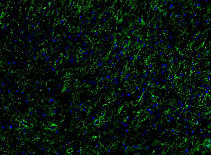
Application: IHC-Fr
Species: Mouse
Site:Hippocampus
Sample: Frozen section
Antibody concentration: 1/200
Antigen retrieval: Not required -

Application: IHC-Fr
Species: Mouse
Site: Cerebral cortex
Sample: Frozen section
Antibody concentration: 1/200
Antigen retrieval: Not required -

☑ Cell treatment (CT)
Western blot analysis of Phospho-Tau (S396) on different lysates with Rabbit anti-Phospho-Tau (S396) antibody (ET1611-68) at 1/2,000 dilution.
Lane 1: Mouse brain tissue lysate
Lane 2: Mouse brain tissue lysate treated with lambda-PP for 30 minutes
Lysates/proteins at 40 µg/Lane.
Predicted band size: 79 kDa
Observed band size: 50-70, 130 kDa
Exposure time: 10 seconds;
4-20% SDS-PAGE gel.
Proteins were transferred to a PVDF membrane and blocked with 5% NFDM/TBST for 1 hour at room temperature. The primary antibody (ET1611-68) at 1/2,000 dilution was used in 5% NFDM/TBST at 4℃ overnight. Goat Anti-Rabbit IgG - HRP Secondary Antibody (HA1001) at 1/50,000 dilution was used for 1 hour at room temperature." -

Immunocytochemistry analysis of PC-12 cells labeling Phospho-Tau (S396) with Rabbit anti-Phospho-Tau (S396) antibody (ET1611-68) at 1/100 dilution.
Cells were fixed in 4% paraformaldehyde for 20 minutes at room temperature, permeabilized with 0.1% Triton X-100 in PBS for 5 minutes at room temperature, then blocked with 1% BSA in 10% negative goat serum for 1 hour at room temperature. Cells were then incubated with Rabbit anti-Phospho-Tau (S396) antibody (ET1611-68) at 1/100 dilution in 1% BSA in PBST overnight at 4 ℃. Goat Anti-Rabbit IgG H&L (iFluor™ 488, HA1121) was used as the secondary antibody at 1/1,000 dilution. PBS instead of the primary antibody was used as the secondary antibody only control. Nuclear DNA was labelled in blue with DAPI.
Beta tubulin (M1305-2, red) was stained at 1/100 dilution overnight at +4℃. Goat Anti-Mouse IgG H&L (iFluor™ 594, HA1126) was used as the secondary antibody at 1/1,000 dilution. -

Phospho-Tau (S396) was immunoprecipitated from 0.2 mg mouse brain tissue lysate with ET1611-68 at 2 µg/10 µl beads. Western blot was performed from the immunoprecipitate using ET1611-68 at 1/1,000 dilution. HRP Conjugated Anti-Rabbit IgG for IP Nano-secondary antibody at 1/5,000 dilution was used for 1 hour at room temperature.
Lane 1: Mouse brain tissue lysate (input)
Lane 2: ET1611-68 IP in mouse brain tissue lysate
Lane 3: Rabbit IgG instead of ET1611-68 in mouse brain tissue ysate
Blocking/Dilution buffer: primary antibody dilution (K1803)
Exposure time: 3 seconds; ECL: K1801
请注意: All products are "FOR RESEARCH USE ONLY AND ARE NOT INTENDED FOR DIAGNOSTIC OR THERAPEUTIC USE"
引文
-
Trehalose Acts as a Mediator: Imbalance in Brain Proteostasis Induced by Polystyrene Nanoplastics via Gut Microbiota Dysbiosis during Early Life
期刊: ACS Applied Nano Materials
DOI: 10.1021/acsnano.5c01639
IF: 15.8
应用: WB
反应种属: Mouse
发表时间: 2025 May
-
Fecal microbiota transplantation attenuates Alzheimer’s disease symptoms in APP/PS1 transgenic mice via inhibition of the TLR4-MyD88-NF-κB signaling pathway-mediated inflammation
期刊: Behavioral and Brain Functions
DOI: 10.1186/s12993-024-00265-8
IF: 3.3
应用: WB
反应种属: Mouse
发表时间: 2025 Jan
-
Frequent fecal microbiota transplantation improves cognitive impairment and pathological changes in Alzheimer's disease FAD4T mice via the microbiota-gut-brain axis
期刊: Heliyon
DOI: 10.1016/j.heliyon.2025.e42925
IF: 3.4
应用: IF,WB
反应种属: Mouse
发表时间: 2025 Feb
-
Blautia coccoides-derived metabolite trimethylamine-N-oxide exacerbates Alzheimer's disease progression via targeting HIF1α signaling
期刊: Gut Microbes
DOI: 10.1080/19490976.2025.2605768
IF: 11
应用: WB
反应种属: Mouse,Human
发表时间: 2025 Dec
-
Highly efficient prime editors for mammalian genome editing based on porcine retrovirus reverse transcriptase
期刊: Trends in Biotechnology
DOI: 10.1016/j.tibtech.2025.07.029
IF: 14.9
应用: WB
反应种属: Pig
发表时间: 2025 Aug
-
DCI improves diabetic encephalopathy by modulating the BDNF/NF-κB/GSK-3β pathway
期刊: Experimental Neurology
DOI: 10.1016/j.expneurol.2025.115236
IF: 4.6
应用: WB
反应种属: Mouse
发表时间: 2025 Apr
-
Fecal microbiota transplantation attenuates Alzheimer’s disease symptoms in APP/PS1 transgenic mice via inhibition of the TLR4-MyD88-NF-κB signaling pathway-mediated inflammation
期刊: Preprint And Has Not Been Certified By Peer Review
DOI:
IF:
应用: WB
反应种属: Mouse
发表时间: 2024 Jan
-
An inhibitor with GSK3β and DYRK1A dual inhibitory properties reduces Tau hyperphosphorylation and ameliorates disease in models of Alzheimer's disease
期刊: Neuropharmacology
DOI:
IF: 4.7
应用: IF-tissue
反应种属: Mouse
发表时间: 2023 Jul
同靶点 & 同通路的产品
Tau Recombinant Rabbit Monoclonal Antibody [PSH16-48] - BSA and Azide free (Capture)
Application: ELISA(Cap)
Reactivity: Human,Mouse,Rat
Conjugate: unconjugated
Tau Recombinant Rabbit Monoclonal Antibody [PSH16-49] - BSA and Azide free (Detector)
Application: ELISA(Det)
Reactivity: Human
Conjugate: unconjugated
Phospho-Tau (T231) Recombinant Rabbit Monoclonal Antibody [SC58-08]
Application: WB,IHC-P,IP,IF-Tissue,IHC-Fr
Reactivity: Human,Mouse,Rat,Cynomolgus monkey,Pig
Conjugate: unconjugated
Tau Recombinant Rabbit Monoclonal Antibody [PSH17-27] - BSA and Azide free
Application: ELISA
Reactivity: Human
Conjugate: unconjugated
Tau Rabbit Polyclonal Antibody
Application: WB,IF-Cell,IHC-P
Reactivity: Human,Mouse,Rat
Conjugate: unconjugated
Phospho-Tau (T231) Recombinant Rabbit Monoclonal Antibody [PSH01-05]
Application: WB,IHC-P
Reactivity: Human,Mouse,Rat
Conjugate: unconjugated
Phospho-Tau (S214) Recombinant Rabbit Monoclonal Antibody [JE66-83]
Application: WB
Reactivity: Human,Mouse,Rat
Conjugate: unconjugated
Phospho-Tau (S198) Recombinant Rabbit Monoclonal Antibody [JE66-79]
Application: WB
Reactivity: Human,Mouse,Rat
Conjugate: unconjugated
Phospho-Tau (T217) Recombinant Rabbit Monoclonal Antibody [PSH09-33]
Application: WB,IF-Cell,IHC-P,IHC-Fr
Reactivity: Human,Mouse,Rat
Conjugate: unconjugated
Phospho-Tau (S404) Recombinant Rabbit Monoclonal Antibody [JE47-88]
Application: WB,IHC-P
Reactivity: Human,Mouse,Rat
Conjugate: unconjugated
Phospho-Tau (S199) Recombinant Rabbit Monoclonal Antibody [JE46-95]
Application: WB
Reactivity: Human,Mouse,Rat
Conjugate: unconjugated
Phospho-Tau (T231) Recombinant Rabbit Monoclonal Antibody [PSH01-04]
Application: WB,IHC-P,IF-Tissue
Reactivity: Human,Mouse,Rat
Conjugate: unconjugated
Phospho-Tau (S199) Recombinant Rabbit Monoclonal Antibody [JE66-80]
Application: WB
Reactivity: Human,Mouse,Rat
Conjugate: unconjugated
Phospho-Tau (S404) Recombinant Rabbit Monoclonal Antibody [JE66-82]
Application: WB,IHC-P
Reactivity: Human,Mouse,Rat
Conjugate: unconjugated
Biotin Conjugated Tau Recombinant Rabbit Monoclonal Antibody [PSH16-49] - Detector
Application: ELISA(Det),ELISA
Reactivity: Human
Conjugate: Biotin
Tau Recombinant Rabbit Monoclonal Antibody [PSH17-26] - BSA and Azide free
Application: ELISA
Reactivity: Human
Conjugate: unconjugated
Phospho-Tau (T181) Recombinant Rabbit Monoclonal Antibody [PSH05-42]
Application: WB,IHC-P
Reactivity: Human,Mouse,Cynomolgus monkey
Conjugate: unconjugated
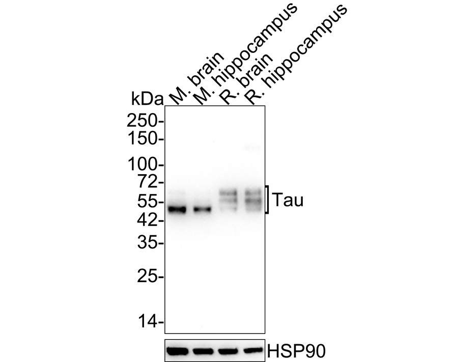
Tau Recombinant Rabbit Monoclonal Antibody [SZ03-03]
Application: WB,IHC-P,IP,IHC-Fr
Reactivity: Human,Mouse,Rat
Conjugate: unconjugated
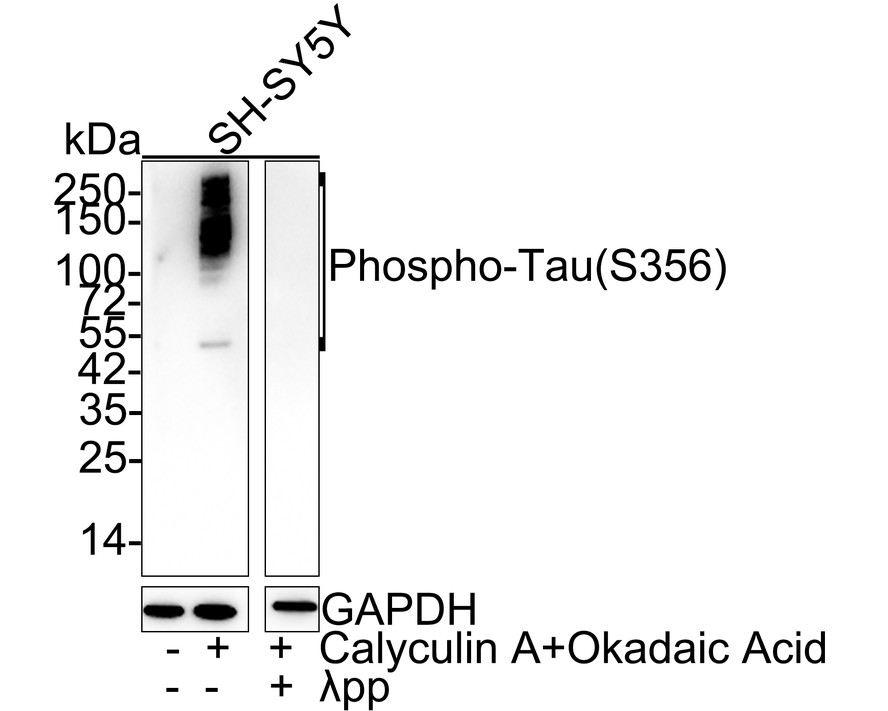
Phospho-Tau (S356) Recombinant Rabbit Monoclonal Antibody [JE46-86]
Application: WB
Reactivity: Human,Mouse,Rat
Conjugate: unconjugated
Tau Recombinant Rabbit Monoclonal Antibody [SD205-09]
Application: WB,IF-Cell,IF-Tissue,IHC-P,FC,IHC-Fr
Reactivity: Human,Mouse,Rat,Cynomolgus monkey,Pig
Conjugate: unconjugated
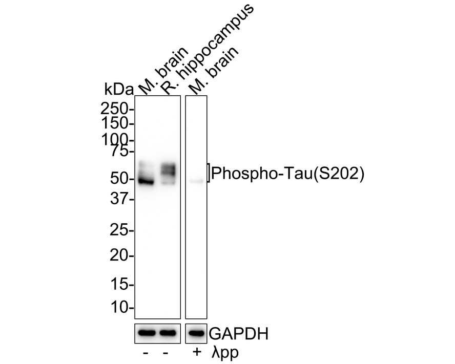
Phospho-Tau (S202) Recombinant Rabbit Monoclonal Antibody [JE66-81]
Application: WB
Reactivity: Human,Mouse,Rat
Conjugate: unconjugated
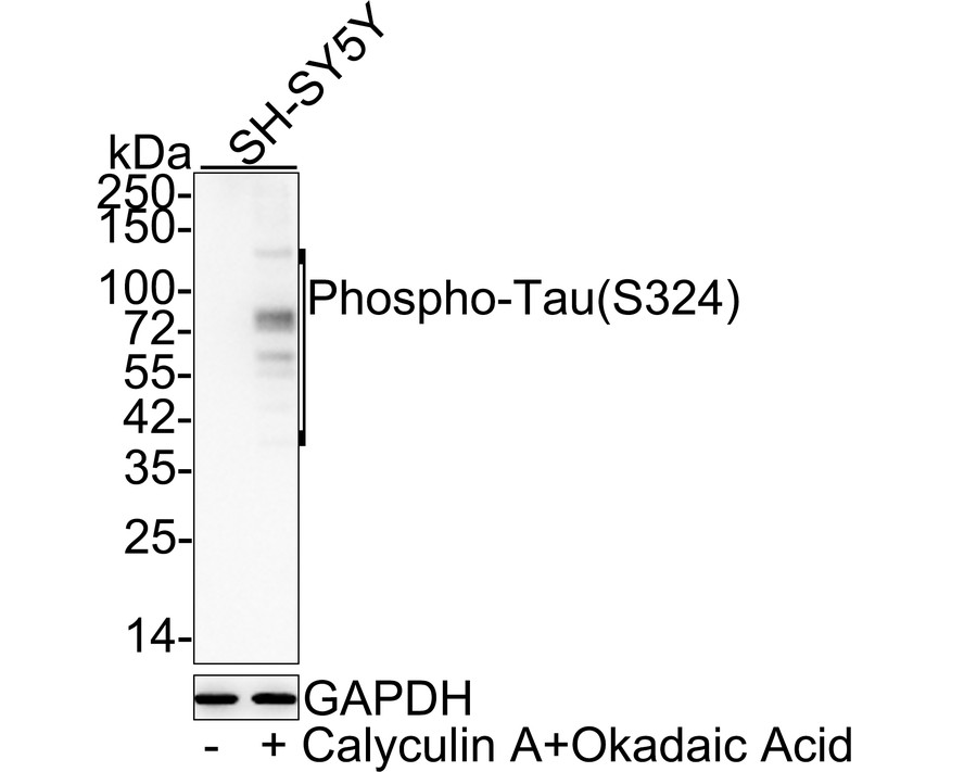
Phospho-Tau (S324) Recombinant Rabbit Monoclonal Antibody [JE30-73]
Application: WB
Reactivity: Human,Mouse,Rat
Conjugate: unconjugated





 浙公网安备 33019202000643号