IL-1 beta Recombinant Mouse Monoclonal Antibody [A7F7-R]
Rmb: 1500
概述
产品名称
IL-1 beta Recombinant Mouse Monoclonal Antibody [A7F7-R]
抗体类型
Recombinant Mouse Monoclonal Antibody
免疫原
Recombinant protein within Human IL-1 beta aa 117-269 / 269.
种属反应性
Human, Mouse, Rat
验证应用
WB, ELISA, IHC-P, IF-Tissue
靶点分子量
Predicted band size: 31 kDa
阳性对照
THP-1 treated with 80nM TPA overnight then replaced with 100ng/mL LPS for 6 hours add 300ng/mL BFA for last 3 hours cell lysate, human tonsil tissue.
偶联
unconjugated
克隆号
A7F7-R
RRID
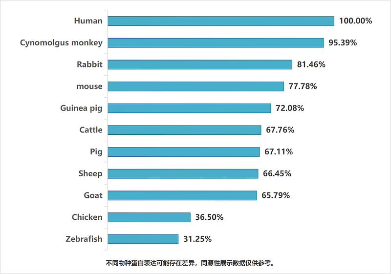
同源性数据
产品特性
形态
Liquid
浓度
存放说明
Shipped at 4℃. Store at +4℃ short term (1-2 weeks). Store at -20℃ long term.
存储缓冲液
PBS (pH7.4), 0.1% BSA, 40% Glycerol. Preservative: 0.05% Sodium Azide.
亚型
IgG1
纯化方式
Protein A affinity purified.
应用稀释度
-
WB
-
1:1,000-1:2,000
-
ELISA
-
1:20,000
-
IHC-P
-
1:100
-
IF-Tissue
-
1:200
靶点
功能
The protein encoded by this gene is a member of the interleukin 1 cytokine family. This cytokine is produced by activated macrophages as a proprotein, which is proteolytically processed to its active form by caspase 1 (CASP1/ICE). This cytokine is an important mediator of the inflammatory response, and is involved in a variety of cellular activities, including cell proliferation, differentiation, and apoptosis. The induction of cyclooxygenase-2 (PTGS2/COX2) by this cytokine in the central nervous system (CNS) is found to contribute to inflammatory pain hypersensitivity. Similarly, IL-1B has been implicated in human osteoarthritis pathogenesis. Patients with severe Coronavirus Disease 2019 (COVID-19) present elevated levels of pro-inflammatory cytokines such as IL-1B in bronchial alveolar lavage fluid samples. The lung damage induced by the Severe acute respiratory syndrome coronavirus 2 (SARS-CoV-2) is to a large extent, a result of the inflammatory response promoted by cytokines such as IL-1B. This gene and eight other interleukin 1 family genes form a cytokine gene cluster on chromosome 2.
背景文献
1. Qiao J et al. Autologous platelet rich plasma injection can be effective in the management of osteoarthritis of the knee: impact on IL-1 beta, TNF-alpha, hs-CRP. J Orthop Surg Res. 2024 Oct
2. Vicens-Artés S et al. Effect of MP-AzeFlu in IL-1 beta-induced IL-6 and proinflammatory cytokines. Immunol Res. 2023 Jun
亚细胞定位
Extracellular exosome, Secreted, Lysosome, Cytosol.
别名
Catabolin antibody
H1 antibody
IFN beta inducing factor antibody
IL 1 antibody
IL 1 beta antibody
IL-1 beta antibody
IL1 antibody
IL1 BETA antibody
IL1B antibody
IL1B_HUMAN antibody
展开图片
-

☑ Cell treatment (CT)
Western blot analysis of IL-1 beta on different lysates with Mouse anti-IL-1 beta antibody (HA601036) at 1/2,000 dilution.
Lane 1: THP-1 cell lysate
Lane 2: THP-1 treated with 80nM TPA overnight then replaced with 100ng/mL LPS for 6 hours add 300ng/mL BFA for last 3 hours cell lysate
Lysates/proteins at 20 µg/Lane.
Predicted band size: 31 kDa
Observed band size: 35/17 kDa
Exposure time: 4 seconds; ECL: K1801;
4-20% SDS-PAGE gel.
Proteins were transferred to a PVDF membrane and blocked with 5% NFDM/TBST for 1 hour at room temperature. The primary antibody (HA601036) at 1/2,000 dilution was used in 5% NFDM/TBST at 4℃ overnight. Goat Anti-Mouse IgG - HRP Secondary Antibody (HA1006) at 1/50,000 dilution was used for 1 hour at room temperature. -

Western blot analysis of IL-1 beta on different proteins with Mouse anti-IL-1 beta antibody (HA601036) at 1/2,000 dilution.
Lane 1: Recombinant mouse IL-1 beta
Lane 2: Recombinant rat IL-1 beta
Lane 3: Recombinant human IL-1 beta
Lysates/proteins at 50 ng/Lane.
Exposure time: 30 seconds;
4-20% SDS-PAGE gel.
Proteins were transferred to a PVDF membrane and blocked with 5% NFDM/TBST for 1 hour at room temperature. The primary antibody (HA601036) at 1/2,000 dilution was used in 5% NFDM/TBST at room temperature for 2 hours. Goat Anti-Mouse IgG - HRP Secondary Antibody (HA1006) at 1/50,000 dilution was used for 1 hour at room temperature. -

Immunohistochemical analysis of paraffin-embedded human tonsil tissue with Mouse anti-IL-1 beta antibody (HA601036) at 1/100 dilution.
The section was pre-treated using heat mediated antigen retrieval with Tris-EDTA buffer (pH 9.0) for 20 minutes. The tissues were blocked in 1% BSA for 20 minutes at room temperature, washed with ddH2O and PBS, and then probed with the primary antibody (HA601036) at 1/100 dilution for 1 hour at room temperature. The detection was performed using an HRP conjugated compact polymer system. DAB was used as the chromogen. Tissues were counterstained with hematoxylin and mounted with DPX. -

IL-1 beta Antibody (HA601036) in indirect ELISA.
Indirect ELISA analysis of IL-1 beta was performed by coating wells of a 96-well plate with 50 µl per well of IL-1 beta antigen diluted in carbonate/bicarbonate buffer, at a concentration of 1 µg/mL overnight at 4℃. Wells of the plate were washed, blocked with StartingBlock blocking buffer, and incubated with 50 µl per well of a mouse IL-1 beta monoclonal antibody starting at a concentration of 20 µg/mL and serially diluting it to a concentration of 1.28 ng/mL for 2 hours at room temperature. The plate was washed and incubated with 50 µl per well of an HRP-conjugated goat anti-mouse IgG secondary antibody at a dilution of 1:10,000 for one hour at room temperature. Detection was performed using an Ultra TMB Substrate for 5 minutes at room temperature in the dark. The reaction was stopped with sulfuric acid and absorbances were read on a spectrophotometer at 450 nm. -

Application: IF-Tissue
Species: Human
Site: tonsil
Sample: Paraffin-embedded section
Antibody concentration: 1/200
请注意: All products are "FOR RESEARCH USE ONLY AND ARE NOT INTENDED FOR DIAGNOSTIC OR THERAPEUTIC USE"
引文
-
PSMB4/MHC-I Signaling in the Cerebrospinal Fluid-Contacting Nucleus Mediates Neuroinflammatory Depression in Mice
期刊: International Journal Of Molecular Sciences
DOI: 10.3390/ijms27114798
IF: 5.6
应用: WB
反应种属: Mouse
发表时间: 2026 May
-
Hydroxysafflor yellow a attenuates oxygen-glucose deprivation/ reoxygenation induced endothelial pyroptosis via PARP-1/NLRP3 pathway
期刊: Frontiers In Pharmacology
DOI: 10.3389/fphar.2026.1811680
IF: 5.4
应用: WB
反应种属: Rat
发表时间: 2026 May
-
Microbiota-Derived SCFAs Mediate the Synergistic Antidepressant Effects of Dajianzhong Decoction and Ketamine via FFAR2-NLRP3-IL-1β Signaling
期刊: Pharmaceuticals
DOI: 10.3390/ph19060877
IF: 5.7
应用: WB
反应种属: Mouse
发表时间: 2026 May
-
Ciclopirox Olamine Inhibits the NLRP3 Inflammasome to Alleviate Inflammatory Diseases
期刊: Advanced Science
DOI: 10.1002/advs.75704
IF: 14.1
应用: WB
反应种属: Human,Mouse
发表时间: 2026 May
-
ANGPTL2 inhibits macrophage pyroptosis and alleviates rheumatoid arthritis progression by regulating mitophagy via IGFBP5
期刊: Cell Death & Disease
DOI: 10.1038/s41419-026-08537-z
IF: 9.6
应用: WB
反应种属: Mouse
发表时间: 2026 Mar
-
Electroacupuncture Ameliorates Cyclophosphamide-Induced Ovarian Impairment in Rats With Diminished Ovarian Reserve and is Associated With Th17/Treg-Related Immune Modulation
期刊: Mediators of Inflammation
DOI: 10.1155/mi/1067286
IF: 4.2
应用: WB
反应种属: Rat
发表时间: 2026 Mar
-
ROS-NLRP3 participates in the pyroptosis response of excretory-secretory products from protoscoleces of Echinococcus granulosus in hepatocytes
期刊: Scientific Reports
DOI: 10.1038/s41598-026-45127-7
IF: 3.9
应用: WB
反应种属: Mouse
发表时间: 2026 Mar
-
SIRT3 mediates mitochondrial protection and attenuates mtROS-TXNIP-NLRP3 signaling activation in dry eye disease
期刊: Experimental Eye Research
DOI: 10.1016/j.exer.2026.110960
IF: 2.7
应用: WB
反应种属: Mouse,Human
发表时间: 2026 Mar
-
APOE-mediated lipid transport prevents C1q-related synaptic loss after spinal cord injury via attenuating macrophage lipid stress
期刊: Journal Of Neuroinflammation
DOI: 10.1186/s12974-026-03756-9
IF: 10.1
应用: WB,IF-Tissue
反应种属: mouse
发表时间: 2026 Mar
-
TRIM25-mediated ubiquitination of KEAP1 and NOX4 underpins the antifibrotic efficacy of sophoricoside in silicosis
期刊: International Journal of Biological Macromolecules
DOI: 10.1016/j.intimp.2026.116905
IF: 8.4
应用: IHC-P,IF-Cell,IF-Tissue,WB
反应种属: Mouse
发表时间: 2026 Jun
-
Immunotherapy-induced sialadenitis: sjögren’s syndrome or a new sialadenitis
期刊: Frontiers In Immunology
DOI: 10.3389/fimmu.2026.1755419
IF: 5.9
应用: WB
反应种属: Human,Mouse
发表时间: 2026 Feb
-
Macrophage membrane-biomimetic nanovehicles delivering amygdalin halt pyroptosis propagation and alleviate endometriosis-associated neuropathic pain
期刊: Materials Today Bio
DOI: 10.1016/j.mtbio.2026.103141
IF: 10.2
应用: WB
反应种属: Mouse,Human
发表时间: 2026 Apr
-
Role of Cellular-Ferroptosis-Mediated HMGB1 Nuclear Translocation in Indium-Tin-Oxide-Nanoparticle-Induced Inflammatory Lung Injury
期刊: Environment & Health
DOI: 10.1021/envhealth.5c00667
IF: 6.3
应用: IHC,IF-tissue
反应种属: Mouse
发表时间: 2026 Apr
-
Programmable Metal-Nucleic Acid Biomineralized Hydrogel for Infected Wound Healing via Mitochondrial Regulation
期刊: Acta Biomaterialia
DOI: 10.1016/j.actbio.2026.04.004
IF: 9.6
应用: WB
反应种属: Mouse,Human
发表时间: 2026 Apr
-
New Cyclic Lipopeptide Compounds for Efficiently Inhibiting Cellular Inflammation and Collagen-Induced Arthritis in Mice
期刊: Journal Of Medicinal Chemistry
DOI: 10.1021/acs.jmedchem.5c02095
IF: 6.8
应用: IHC
反应种属: Mouse
发表时间: 2025 Sept
-
Key copper homeostasis genes and inflammatory mechanisms in ischemic stroke: A bioinformatics and experimental study
期刊: Functional & Integrative Genomics
DOI: 10.1007/s10142-025-01692-0
IF: 3.1
应用: WB
反应种属: Rat
发表时间: 2025 Sept
-
Study of Lycium barbarum in the Treatment of Osteoradionecrosis of the Jaw: An Integration of Network Pharmacology With Experimental Validation
期刊: International Journal of Pharmacology
DOI: 10.31083/IJP44912
IF: 0.2
应用: IHC
反应种属: Rat
发表时间: 2025 Oct
-
CircCdh7 induces astrogliosis and neuroinflammation to trigger hypertensive effects in the rostral ventrolateral medulla
期刊: Cell & Bioscience
DOI: 10.1186/s13578-025-01480-0
IF: 6.2
应用: WB
反应种属: Rat
发表时间: 2025 Oct
-
Cardioprotective Effect and Mechanism of Panaxadiol Saponin Component in Radiation-Induced Heart Disease in Mice
期刊: Chinese Journal Of Integrative Medicine
DOI: 10.1007/s11655-025-4144-y
IF: 2.5
应用: WB
反应种属: Mouse
发表时间: 2025 Oct
-
Human cytomegalovirus protein US18 promotes NLRP3 inflammasome activation
期刊: Biochemical And Biophysical Research Communications
DOI: 10.1016/j.bbrc.2025.152110
IF: 2.5
应用: WB
反应种属: Human
发表时间: 2025 May
-
Evodiamine Promotes Autophagy and Alleviates Oxidative Stress in Dry Eye Disease Through the p53/mTOR Pathway
期刊: Investigative Ophthalmology & Visual Science
DOI: 10.1167/iovs.66.3.44
IF: 4.7
应用: WB
反应种属: Mouse,Human
发表时间: 2025 Mar
-
Facile Nanocomposite Hydrogel Scaffold with Sustained Drug Release and Osteo-Immunomodulatory Effects to Enhance Bone Regeneration
期刊: ACS Applied Materials & Interfaces
DOI: 10.1021/acsami.4c20390
IF: 8.3
应用: IHC-P
反应种属: Rat
发表时间: 2025 Mar
-
Multi-omics joint analysis: Pachymic acid ameliorated non-alcoholic fatty liver disease by regulating gut microbiota
期刊: Food Research International
DOI: 10.1016/j.foodres.2025.116178
IF: 7
应用: WB
反应种属: Mouse
发表时间: 2025 Mar
-
Elevated Acetylation of MFN2 is Accompanied by the Disruption of Mitochondrial Energy Metabolism and Inflammation in a Mouse Model of Depression
期刊: MOLECULAR NEUROBIOLOGY
DOI: 10.1007/s12035-025-05222-8
IF: 4.3
应用: WB
反应种属: Mouse
发表时间: 2025 Jul
-
Aldometanib mitigates LPS-induced Lung fibroblasts injury through the activation of AMPK-mediated signaling pathways
期刊: Toxicon
DOI: 10.1016/j.toxicon.2025.108489
IF: 2.4
应用: WB
反应种属: Human
发表时间: 2025 Jul
-
The regulatory role of FABP4 in macrophage polarization and lipid accumulation in silicosis
期刊: Cellular Signalling
DOI: 10.1016/j.cellsig.2025.112041
IF: 3.7
应用: WB
反应种属: Human
发表时间: 2025 Jul
-
Low-level laser therapy alleviates periodontal age-related inflammation in diabetic mice via the GLUT1/mTOR pathway
期刊: Journal Of Lasers In Medical Sciences
DOI:
IF: 2.1
应用: IHC-P
反应种属: Mouse
发表时间: 2024 Jan
Alternative Products

IL-1 beta Recombinant Rabbit Monoclonal Antibody [JJ087-3]
Application: WB,IF-Cell,IHC-P
Reactivity: Human
Conjugate: unconjugated

Human IL-1 beta Recombinant Rabbit Monoclonal Antibody [PS01-39]
Application: WB
Reactivity: Human
Conjugate: unconjugated

Human IL-1 beta Recombinant Rabbit Monoclonal Antibody [PS00-82]
Application: WB
Reactivity: Human
Conjugate: unconjugated
同靶点 & 同通路的产品

Human IL-1 beta Recombinant Rabbit Monoclonal Antibody [PSH16-86] - BSA and Azide free (Detector)
Application: ELISA(Det)
Reactivity: Human,Dog,Cynomolgus monkey
Conjugate: unconjugated

Biotin Conjugated Human IL-1 beta Recombinant Rabbit Monoclonal Antibody [PSH16-86] - Detector
Application: ELISA(Det),ELISA
Reactivity: Human,Dog,Cynomolgus monkey
Conjugate: Biotin

IL-1 beta Mouse Monoclonal Antibody [A7F9]
Application: WB,ELISA,IF-Cell
Reactivity: Human,Mouse,Rat
Conjugate: unconjugated

Human IL-1 beta Recombinant Rabbit Monoclonal Antibody [PSH16-87] - BSA and Azide free (Capture)
Application: ELISA(Cap)
Reactivity: Human,Dog
Conjugate: unconjugated
IL-1 beta Recombinant Rabbit Monoclonal Antibody [JJ087-3]
Application: WB,IF-Cell,IHC-P
Reactivity: Human
Conjugate: unconjugated

Human IL-1 beta Recombinant Rabbit Monoclonal Antibody [PSH16-85] - BSA and Azide free (Capture)
Application: ELISA(Cap)
Reactivity: Human,Dog,Cynomolgus monkey
Conjugate: unconjugated

IL-1 beta Recombinant Mouse Monoclonal Antibody [A7F7-R] - BSA and Azide free
Application: WB,ELISA,IHC-P,IF-Tissue
Reactivity: Human,Mouse,Rat
Conjugate: unconjugated

Human IL-1 beta Recombinant Rabbit Monoclonal Antibody [PS00-82] - BSA and Azide free (Detector)
Application: ELISA(Det)
Reactivity: Human,Dog
Conjugate: unconjugated

Human IL-1 beta Recombinant Rabbit Monoclonal Antibody [PS01-39] - BSA and Azide free (Capture)
Application: ELISA(Cap)
Reactivity: Human,Dog
Conjugate: unconjugated

 浙公网安备 33019202000643号