Beta Catenin Recombinant Rabbit Monoclonal Antibody [SA30-04]


Rmb: 980 特惠 1500
概述
产品名称
Beta Catenin Recombinant Rabbit Monoclonal Antibody [SA30-04]
抗体类型
Recombinant Rabbit monoclonal Antibody
免疫原
Synthetic peptide within human Beta-Catenin aa 30-70.
种属反应性
Human, Mouse, Rat
验证应用
WB, IHC-P, IF-Tissue, IP, mIHC, IF-Cell, IHC-Fr, FC
靶点分子量
Predicted band size: 85 kDa
阳性对照
SW480 cell lysate, A431 cell lysate, HT-29 cell lysates, NIH/3T3 cell lysate, rat brain tissue lysate, mouse pancreas, mouse liver, human colon cancer tissue, mouse colon tissue, A431, C6.
偶联
unconjugated
克隆号
SA30-04
RRID
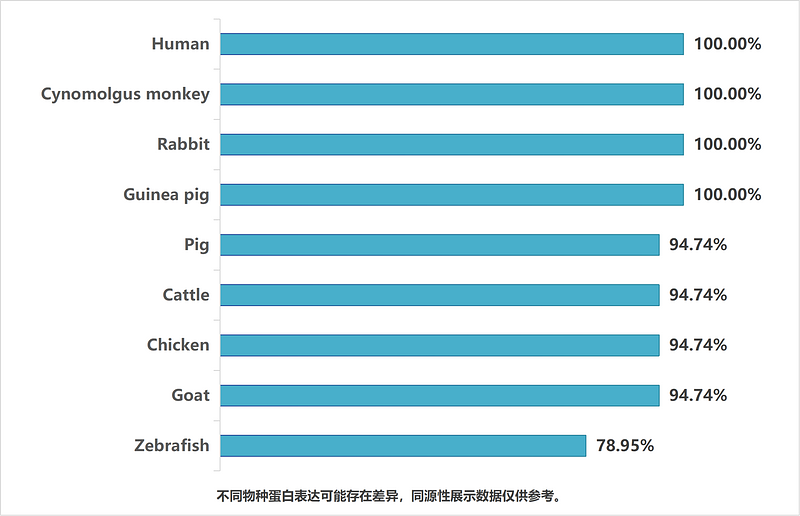
同源性数据
产品特性
形态
Liquid
浓度
存放说明
Shipped at 4℃. Store at +4℃ short term (1-2 weeks). Store at -20℃ long term.
存储缓冲液
1*TBS (pH7.4), 0.05% BSA, 40% Glycerol. Preservative: 0.05% Sodium Azide.
亚型
IgG
纯化方式
Protein A affinity purified.
应用稀释度
-
WB
-
1:1,000-1:2,000
-
IHC-P
-
1:200-1:1,000
-
IF-Tissue
-
1:100
-
IP
-
1-2μg/sample
-
mIHC
-
1:2,000
-
IF-Cell
-
1:100
-
IHC-Fr
-
1:200
-
FC
-
1:1,000
靶点
功能
Catenin beta-1, also known as beta-catenin (β-catenin), is a protein that in humans is encoded by the CTNNB1 gene. Beta-catenin is a dual function protein, involved in regulation and coordination of cell–cell adhesion and gene transcription. In humans, the CTNNB1 protein is encoded by the CTNNB1 gene. In Drosophila, the homologous protein is called armadillo. β-catenin is a subunit of the cadherin protein complex and acts as an intracellular signal transducer in the Wnt signaling pathway. Mutations and overexpression of β-catenin are associated with many cancers, including hepatocellular carcinoma, colorectal carcinoma, lung cancer, malignant breast tumors, ovarian and endometrial cancer. Alterations in the localization and expression levels of beta-catenin have been associated with various forms of heart disease, including dilated cardiomyopathy. β-catenin is regulated and destroyed by the beta-catenin destruction complex, and in particular by the adenomatous polyposis coli (APC) protein, encoded by the tumour-suppressing APC gene. Therefore, genetic mutation of the APC gene is also strongly linked to cancers, and in particular colorectal cancer resulting from familial adenomatous polyposis (FAP).
背景文献
1. Liu J et al. Wnt/beta-catenin signalling: function, biological mechanisms, and therapeutic opportunities. Signal Transduct Target Ther. 2022 Jan
2. Yu F et al. Wnt/beta-catenin signaling in cancers and targeted therapies. Signal Transduct Target Ther. 2021 Aug
序列相似性
Belongs to the beta-catenin family.
组织特异性
Expressed in several hair follicle cell types: basal and peripheral matrix cells, and cells of the outer and inner root sheaths. Expressed in colon. Present in cortical neurons (at protein level). Expressed in breast cancer tissues (at protein level).
翻译后修饰
Phosphorylation at Ser-552 by AMPK promotes stabilizion of the protein, enhancing TCF/LEF-mediated transcription (By similarity). Phosphorylation by GSK3B requires prior phosphorylation of Ser-45 by another kinase. Phosphorylation proceeds then from Thr-41 to Ser-37 and Ser-33. Phosphorylated by NEK2. EGF stimulates tyrosine phosphorylation. Phosphorylation on Tyr-654 decreases CDH1 binding and enhances TBP binding. Phosphorylated on Ser-33 and Ser-37 by HIPK2 and GSK3B, this phosphorylation triggers proteasomal degradation. Phosphorylation on Ser-191 and Ser-246 by CDK5. Phosphorylation by CDK2 regulates insulin internalization. Phosphorylation by PTK6 at Tyr-64, Tyr-142, Tyr-331 and/or Tyr-333 with the predominant site at Tyr-64 is not essential for inhibition of transcriptional activity.; Ubiquitinated by the SCF(BTRC) E3 ligase complex when phosphorylated by GSK3B, leading to its degradation. Ubiquitinated by a E3 ubiquitin ligase complex containing UBE2D1, SIAH1, CACYBP/SIP, SKP1, APC and TBL1X, leading to its subsequent proteasomal degradation. Ubiquitinated and degraded following interaction with SOX9 (By similarity).; S-nitrosylation at Cys-619 within adherens junctions promotes VEGF-induced, NO-dependent endothelial cell permeability by disrupting interaction with E-cadherin, thus mediating disassembly adherens junctions.; O-glycosylation at Ser-23 decreases nuclear localization and transcriptional activity, and increases localization to the plasma membrane and interaction with E-cadherin CDH1.; Deacetylated at Lys-49 by SIRT1.
亚细胞定位
Cytoplasm, Nucleus, Cell membrane, Cell junction
别名
β Catenin
Beta catenin antibody
Beta-catenin antibody
Cadherin associated protein antibody
Catenin (cadherin associated protein), beta 1, 88kDa antibody
Catenin beta 1 antibody
Catenin beta-1 antibody
CATNB antibody
CHBCAT antibody
CTNB1_HUMAN antibody
展开图片
-

Western blot analysis of Beta Catenin on different lysates with Rabbit anti-Beta Catenin antibody (ET1601-5) at 1/2,000 dilution.
Lane 1: SW480 cell lysate
Lane 2: A431 cell lysate
Lane 3: HT-29 cell lysate
Lysates/proteins at 20 µg/Lane.
Predicted band size: 85 kDa
Observed band size: 100 kDa
Exposure time: 3 minutes;
4-20% SDS-PAGE gel.
Proteins were transferred to a PVDF membrane and blocked with 5% NFDM/TBST for 1 hour at room temperature. The primary antibody (ET1601-5) at 1/2,000 dilution was used in 5% NFDM/TBST at 4℃ overnight. Goat Anti-Rabbit IgG - HRP Secondary Antibody (HA1001) at 1/50,000 dilution was used for 1 hour at room temperature. -

☑ Knockdown (KD)
Western blot analysis of Beta Catenin on different lysates with Rabbit anti-Beta Catenin antibody (ET1601-5) at 1/2,000 dilution.
Lane 1: THP-1-si NT cell lysate
Lane 2: THP-1-si Beta Catenin cell lysate
Lysates/proteins at 10 µg/Lane.
Predicted band size: 85 kDa
Observed band size: 85 kDa
Exposure time: 3 minutes; ECL: K1801;
4-20% SDS-PAGE gel.
Proteins were transferred to a PVDF membrane and blocked with 5% NFDM/TBST for 1 hour at room temperature. The primary antibody (ET1601-5) at 1/2,000 dilution was used in primary antibody dilution at 4℃ overnight. Goat Anti-Rabbit IgG - HRP Secondary Antibody (HA1001) at 1/50,000 dilution was used for 1 hour at room temperature. -

☑ Knockdown (KD)
Western blot analysis of Beta Catenin on different lysates with Rabbit anti-Beta Catenin antibody (ET1601-5) at 1/2,000 dilution.
Lane 1: HAP1-parental cell lysate
Lane 2: HAP1-Beta Catenin KD cell lysate
Lysates/proteins at 10 µg/Lane.
Predicted band size: 85 kDa
Observed band size: 85 kDa
Exposure time: 40 seconds; ECL: K1801;
4-20% SDS-PAGE gel.
Proteins were transferred to a PVDF membrane and blocked with 5% NFDM/TBST for 1 hour at room temperature. The primary antibody (ET1601-5) at 1/2,000 dilution was used in K1803 at 4℃ overnight. Goat Anti-Rabbit IgG - HRP Secondary Antibody (HA1001) at 1/50,000 dilution was used for 1 hour at room temperature. -

Fluorescence multiplex immunohistochemical analysis of mouse pancreas (Formalin/PFA-fixed paraffin-embedded sections). Panel A: the merged image of anti-β-catenin (ET1601-5, Red), anti-Glucagon (ET1702-20, Green), anti-Insulin (ET1601-12, White), anti-CK19 (ET1601-6, Magenta) and anti-aSMA (ET1607-53, Yellow) on mouse pancreas. HRP Conjugated UltraPolymer Goat Polyclonal Antibody HA1119/HA1120 was used as a secondary antibody. The immunostaining was performed with the Sequential Immuno-staining Kit (IRISKit™MH010101, www.luminiris.cn). The section was incubated in five rounds of staining: in the order of ET1601-5 (1/2,000 dilution), ET1702-20 (1/6,000 dilution), ET1601-12 (1/8,000 dilution), ET1601-6 (1/5,000 dilution) and ET1607-53 (1/10,000 dilution) for 20 mins at room temperature. Each round was followed by a separate fluorescent tyramide signal amplification system. Heat mediated antigen retrieval with Tris-EDTA buffer (pH 9.0) for 30 mins at 95℃. DAPI (blue) was used as a nuclear counter stain. Image acquisition was performed with Olympus VS200 Slide Scanner.
-

Fluorescence multiplex immunohistochemical analysis of mouse liver (Formalin/PFA-fixed paraffin-embedded sections). Panel A: the merged image of anti-β-catenin (ET1601-5, Tangerine), anti-αSMA (ET1607-53, Yellow), anti-SOX9 (ET1611-56, Green), anti-Albumin (ET1702-55, Cyan) anti-GS (EM1902-39, Magenta) and anti-CK19 (ET1601-6, Orange) on mouse liver. HRP Conjugated UltraPolymer Goat Polyclonal Antibody HA1119/HA1120 was used as a secondary antibody. The immunostaining was performed with the Sequential Immuno-staining Kit (IRISKit™MH010101, www.luminiris.cn). The section was incubated in six rounds of staining: in the order of ET1601-5 (1/2,000 dilution), ET1607-53 (1/3,000 dilution), ET1611-56 (1/1,500 dilution), ET1702-55 (1/3,000 dilution), EM1902-39 (1/2,000 dilution) and ET1601-6 (1/3,000 dilution) for 20 mins at room temperature. Each round was followed by a separate fluorescent tyramide signal amplification system. Heat mediated antigen retrieval with Tris-EDTA buffer (pH 9.0) for 30 mins at 95℃. DAPI (blue) was used as a nuclear counter stain. Image acquisition was performed with Olympus VS200 Slide Scanner.
-

Fluorescence multiplex immunohistochemical analysis of mouse pancreas (Formalin/PFA-fixed paraffin-embedded sections). Panel A: the merged image of anti-Beta Catenin (ET1601-5, Violet), anti-Glucagon (ET1702-20, Green) and anti-Insulin (ET1601-12, White) on pancreas. HRP Conjugated UltraPolymer Goat Polyclonal Antibody HA1119/HA1120 was used as a secondary antibody. The immunostaining was performed with the Sequential Immuno-staining Kit (IRISKit™MH010101, www.luminiris.cn). The section was incubated in three rounds of staining: in the order of ET1601-5 (1/2,000 dilution), ET1702-20 (1/6,000 dilution) and ET1601-12 (1/8,000 dilution) for 20 mins at room temperature. Each round was followed by a separate fluorescent tyramide signal amplification system. Heat mediated antigen retrieval with Tris-EDTA buffer (pH 9.0) for 30 mins at 95℃. DAPI (blue) was used as a nuclear counter stain. Image acquisition was performed with Zeiss Observer 7 Inverted Fluorescence Microscope.
-

Fluorescence multiplex immunohistochemical analysis of mouse liver (Formalin/PFA-fixed paraffin-embedded sections). Panel A: the merged image of anti-Th (ET1611-12, Green), anti-HNF4a (HA721006, Magenta), anti-CK19 (ET1601-6, Cyan), anti-α-sma (ET1607-53, Red) and anti-β-catenin (ET1601-5, Yellow) on liver. HRP Conjugated UltraPolymer Goat Polyclonal Antibody HA1119/HA1120 was used as a secondary antibody. The immunostaining was performed with the Sequential Immuno-staining Kit (IRISKit™MH010101, www.luminiris.cn). The section was incubated in three rounds of staining: in the order of ET1611-12 (1/1,000 dilution), HA721006 (1/2,000 dilution), ET1601-6 (1/3,000 dilution), ET1607-53 (1/10,000 dilution) and ET1601-5 (1/2,000 dilution) for 20 mins at room temperature. Each round was followed by a separate fluorescent tyramide signal amplification system. Heat mediated antigen retrieval with Tris-EDTA buffer (pH 9.0) for 30 mins at 95℃. DAPI (blue) was used as a nuclear counter stain. Image acquisition was performed with Olympus VS200 Slide Scanner.
-

Immunohistochemical analysis of paraffin-embedded human colon cancer tissue with Rabbit anti-Beta Catenin antibody (ET1601-5) at 1/1,000 dilution.
The section was pre-treated using heat mediated antigen retrieval with sodium citrate buffer (pH 6.0) (high pressure) for 2 minutes. The tissues were blocked in 1% BSA for 20 minutes at room temperature, washed with ddH2O and PBS, and then probed with the primary antibody (ET1601-5) at 1/1,000 dilution for 1 hour at room temperature. The detection was performed using an HRP conjugated compact polymer system. DAB was used as the chromogen. Tissues were counterstained with hematoxylin and mounted with DPX. -

Immunohistochemical analysis of paraffin-embedded mouse colon tissue with Rabbit anti-Beta Catenin antibody (ET1601-5) at 1/1,000 dilution.
The section was pre-treated using heat mediated antigen retrieval with sodium citrate buffer (pH 6.0) (high pressure) for 2 minutes. The tissues were blocked in 1% BSA for 20 minutes at room temperature, washed with ddH2O and PBS, and then probed with the primary antibody (ET1601-5) at 1/1,000 dilution for 1 hour at room temperature. The detection was performed using an HRP conjugated compact polymer system. DAB was used as the chromogen. Tissues were counterstained with hematoxylin and mounted with DPX. -

Immunohistochemical analysis of paraffin-embedded rat brain tissue with Rabbit anti-Beta Catenin antibody (ET1601-5) at 1/200 dilution.
The section was pre-treated using heat mediated antigen retrieval with sodium citrate buffer (pH 6.0) (high pressure) for 2 minutes. The tissues were blocked in 1% BSA for 20 minutes at room temperature, washed with ddH2O and PBS, and then probed with the primary antibody (ET1601-5) at 1/200 dilution for 1 hour at room temperature. The detection was performed using an HRP conjugated compact polymer system. DAB was used as the chromogen. Tissues were counterstained with hematoxylin and mounted with DPX. -

Immunofluorescence analysis of paraffin-embedded human colon cancer tissue labeling Beta Catenin with Rabbit anti-Beta Catenin antibody (ET1601-5) at 1/100 dilution.
The section was pre-treated using heat mediated antigen retrieval with Tris-EDTA buffer (pH 9.0) for 20 minutes. The tissues were blocked in 10% negative goat serum for 1 hour at room temperature, washed with PBS, and then probed with the primary antibody (ET1601-5, green) at 1/100 dilution overnight at 4 ℃, washed with PBS. Goat Anti-Rabbit IgG H&L (iFluor™ 488, HA1121) was used as the secondary antibody at 1/1,000 dilution. Nuclei were counterstained with DAPI (blue). -

Immunofluorescence analysis of paraffin-embedded mouse colon tissue labeling Beta Catenin with Rabbit anti-Beta Catenin antibody (ET1601-5) at 1/100 dilution.
The section was pre-treated using heat mediated antigen retrieval with Tris-EDTA buffer (pH 9.0) for 20 minutes. The tissues were blocked in 10% negative goat serum for 1 hour at room temperature, washed with PBS, and then probed with the primary antibody (ET1601-5, green) at 1/100 dilution overnight at 4 ℃, washed with PBS. Goat Anti-Rabbit IgG H&L (iFluor™ 488, HA1121) was used as the secondary antibody at 1/1,000 dilution. Nuclei were counterstained with DAPI (blue). -

Immunocytochemistry analysis of A431 cells labeling Beta Catenin with Rabbit anti-Beta Catenin antibody (ET1601-5) at 1/100 dilution.
Cells were fixed in 4% paraformaldehyde for 20 minutes at room temperature, permeabilized with 0.1% Triton X-100 in PBS for 5 minutes at room temperature, then blocked with 1% BSA in 10% negative goat serum for 1 hour at room temperature. Cells were then incubated with Rabbit anti-Beta Catenin antibody (ET1601-5) at 1/100 dilution in 1% BSA in PBST overnight at 4 ℃. Goat Anti-Rabbit IgG H&L (iFluor™ 488, HA1121) was used as the secondary antibody at 1/1,000 dilution. PBS instead of the primary antibody was used as the secondary antibody only control. Nuclear DNA was labelled in blue with DAPI. Beta tubulin (M1305-2, red) was stained at 1/100 dilution overnight at +4℃. Goat Anti-Mouse IgG H&L (iFluor™ 594, HA1126) was used as the secondary antibody at 1/1,000 dilution. -

Immunocytochemistry analysis of C6 cells labeling Beta Catenin with Rabbit anti-Beta Catenin antibody (ET1601-5) at 1/100 dilution.
Cells were fixed in 4% paraformaldehyde for 20 minutes at room temperature, permeabilized with 0.1% Triton X-100 in PBS for 5 minutes at room temperature, then blocked with 1% BSA in 10% negative goat serum for 1 hour at room temperature. Cells were then incubated with Rabbit anti-Beta Catenin antibody (ET1601-5) at 1/100 dilution in 1% BSA in PBST overnight at 4 ℃. Goat Anti-Rabbit IgG H&L (iFluor™ 488, HA1121) was used as the secondary antibody at 1/1,000 dilution. PBS instead of the primary antibody was used as the secondary antibody only control. Nuclear DNA was labelled in blue with DAPI. Beta tubulin (M1305-2, red) was stained at 1/100 dilution overnight at +4℃. Goat Anti-Mouse IgG H&L (iFluor™ 594, HA1126) was used as the secondary antibody at 1/1,000 dilution. -

Immunofluorescence analysis of frozen mouse colon tissue with Rabbit anti-Beta Catenin antibody (ET1601-5) at 1/200 dilution.
The section was pre-treated using heat mediated antigen retrieval with sodium citrate buffer (pH 6.0) for about 2 minutes in microwave oven. The tissues were blocked in 10% negative goat serum for 1 hour at room temperature, washed with PBS, and then probed with the primary antibody (ET1601-5, green) at 1/200 dilution overnight at 4 ℃, washed with PBS. Goat Anti-Rabbit IgG H&L (iFluor™ 488, HA1121) was used as the secondary antibody at 1/1,000 dilution. Nuclei were counterstained with DAPI (blue). -

Beta Catenin was immunoprecipitated from 0.2 mg rat brain tissue lysate with ET1601-5 at 2 µg/10 µl beads. Western blot was performed from the immunoprecipitate using ET1601-5 at 1/1,000 dilution. Anti-Rabbit IgG for IP Nano-secondary antibody (NBI01H) at 1/5,000 dilution was used for 1 hour at room temperature.
Lane 1: Rat brain tissue lysate (input)
Lane 2: ET1601-5 IP in rat brain tissue lysate
Lane 3: Rabbit IgG instead of ET1601-5 in rat brain tissue lysate
Blocking/Dilution buffer: 5% NFDM/TBST
Exposure time: 2 seconds; ECL: K1801 -

Flow cytometric analysis of A431 cells labeling Beta Catenin.
Cells were fixed and permeabilized. Then stained with the primary antibody (ET1601-5, 1/1,000) (red) compared with Rabbit IgG Isotype Control (green). After incubation of the primary antibody at +4℃ for an hour, the cells were stained with a iFluor™ 488 conjugate-Goat anti-Rabbit IgG Secondary antibody (HA1121) at 1/1,000 dilution for 30 minutes at +4℃. Unlabelled sample was used as a control (cells without incubation with primary antibody; black).
请注意: All products are "FOR RESEARCH USE ONLY AND ARE NOT INTENDED FOR DIAGNOSTIC OR THERAPEUTIC USE"
引文
-
PFAS exacerbates diabetic wound healing by targeting GRP94 glycosylation, antagonized by ligustilide
期刊: Journal Of Hazardous Materials
DOI: 10.1016/j.jhazmat.2026.142464
IF: 10.6
应用: WB
反应种属: Rat
发表时间: 2026 May
-
Urolithin A supplementation alleviates osteogenic disfunction and promotes bone fracture healing in inflammatory environments
期刊: Food & Nutrition Research
DOI: 10.29219/fnr.v70.13033
IF: 4.5
应用: WB
反应种属: Mouse
发表时间: 2026 May
-
LINC01996 suppresses non-small cell lung cancer proliferation and metastasis by orchestrating the miR-12115/CNRIP1/Ras signaling axis
期刊: Clinical & Experimental Metastasis
DOI: 10.1007/s10585-026-10399-w
IF: 3.2
应用: WB
反应种属: Human
发表时间: 2026 Mar
-
Magnesium Deficiency Accelerates Gut Aging and Increases Susceptibility to Colitis
期刊: Aging Cell
DOI: 10.1111/acel.70446
IF: 7.1
应用: WB
反应种属: Mouse
发表时间: 2026 Mar
-
Potential mechanisms underlying kaempferol-promoted osteoblast proliferation and osteogenic differentiation: A network pharmacology and experimental validation study
期刊: Letters in Drug Design & Discovery
DOI: 10.1016/j.lddd.2026.100432
IF: 1.6
应用: WB
反应种属: Mouse
发表时间: 2026 Jun
-
IGF2BP3 promotes epithelial ovarian cancer progression by regulating FASN expression
期刊: Scientific Reports
DOI: 10.1038/s41598-026-57059-3
IF: 4.9
应用: WB
反应种属: Human
发表时间: 2026 Jun
-
Naringenin inhibits endothelial-mesenchymal transition and alleviates myocardial fibrosis in rats by regulating the AKT/GSK3β/β-catenin pathway
期刊: Molecular And Cellular Biochemistry
DOI: 10.1007/s11010-026-05603-0
IF: 4.7
应用: WB
反应种属: Rat
发表时间: 2026 Jun
-
SPP1 Drives Colorectal Cancer Liver Metastasis and Immunotherapy Resistance by Stimulating CXCL12 Production in Cancer-Associated Fibroblasts
期刊: Cancer Research
DOI: 10.1158/0008-5472.CAN-24-4916
IF: 16.6
应用: WB,IHC
反应种属: Human
发表时间: 2026 Jan
-
Mitofusin 2 Alleviates Liver Fibrogenesis by Suppressing β-Catenin Nuclear Translocation
期刊: Current Medical Science
DOI: 10.1007/s11596-026-00167-y
IF: 1.5
应用: WB
反应种属: Mouse
发表时间: 2026 Feb
-
MaR1 and NGF combine to inhibit autophagy through the GSK-3β/β-catenin pathway to promote sciatic nerve repair
期刊: Journal Of Translational Medicine
DOI: 10.1186/s12967-026-07804-z
IF: 7.5
应用: WB,IHC
反应种属: Rat
发表时间: 2026 Feb
-
Hippocampal expression of Wnt7a and β-catenin in depression: evidence from chronic unpredictable mild stress
期刊: Peer J
DOI: 10.7717/peerj.20837
IF: 2.4
应用: WB
反应种属: Rat
发表时间: 2026 Feb
-
Rhapontici Radix Extract Inhibits Colorectal Intraepithelial Neoplasia by Regulating the YAP/PI3K-AKT Signaling Pathway: Evidence from Animal Models, Organoids, and Cytological Studies
期刊: Biomedicines
DOI: 10.3390/biomedicines14050956
IF: 3.9
应用: IF-cell,WB
反应种属: Mouse
发表时间: 2026 Apr
-
Ultrasound-responsive piezoelectric hydrogels accelerate bone defect repair by regulating mitochondrial OXPHOS via activating AKT/GSK3β/β-catenin signaling axis
期刊: Chemical Engineering Journal
DOI: 10.1016/j.cej.2025.168176
IF: 13.2
应用: WB
反应种属: Mouse
发表时间: 2025 Sept
-
Mechanistic studies of miR-582-3p targeting of PTPRCAP affecting lung adenocarcinoma via the Wnt/β-catenin pathway
期刊: Frontiers in Oncology
DOI: 10.3389/fonc.2025.1652176
IF: 3.3
应用: WB
反应种属: Human
发表时间: 2025 Sept
-
Evodiamine Inhibits Colorectal Cancer by Downregulating ASS1 via Wnt/β-Catenin/c-MYC Pathway to Block Arginine Synthesis
期刊: Pharmaceuticals
DOI: 10.3390/ph18111736
IF: 4.8
应用: WB
反应种属: Mouse,Human
发表时间: 2025 Nov
-
Sustainability of functional hair follicle activity: Impact of spatially anchored multifunctional tetrahedral framework nucleic acids
期刊: Bioactive Materials
DOI: 10.1016/j.bioactmat.2025.11.006
IF: 20.3
应用: WB
反应种属: Mouse,Rat
发表时间: 2025 Nov
-
Ganetespib as an inhibitor of heat shock protein 90 alleviates pulmonary fibrosis by inhibiting the Wnt/β-catenin signaling pathway
期刊: International Immunopharmacology
DOI: 10.1016/j.intimp.2025.115837
IF: 4.7
应用: WB
反应种属: Mouse
发表时间: 2025 Nov
-
Design of photoactivatable methylene blue-bufalin conjugate for GPX4-targeted degradation to induce ferroptosis-like death in breast cancer therapy
期刊: Bioorganic Chemistry
DOI: 10.1016/j.bioorg.2025.108629
IF: 4.5
应用: WB
反应种属: Human
发表时间: 2025 May
-
Mg–Sc alloy promotes angiogenesis–osteogenesis coupling and ameliorates steroid-induced osteonecrosis of the femoral head
期刊: Rare Metals
DOI: 10.1007/s12598-025-03322-x
IF: 9.6
应用: WB
反应种属: Mouse
发表时间: 2025 May
-
Decoupling of Density-Dependent Migration/Proliferation Dichotomy on Surface Potential Gradient
期刊: ACS Applied Materials & Interfaces
DOI: 10.1021/acsami.4c18787
IF: 8.3
应用: WB
反应种属: Human
发表时间: 2025 Mar
-
Tumor suppressor SLC9A2 inhibits colorectal cancer metastasis and reverses immunotherapy resistance by suppressing angiogenesis
期刊: Journal Of Experimental & Clinical Cancer Research
DOI: 10.1186/s13046-025-03422-7
IF: 12.8
应用: IHC
反应种属: Mouse,Human
发表时间: 2025 Jun
-
An unrecognized mechanism of self-protection in degenerating neurons mediated by astrocytic YAP through Wnts/β-catenin/EAAT2 signaling in C9orf72-poly-GA mice
期刊: Theranostics
DOI: 10.7150/thno.113599
IF: 13.3
应用: WB
反应种属: Mouse
发表时间: 2025 Jul
-
STING1 targets MYH9 to drive adipogenesis through the AKT/GSK3β/β-catenin pathway
期刊: Biochemical And Biophysical Research Communications
DOI:
IF: 2.5
应用: WB
反应种属: Mouse
发表时间: 2025 Jan
-
Magnetic Nanoactuator-Protein Fiber Coated Hydrogel Dressing for Well-Balanced Skin Wound Healing and Tissue Regeneration
期刊: ACS Applied Nano Materials
DOI:
IF: 15.8
应用: WB
反应种属: Mouse,Human
发表时间: 2025 Jan
-
STING1 targets MYH9 to drive adipogenesis through the AKT/GSK3β/β-catenin pathway
期刊: BIOCHEMICAL AND BIOPHYSICAL RESEARCH COMMUNICATIONS
DOI: 10.1016/j.bbrc.2025.151352
IF: 2.2
应用: WB
反应种属: Mouse
发表时间: 2025 Jan
-
Dandelion alleviates Helicobacter pylori associated gastritis by inhibiting proteolytic activity of HtrA
期刊: Journal Of Ethnopharmacology
DOI: 10.1016/j.jep.2025.121081
IF: 5.4
应用: WB
反应种属: Mouse
发表时间: 2025 Dec
-
Gut microbiota-derived indole-3-Acetic Acid attenuates skeletal fluorosis via AHR-mediated suppression of Wnt/β-Catenin signaling
期刊: Ecotoxicology And Environmental Safety
DOI: 10.1016/j.ecoenv.2025.119520
IF: 6.1
应用: WB
反应种属: Mouse
发表时间: 2025 Dec
-
The interaction between nanoscale MIL-53(Fe) and Fzd6 protein drives enhanced bone regeneration
期刊: Materials & Design
DOI: 10.1016/j.matdes.2025.115248
IF: 7.9
应用: WB,IHC
反应种属: Rat,Human
发表时间: 2025 Dec
-
KCTD10 inhibits lung cancer metastasis and angiogenesis via ubiquitin-mediated β-catenin degradation
期刊: Frontiers In Immunology
DOI: 10.3389/fimmu.2025.1630311
IF: 5.9
应用: WB
反应种属: Human,Mouse
发表时间: 2025 Aug
-
Treatment of rheumatoid arthritis via tetrahedral framework nucleic acid‑based efficient delivery of Jakinib: Synergistic anti-inflammatory and osteogenic effects
期刊: Chemical Engineering Journal
DOI: 10.1016/j.cej.2025.162405
IF: 13.3
应用: WB
反应种属: Rat
发表时间: 2025 Apr
-
Exosomes Extracted from Human Umbilical Cord MSCs Contribute to Osteoarthritic Cartilage and Chondrocytes Repair Through Enhancing Autophagy While Suppressing the Wnt/β-Catenin Pathway
期刊: Tissue Engineering And Regenerative Medicine
DOI: 10.1007/s13770-025-00716-x
IF: 4.4
应用: WB,IHC-P
反应种属: Human,Rat
发表时间: 2025 Apr
-
Identifying the signature of NAD+ metabolism-related genes for immunotherapy of gastric cancer
期刊: Heliyon
DOI: 10.1016/j.heliyon.2024.e38823
IF: 3.4
应用: WB
反应种属: Human
发表时间: 2024 Oct
-
Role and mechanism of Actein on condylar bone metabolism in APOE deletion-induced osteoporotic mice
期刊: Bone
DOI:
IF: 3.5
应用: IHC-P
反应种属: Mouse
发表时间: 2024 Oct
-
Reduced Proline-Rich Tyrosine Kinase 2 Promotes Tumor Metastasis by Activating Epithelial–Mesenchymal Transition in Colorectal Cancer
期刊: Digestive Diseases And Sciences
DOI:
IF: 2.5
应用: WB
反应种属: Human
发表时间: 2024 Oct
-
The important role of the Wnt/β-catenin signaling pathway in small molecules mediated gingival mesenchymal stem cells transdifferentiate into neuron-like cells
期刊: Archives Of Oral Biology
DOI:
IF: 2.2
应用: WB
反应种属: Human
发表时间: 2024 Oct
-
Family with sequence similarity 83, member A (FAM83A) inhibits ferroptosis via the Wnt/β-catenin pathway in lung squamous cell cancer
期刊: Cell Death Discovery
DOI: 10.1038/s41420-024-02101-4
IF: 6.1
应用: WB
反应种属: Human
发表时间: 2024 Jul
-
CISD2 downregulation participates in the ferroptosis process of human ovarian SKOV-3 cells through modulating the wild type p53-mediated GLS2/SAT1/SLC7A11 and Gpx4/TRF signaling pathway
期刊: Tissue & Cell
DOI:
IF: 2.7
应用: WB
反应种属: Human
发表时间: 2024 Jul
-
Cell-Surface GRP78-Targeted Chimeric Antigen Receptor T Cells Eliminate Lung Cancer Tumor Xenografts
期刊: International Journal Of Molecular Sciences
DOI:
IF: 5.6
应用: IF-cell
反应种属: Mouse
发表时间: 2024 Jan
-
LepR-expressing cells are a critical population in periodontal healing post periodontitis
期刊: Journal Of Bone And Mineral Research
DOI:
IF: 6.4
应用: WB
反应种属: Mouse
发表时间: 2024 Jan
-
Propranolol inhibits EMT and metastasis in breast cancer through miR-499-5p-mediated Sox6
期刊: Journal Of Cancer Research And Clinical Oncology
DOI: 10.1007/s00432-023-05599-w
IF: 2.7
应用: WB
反应种属: Mouse
发表时间: 2024 Jan
-
Hyperuricemia Induces Reproductive Damage in Male Rats through Zinc Homeostasis Imbalance and Oxidative Stress
期刊: Andrology
DOI:
IF: 2.5
应用: WB
反应种属: Rat
发表时间: 2024 Jan
-
Long non‐coding RNA LncTUG1 regulates favourable compression force‐induced cementocytes mineralization via PU. 1/TLR4/SphK1 signalling
期刊: Cell Proliferation
DOI:
IF: 8.5
应用: WB
反应种属: Mouse
发表时间: 2024 Feb
-
Age-, sex- and proximal-distal-resolved multi-omics identifies regulators of intestinal aging in non-human primates
期刊: Nature Aging
DOI:
IF: 16.6
应用: WB
反应种属: Mouse
发表时间: 2024 Feb
-
MicroRNA-29c-tetrahedral framework nucleic acids: Towards osteogenic differentiation of mesenchymal stem cells and bone regeneration in critical-sized calvarial defects
期刊: Cell Proliferation
DOI: 10.1111/cpr.13624
IF: 5.9
应用: WB
反应种属: Rat
发表时间: 2024 Feb
-
A Multifunctional miRNA Delivery System Based on Tetrahedral Framework Nucleic Acids for Regulating Inflammatory Periodontal Ligament Stem Cells and Attenuating Periodontitis Bone Loss
期刊: ACS Applied Materials & Interfaces
DOI:
IF: 8.3
应用: WB
反应种属: Rat
发表时间: 2024 Dec
-
Discovery of a Novel Non-invasive AR PROTAC Degrader for the Topical Treatment of Androgenetic Alopecia
期刊: Journal Of Medicinal Chemistry
DOI:
IF: 6.8
应用: WB
反应种属: Mouse
发表时间: 2024 Dec
-
Pulsed electromagnetic fields potentiate bone marrow mesenchymal stem cell chondrogenesis by regulating the Wnt/β-catenin signaling pathway
期刊: Journal Of Translational Medicine
DOI:
IF: 6.1
应用: WB
反应种属: Mouse
发表时间: 2024 Aug
-
CDC42‑mediated Wnt signaling facilitates odontogenic differentiation of DPCs during tooth root elongation
期刊: Stem Cell Research & Therapy
DOI:
IF: 7.5
应用: WB
反应种属: Mouse,Rat
发表时间: 2023 Sept
-
GAD1-mediated GABA elicits aggressive characteristics of human oral cancer cells
期刊: Biochemical And Biophysical Research Communications
DOI: 10.1016/j.bbrc.2023.09.041
IF: 3.1
应用:
反应种属:
发表时间: 2023 Sept
-
Co-targeting of ACK1 and KIT triggers additive anti-proliferative and-migration effects in imatinib-resistant gastrointestinal stromal tumors
期刊: Biochimica Et Biophysica Acta-Molecular Basis Of Disease
DOI:
IF: 6.2
应用: WB
反应种属: Human
发表时间: 2023 Jun
-
Knockdown of LMNA inhibits Akt/β-catenin-mediated cell invasion and migration in clear cell renal cell carcinoma cells
期刊: Cell Adhesion & Migration
DOI:
IF: 3.2
应用: WB
反应种属: Human
发表时间: 2023 Dec
-
Proliferation Inhibitory Activity of Quinones from Blaps rynchopetera Defense Secretion on Colorectal Tumor Cells
期刊: Chinese Journal Of Integrative Medicine
DOI:
IF: 2.9
应用: WB
反应种属: Human
发表时间: 2023 Aug
-
Circular RNA circRNA_0067934 promotes glioma development by modulating the microRNA miR-7/ Wnt/β-catenin axis
期刊: Bioengineered
DOI:
IF: 3.27
应用: WB,IF-cell
反应种属: Human
发表时间: 2022 Mar
-
Mechanisms of sphingosine-1-phosphate (S1P) signaling on excessive stress-induced root resorption during orthodontic molar intrusion
期刊: Clinical Oral Investigations
DOI:
IF: 3.606
应用: WB
反应种属: Rat
发表时间: 2022 Jan
-
A grooved porous hydroxyapatite scaffold induces osteogenic differentiation via regulation of PKA activity by upregulating miR‐129‐5p expression
期刊: Journal Of Periodontal Research
DOI:
IF: 3.5
应用: WB
反应种属: Mouse
发表时间: 2022 Dec
-
SDF-1 induces directional chemotaxis of BMSCs at the intervertebral fusion site and promotes osteogenic differentiation by regulating Wnt/β-catenin in the bone marrow chimera spinal intervertebral fusion mouse model
期刊: Turkish Journal Of Biology
DOI:
IF: 2.2
应用: WB
反应种属: Mouse
发表时间: 2022 Dec
-
Long noncoding RNA expression profiles in intermittent parathyroid hormone induced cementogenesis. Genomics, 113(1 Pt 1), 217–228.
期刊: Genomics
DOI:
IF: 6.205
应用: WB
反应种属: Mouse
发表时间: 2021 Jan
-
The role of sphingosine-1-phosphate signaling pathway in cementocyte mechanotransduction. Biochemical and biophysical research communications, 523(3), 595–601.
期刊: Biochemical And Biophysical Research Communications
DOI:
IF: 2.709
应用: WB
反应种属: Mouse
发表时间: 2020 Mar
-
Parathyroid hormone increases alveolar bone homoeostasis during orthodontic tooth movement in rats with periodontitis via crosstalk between STAT3 and β-catenin
期刊: International Journal Of Oral Science
DOI:
IF: 3.05
应用: WB
反应种属: Rat
发表时间: 2020 Dec
-
Frequent B7-H3 overexpression in craniopharyngioma.
期刊: Biochemical And Biophysical Research Communications
DOI:
IF: 2.705
应用: WB
反应种属: Human
发表时间: 2019 Jun
-
Tex10 promotes stemness and EMT phenotypes in esophageal squamous cell carcinoma via the Wnt/β-catenin pathway
期刊: Oncology Reports
DOI:
IF: 3.041
应用: WB
反应种属: Human
发表时间: 2019 Dec
同靶点 & 同通路的产品

Beta Catenin Recombinant Antibody - Rat IgG1 (Chimeric) - BSA and Azide free
Application: WB,IHC-Fr,IHC-P,IF-Cell,mIHC
Reactivity: Human,Mouse,Rat
Conjugate: unconjugated

Phospho-Beta Catenin (T41/S45) Recombinant Rabbit Monoclonal Antibody [JE54-02]
Application: WB,IHC-P
Reactivity: Human,Mouse,Rat
Conjugate: unconjugated

Beta Catenin Recombinant Mouse Monoclonal Antibody [A6-F8-R] - BSA and Azide free
Application: WB,IF-Cell,IHC-P,FC
Reactivity: Human,Mouse,Rat
Conjugate: unconjugated

Phospho-Beta Catenin (S33 + S37) Recombinant Rabbit Monoclonal Antibody [JE59-59]
Application: WB
Reactivity: Human,Rat,Mouse
Conjugate: unconjugated

Beta Catenin Recombinant Rabbit Monoclonal Antibody [SA30-04] - BSA and Azide free
Application: WB,IHC-P,IF-Tissue,IP,IF-Cell,IHC-Fr,FC
Reactivity: Human,Mouse,Rat
Conjugate: unconjugated

Beta Catenin Recombinant Antibody - Rat IgG1 (Chimeric)
Application: WB,IHC-Fr,IHC-P,IF-Cell,mIHC
Reactivity: Human,Mouse,Rat
Conjugate: unconjugated

Beta Catenin Mouse Monoclonal Antibody [A6-F8]
Application: WB,IF-Cell,IHC-P,FC,IF-Tissue,ChIP
Reactivity: Human,Mouse,Rat
Conjugate: unconjugated

Beta Catenin Mouse Monoclonal Antibody [10-C0-B7]
Application: WB,IF-Cell,IHC-P,FC
Reactivity: Human,Mouse,Rat
Conjugate: unconjugated

Beta Catenin Rabbit Polyclonal Antibody
Application: WB,IF-Cell
Reactivity: Human,Mouse,Rat,Zebrafish
Conjugate: unconjugated

Phospho-Beta Catenin (S552) Recombinant Rabbit Monoclonal Antibody [PSH08-72] - BSA and Azide free
Application: WB,IF-Cell,IHC-P,FC
Reactivity: Human,Mouse,Rat
Conjugate: unconjugated

Beta Catenin Recombinant Antibody - Mouse IgG1 (Chimeric)
Application: mIHC
Reactivity: Human
Conjugate: unconjugated

Beta Catenin Recombinant Antibody - Rat IgG1 (Chimeric)
Application: mIHC
Reactivity: Human
Conjugate: unconjugated

Phospho-Beta Catenin (S552) Recombinant Rabbit Monoclonal Antibody [PSH08-72]
Application: WB,IF-Cell,IHC-P,FC
Reactivity: Human,Mouse,Rat
Conjugate: unconjugated

Beta Catenin Rabbit Polyclonal Antibody
Application: WB,IHC-P,FC,IF-Cell,IF-Tissue
Reactivity: Human,Mouse,Rat
Conjugate: unconjugated

iFluor™ 488 Conjugated Beta Catenin Recombinant Rabbit Monoclonal Antibody [SA30-04]
Application: IF-Tissue,IF-Cell
Reactivity: Human,Mouse
Conjugate: iFluor™ 488

Beta Catenin Recombinant Mouse Monoclonal Antibody [A6-F8-R]
Application: WB,IF-Cell,IHC-P,FC
Reactivity: Human,Mouse,Rat
Conjugate: unconjugated

 浙公网安备 33019202000643号