beta Actin Recombinant Rabbit Monoclonal Antibody [PSH03-63]


Catalog# HA722023
beta Actin Recombinant Rabbit Monoclonal Antibody [PSH03-63]
-
WB
-
IF-Cell
-
FC
-
IP
-
Human
-
Mouse
-
Rat
-
Rabbit
-
Goat
-
Zebrafish
-
Dog
-
Monkey
-
Pig
-
Chicken
-
Cow
-
Hamster
-
Green monkey
概述
产品名称
beta Actin Recombinant Rabbit Monoclonal Antibody [PSH03-63]
抗体类型
Recombinant Rabbit monoclonal Antibody
免疫原
Synthetic peptide within N-terminal residues of β-Actin.
种属反应性
Human, Mouse, Rat, Rabbit, Goat, Zebrafish, Dog, Monkey, Pig, Chicken, Cow, Hamster, Green monkey
验证应用
WB, IF-Cell, FC, IP
分子量
Predicted band size: 42 kDa
阳性对照
HeLa cell lysate, HepG2 cell lysate, MCF7 cell lysate, A431 cell lysate, Jurkat cell lysate, HEK-293 cell lysate, RAW264.7 cell lysate, C2C12 cell lysate, PC-12 cell lysate, mouse testis tissue lysate, mouse spleen tissue lysate, rat testis tissue lysate, rat kidney tissue lysate, HeLa, NIH/3T3, C6.
偶联
unconjugated
克隆号
PSH03-63
RRID
产品特性
形态
Liquid
浓度
1ug/ul
存放说明
Store at +4℃ after thawing. Aliquot store at -20℃. Avoid repeated freeze / thaw cycles.
存储缓冲液
PBS (pH7.4), 0.1% BSA, 40% Glycerol. Preservative: 0.05% Sodium Azide.
亚型
IgG
纯化方式
Protein A affinity purified.
应用稀释度
-
WB
-
1:20,000-1:640,000
-
IF-Cell
-
1:100-1:250
-
FC
-
1:1,000
-
IP
-
1-2μg/sample
发表文章中的应用
发表文章中的种属
| human | See 4 publications below |
| Human | See 4 publications below |
| Mouse | See 3 publications below |
靶点
功能
Actins are highly conserved proteins involved in cell motility, structure and integrity. Actin has been found to be expressed in at least six isomeric forms. It is expressed in heart and skeletal striated muscle tissue, and in certain smooth muscle tissues, regulating contractile potentials for these cells. It is also expressed in the cytoplasm of non-muscle cells, functioning to control cell structure and motility. Beta actin is usually used as a loading control, for among others, the integrity of cells, protein degradation, in Western Blotting.
背景文献
1. Ponte P., Ng S.Y., Engel J., Gunning P., Kedes L."Evolutionary conservation in the untranslated regions of actin mRNAs: DNA sequence of a human beta-actin cDNA." Nucleic Acids Res. 12:1687-1696(1984) Ohmori H., Toyama S., Toyama S."Direct proof that the primary site of action of cytochalasin on cell motility processes is actin."J. Cell Biol. 116:933-941(1992)
亚细胞定位
Cytoskeleton.
别名
A26C1A antibody
A26C1B antibody
ACTB antibody
ACTB_HUMAN antibody
Actin beta antibody
Actin cytoplasmic 1 antibody
Actin, cytoplasmic 1, N-terminally processed antibody
Actx antibody
b actin antibody
Beta cytoskeletal actin antibody
展开图片
-
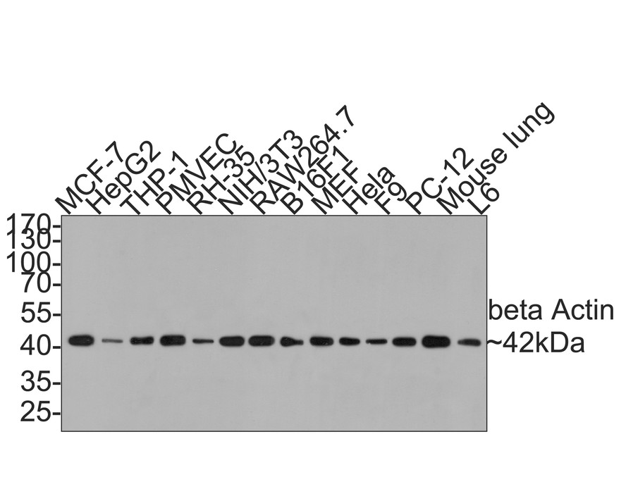
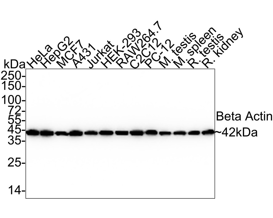
Western blot analysis of beta Actin on different lysates with Rabbit anti-beta Actin antibody (HA722023) at 1/20,000 dilution.
Lane 1: HeLa cell lysate (10 µg/Lane)
Lane 2: HepG2 cell lysate (10 µg/Lane)
Lane 3: MCF7 cell lysate (10 µg/Lane)
Lane 4: A431 cell lysate (10 µg/Lane)
Lane 5: Jurkat cell lysate (10 µg/Lane)
Lane 6: HEK-293 cell lysate (10 µg/Lane)
Lane 7: RAW264.7 cell lysate (10 µg/Lane)
Lane 8: C2C12 cell lysate (10 µg/Lane)
Lane 9: PC-12 cell lysate (10 µg/Lane)
Lane 10: Mouse testis tissue lysate (10 µg/Lane)
Lane 11: Mouse spleen tissue lysate (10 µg/Lane)
Lane 12: Rat testis tissue lysate (10 µg/Lane)
Lane 13: Rat kidney tissue lysate (10 µg/Lane)
Predicted band size: 42 kDa
Observed band size: 42 kDa
Exposure time: 3 seconds;
4-20% SDS-PAGE gel.
Proteins were transferred to a PVDF membrane and blocked with 5% NFDM/TBST for 1 hour at room temperature. The primary antibody (HA722023) at 1/20,000 dilution was used in 5% NFDM/TBST at 4℃ overnight. Goat Anti-Rabbit IgG - HRP Secondary Antibody (HA1001) at 1/50,000 dilution was used for 1 hour at room temperature. -
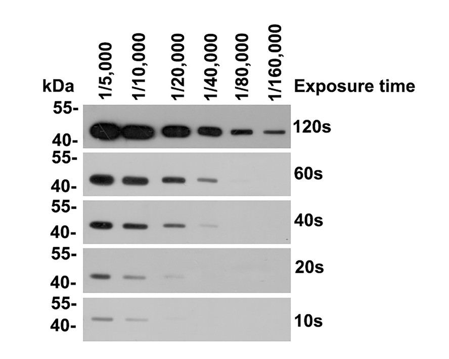
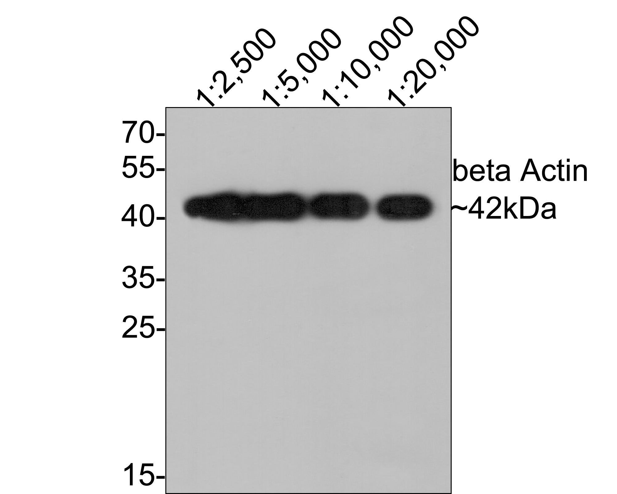
Western blot analysis of beta Actin on HepG2 cell lysates with Rabbit anti-beta Actin antibody (HA722023) at different dilutions.
Lysates/proteins at 20 µg/Lane.
Predicted band size: 42 kDa
Observed band size: 42 kDa
Exposure time: 30 seconds;
4-20% SDS-PAGE gel.
Proteins were transferred to a PVDF membrane and blocked with 5% NFDM/TBST for 1 hour at room temperature. The primary antibody (HA722023) at different dilutions was used in 5% NFDM/TBST at 4℃ overnight. Goat Anti-Rabbit IgG - HRP Secondary Antibody (HA1001) at 1/50,000 dilution was used for 1 hour at room temperature. -
Immunocytochemistry analysis of HeLa cells labeling beta Actin with Rabbit anti-beta Actin antibody (HA722023) at 1/250 dilution.
Cells were fixed in 4% paraformaldehyde for 20 minutes at room temperature, permeabilized with 0.1% Triton X-100 in PBS for 5 minutes at room temperature, then blocked with 1% BSA in 10% negative goat serum for 1 hour at room temperature. Cells were then incubated with Rabbit anti-beta Actin antibody (HA722023) at 1/250 dilution in 1% BSA in PBST overnight at 4 ℃. Goat Anti-Rabbit IgG H&L (iFluor™ 488, HA1121) was used as the secondary antibody at 1/1,000 dilution. PBS instead of the primary antibody was used as the secondary antibody only control. Nuclear DNA was labelled in blue with DAPI.
Beta tubulin (M1305-2, red) was stained at 1/100 dilution overnight at +4℃. Goat Anti-Mouse IgG H&L (iFluor™ 594, HA1126) was used as the secondary antibody at 1/1,000 dilution. -
Flow cytometric analysis of HeLa cells labeling beta Actin.
Cells were fixed and permeabilized. Then stained with the primary antibody (HA722023, 1μg/mL) (red) compared with Rabbit IgG Isotype Control (green). After incubation of the primary antibody at +4℃ for an hour, the cells were stained with a iFluor™ 488 conjugate-Goat anti-Rabbit IgG Secondary antibody (HA1121) at 1/1,000 dilution for 30 minutes at +4℃. Unlabelled sample was used as a control (cells without incubation with primary antibody; black). -
Immunocytochemistry analysis of NIH/3T3 cells labeling beta Actin with Rabbit anti-beta Actin antibody (HA722023) at 1/100 dilution.
Cells were fixed in 4% paraformaldehyde for 20 minutes at room temperature, permeabilized with 0.1% Triton X-100 in PBS for 5 minutes at room temperature, then blocked with 1% BSA in 10% negative goat serum for 1 hour at room temperature. Cells were then incubated with Rabbit anti-beta Actin antibody (HA722023) at 1/100 dilution in 1% BSA in PBST overnight at 4 ℃. Goat Anti-Rabbit IgG H&L (iFluor™ 488, HA1121) was used as the secondary antibody at 1/1,000 dilution. PBS instead of the primary antibody was used as the secondary antibody only control. Nuclear DNA was labelled in blue with DAPI.
Beta tubulin (M1305-2, red) was stained at 1/100 dilution overnight at +4℃. Goat Anti-Mouse IgG H&L (iFluor™ 594, HA1126) was used as the secondary antibody at 1/1,000 dilution. -
Flow cytometric analysis of NIH/3T3 cells labeling beta Actin.
Cells were fixed and permeabilized. Then stained with the primary antibody (HA722023, 1μg/mL) (red) compared with Rabbit IgG Isotype Control (green). After incubation of the primary antibody at +4℃ for an hour, the cells were stained with a iFluor™ 488 conjugate-Goat anti-Rabbit IgG Secondary antibody (HA1121) at 1/1,000 dilution for 30 minutes at +4℃. Unlabelled sample was used as a control (cells without incubation with primary antibody; black). -
Immunocytochemistry analysis of L6 cells labeling beta Actin with Rabbit anti-beta Actin antibody (HA722023) at 1/100 dilution.
Cells were fixed in 4% paraformaldehyde for 20 minutes at room temperature, permeabilized with 0.1% Triton X-100 in PBS for 5 minutes at room temperature, then blocked with 1% BSA in 10% negative goat serum for 1 hour at room temperature. Cells were then incubated with Rabbit anti-beta Actin antibody (HA722023) at 1/100 dilution in 1% BSA in PBST overnight at 4 ℃. Goat Anti-Rabbit IgG H&L (iFluor™ 488, HA1121) was used as the secondary antibody at 1/1,000 dilution. PBS instead of the primary antibody was used as the secondary antibody only control. Nuclear DNA was labelled in blue with DAPI.
Beta tubulin (M1305-2, red) was stained at 1/100 dilution overnight at +4℃. Goat Anti-Mouse IgG H&L (iFluor™ 594, HA1126) was used as the secondary antibody at 1/1,000 dilution. -
Flow cytometric analysis of C6 cells labeling beta Actin.
Cells were fixed and permeabilized. Then stained with the primary antibody (HA722023, 1μg/mL) (red) compared with Rabbit IgG Isotype Control (green). After incubation of the primary antibody at +4℃ for an hour, the cells were stained with a iFluor™ 488 conjugate-Goat anti-Rabbit IgG Secondary antibody (HA1121) at 1/1,000 dilution for 30 minutes at +4℃. Unlabelled sample was used as a control (cells without incubation with primary antibody; black). -
beta Actin was immunoprecipitated from 0.2 mg NIH/3T3 cell lysate with HA722023 at 2 µg/25 µl agarose. Western blot was performed from the immunoprecipitate using HA722023 at 1/5,000 dilution. Anti-Rabbit IgG for IP Nano-secondary antibody (NBI01H) at 1/5,000 dilution was used for 1 hour at room temperature.
Lane 1: NIH/3T3 cell lysate (input)
Lane 2: HA722023 IP in NIH/3T3 cell lysate
Lane 3: Rabbit IgG instead of HA722023 in NIH/3T3 cell lysate
Blocking/Dilution buffer: 5% NFDM/TBST
Exposure time: 1 minute 2 seconds; ECL: K1801
Please note: All products are "FOR RESEARCH USE ONLY AND ARE NOT INTENDED FOR DIAGNOSTIC OR THERAPEUTIC USE"
引文
-
Design and synthesis of N-aryl-2-trifluoromethyl-quinazoline-4-amine derivatives as potential Werner-dependent antiproliferative agents
Author: Li Huimin,et al
PMID: 38739229
应用: WB
反应种属: Human
发表时间: 2024 May
-
 Citation
-
IGFBP7 promotes gastric cancer by facilitating epithelial-mesenchymal transition of gastric cells
Author: Wang Jinqing,et al
PMID: NO PMID20240511

应用: WB
反应种属: Human
发表时间: 2024 May
-
Citation
-
Enhancing the therapeutic efficacy of gefitinib on subcutaneously transplanted SKOV3 ovarian cancer tumors in nude mice via ultrasound‑stimulated microbubble cavitation
Author: Chen Jianghong,et al
PMID: NOPMID20240713

应用: WB
反应种属: Mouse
发表时间: 2024 Jul
-
Citation
-
Mangiferin relieves CCl4-induced liver fibrosis in mice
Author:
PMID: 36914687

应用: WB
反应种属: mouse
发表时间: 2023 Mar
-
Citation
-
Dynamic changes in human THP-1-derived M1-to-M2 macrophage polarization during Thelazia callipaeda MIF induction
Author:
PMID: 36713445

应用: WB
反应种属: human
发表时间: 2023 Jan
-
Citation
-
FBXO6 regulates the antiviral immune responses via mediating alveolar macrophages survival
Author:
PMID: 36217277

应用: WB
反应种属: human
发表时间: 2023 Jan
-
Citation
-
Study on the alleviation of Fengshi Gutong capsule on rheumatoid arthritis through integrating network pharmacology and experimental exploration
Author:
PMID: 34329717

应用: WB
反应种属: mouse
发表时间: 2021 Nov
-
Citation
-
The water extract of Sophorae tonkinensis Radix et Rhizoma alleviates non-alcoholic fatty liver disease and its mechanism
Author:
PMID: 32702591

应用: WB
反应种属: Mouse
发表时间: 2020 Oct
-
Citation
-
The diferential efects of isofurane and sevofurane on neonatal mice
Author:
PMID: 33168900

应用: WB
反应种属: Mouse
发表时间: 2020 Nov
-
Citation
-
Dynamics of transmissible gastroenteritis virus internalizationunraveled by single-virus tracking in live cells
Author:
PMID: 32017270

应用: WB
反应种属: Pig
发表时间: 2020 Mar
-
Citation
-
Somatostatin stimulates colonic MUC2 expression through SSTR5-Notch-Hes1 signaling pathway
Author:
PMID: 31733832

应用: WB
反应种属: human
发表时间: 2020 Jan
-
Citation
-
Chicken optineurin suppresses MDA5-mediated interferon b production
Author:
PMID: 33357711

应用: WB
反应种属: Chicken
发表时间: 2020 Jan
-
Citation
-
SUV39H1 is a New Client Protein of Hsp90 Degradated by Chaetocin as a Novel C-Terminal Inhibitor of Hsp90
Author:
PMID: 33162400
应用: WB
反应种属: Human
发表时间: 2020 Jan
-
Citation
-
Parathyroid hormone increases alveolar bone homoeostasis during orthodontic tooth movement in rats with periodontitis via crosstalk between STAT3 and β-catenin
Author:
PMID: 33380723

应用: WB
反应种属: Rat
发表时间: 2020 Dec
-
Citation
-
N6-(2-hydroxyethyl)-Adenosine Induces Apoptosis via ER Stress and Autophagy of Gastric Carcinoma Cells In Vitro and In Vivo
Author:
PMID: 32823628

应用: WB
反应种属: Human
发表时间: 2020 Aug
-
Citation
-
Acid-induced autophagy protects human gastric cancer cells from apoptosis by activating Erk1/2 pathway
Author:
PMID: 35116899

应用: WB
反应种属: human
发表时间: 2019 Aug
-
Citation
同靶点&同通路的产品

beta Actin Rabbit Polyclonal Antibody
Application: WB,IHC-P
Reactivity: Human,Mouse,Rat
Conjugate: unconjugated
beta Actin Mouse Monoclonal Antibody [A2-F6]
Application: WB,IF-Cell,IHC-P,FC
Reactivity: Human,Mouse,Rat
Conjugate: unconjugated
beta Actin Rabbit Polyclonal Antibody
Application: WB,IF-Cell,IHC-P,FC
Reactivity: Human,Mouse,Rat,Zebrafish,Bamboo
Conjugate: unconjugated

HRP Conjugated beta Actin Recombinant Rabbit Monoclonal Antibody [JF53-10]
Application: WB
Reactivity: Human,Mouse,Rat,Zebrafish,Monkey,Hamster,Arabidopsis thaliana,Rice
Conjugate: HRP
beta Actin Mouse Monoclonal Antibody [B4-B2]
Application: WB,IF-Cell,IHC-P,FC
Reactivity: Human,Mouse,Rat
Conjugate: unconjugated

HRP Conjugated beta Actin Mouse Monoclonal Antibody [A2-F6]
Application: WB
Reactivity: Human,Mouse,Rat
Conjugate: HRP
HRP Conjugated beta Actin Recombinant Mouse Monoclonal Antibody [A2-F6-R]
Application: WB
Reactivity: Human,Mouse,Rat
Conjugate: HRP