概述
产品名称
CD20 Recombinant Rabbit Monoclonal Antibody [PD00-02]
抗体类型
Recombinant Rabbit monoclonal Antibody
免疫原
Synthetic peptide within Human CD20 aa 210-297 (Cytoplasmic).
种属反应性
Human, Mouse
验证应用
IHC-P, mIHC, WB, IF-Cell, IP
分子量
Predicted band size: 33 kDa
阳性对照
Human tonsil tissue, human lymph nodes tissue, human spleen tissue, human small cell lung cancer, human non-small cell lung cancer, human prostate cancer, human non-small cell lung cancer, mouse spleen tissue, Raji cell lysate, Ramos cell lysate, Daudi cell lysate.
偶联
unconjugated
克隆号
PD00-02
RRID
产品特性
形态
Liquid
浓度
1ug/ul
存放说明
Store at +4℃ after thawing. Aliquot store at -20℃. Avoid repeated freeze / thaw cycles.
存储缓冲液
PBS (pH7.4), 0.1% BSA, 40% Glycerol. Preservative: 0.05% Sodium Azide.
亚型
IgG
纯化方式
Protein A affinity purified.
应用稀释度
-
IHC-P
-
1:1,000-1:2,000
-
mIHC
-
1:1,500-1:3,000
-
WB
-
1:2,000
-
IF-Cell
-
1:500
-
IP
-
1-2μg/sample
发表文章中的应用
| IF-tissue | See 1 publications below |
发表文章中的种属
| Human | See 1 publications below |
靶点
功能
The CD20 antigen is a membrane-embedded, non-glycosylated phosphoprotein, 33-37 kDa. CD20 functions as a Ca2+-permeable cation channel, involved in the regulation of B-cell activation, proliferation and differentiation. CD20 appears on the surface of the pre-B lymphocyte between the time of light chain rearrangement and expression of intact surface immunoglobulin and is lost just before terminal B-cell differentiation into plasma cells. CD20 is virtually specific for normal B-cells. A weak expression has been demonstrated in a subpopulation of T-cells, but not in any other cell type. CD20 is expressed in the large majority of cases of B-cell leukaemia/lymphoma. Early stage precursor B lymphoblastic leukaemia/lymphoma may be negative, and chronic lymphocytic leukaemia/small cell lymphoma may show a weak staining. Plasma cell neoplasms are as a rule CD20 negative. T-cell lymphomas are almost always negative, but CD20 has been demonstrated in few cases of various types of T-cell lymphoma. In Hodgkin lymphoma, the nodular lymphocyte-predominant subtype shows CD20 staining of L&H cells in most cases, while Reed-Sternberg cells in the other subtypes reveal CD20 positivity in about 40, albeit in a minority of neoplastic cells. Acute myeloid leukaemia is CD20 positive in few cases, while blastic transformation in chronic myeloid leukaemia is accompanied by CD20 positivity in about 30%. Thymoma may reveal CD20 positivity in a spindle cell component. In patients treated with retuximab (a humanized anti-CD20 antibody) for malignant B-cell lymphoma, the CD20 epitopes disappear (both in normal and neoplastic B-cells) as a result of down-modulation of CD20 m-RNA in the cells. This process is potentially reversible. Together with CD79a, CD20 is one of the most important markers for the identification of B-cell neoplasms as outlined above. Tonsil and appendix are appropriate controls: The mantle zone B-cells and the germinal centre B-cells must show a very strong staining reaction. No other cells should stain.
背景文献
1. Jiang D. et. al. Pyruvate dehydrogenase kinase 4-mediated metabolic reprogramming is involved in rituximab resistance in diffuse large B-cell lymphoma by affecting the expression of MS4A1/CD20. Cancer Sci. 2021 Sep
2. Pavlasova G. et. al. The regulation and function of CD20: an "enigma" of B-cell biology and targeted therapy. Haematologica. 2020 Jun
亚细胞定位
Cell membrane.
别名
APY antibody
ATOPY antibody
B lymphocyte antigen CD20 antibody
B Lymphocyte Cell Surface Antigen B1 antibody
B-lymphocyte antigen CD20 antibody
B-lymphocyte cell-surface antigen B1 antibody
B-lymphocyte surface antigen B1 antibody
B1 antibody
Bp 35 antibody
Bp35 antibody
展开APY antibody
ATOPY antibody
B lymphocyte antigen CD20 antibody
B Lymphocyte Cell Surface Antigen B1 antibody
B-lymphocyte antigen CD20 antibody
B-lymphocyte cell-surface antigen B1 antibody
B-lymphocyte surface antigen B1 antibody
B1 antibody
Bp 35 antibody
Bp35 antibody
CD 20 antibody
CD20 antibody
CD20 antigen antibody
CD20 receptor antibody
CD20_HUMAN antibody
CVID 5 antibody
CVID5 antibody
Fc epsilon receptor I beta chain antibody
Fc Fragment of IgE high affinity I receptor for beta polypeptide antibody
FCER1B antibody
High affinity immunoglobulin epsilon receptor subunit beta antibody
IgE Fc receptor subunit beta antibody
IGEL antibody
IGER antibody
IGHER antibody
LEU 16 antibody
Leu-16 antibody
LEU16 antibody
leukocyte surface antigen Leu 16 antibody
Leukocyte surface antigen Leu-16 antibody
Ly44 antibody
Membrane spanning 4 domains A1 antibody
Membrane spanning 4 domains subfamily A member 2 antibody
membrane-spanning 4-domains A1 antibody
Membrane-spanning 4-domains subfamily A member 1 antibody
MGC3969 antibody
MS4A1 antibody
MS4A2 antibody
S7 antibody
折叠图片
-

Fluorescence multiplex immunohistochemical analysis of Tertiary Lymphoid Structures in Human Small Cell Lung Cancer (Formalin/PFA-fixed paraffin-embedded sections). Panel A: the merged image of anti-CD20 (HA721138, green), anti-PD-L1 (HA721176, cyan), anti-CD56 (ET1702-43, magenta) and anti-CD3 (HA720082, yellow) on tertiary lymphoid structures. Panel B: anti- CD20 stained on B cells. Panel C: anti-PD-L1 stained on dendritic cells and macrophages cells. Panel D: anti-CD56 stained on NKT cells. Panel E: anti-CD3 stained on T cells. HRP Conjugated UltraPolymer Goat Polyclonal Antibody HA1119/HA1120 was used as a secondary antibody. The immunostaining was performed with the Sequential Immuno-staining Kit (IRISKit™MH010101, www.luminiris.cn). The section was incubated in four rounds of staining: in the order of HA721138 (1/1,500 dilution), HA721176 (1/1,000 dilution), ET1702-43 (1/1,000 dilution), and HA720082 (1/500 dilution) for 20 mins at room temperature. Each round was followed by a separate fluorescent tyramide signal amplification system. Heat mediated antigen retrieval with Tris-EDTA buffer (pH 9.0) for 30 mins at 95℃. DAPI (blue) was used as a nuclear counter stain. Image acquisition was performed with Olympus VS200 Slide Scanner.
-

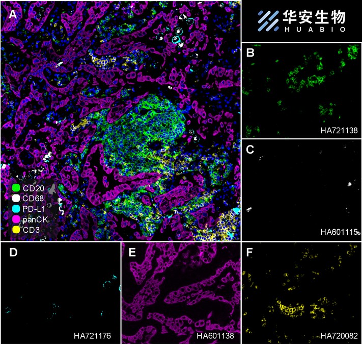
Fluorescence multiplex immunohistochemical analysis of the human non-small cell lung cancer (Formalin/PFA-fixed paraffin-embedded sections). Panel A: the merged image of anti-CD20 (HA721138, green), anti-CD68 (HA601115, gray), anti-PD-L1 (HA721176, cyan), anti-panCK (HA601138, magenta) and anti-CD3 (HA720082, yellow) on human non-small cell lung cancer. Panel B: anti- CD20 stained on B cells. Panel C: anti-CD68 stained on macrophage M1 and macrophage M2. Panel D: anti-PD-L1 stained on dendritic cells and macrophages cells. Panel E: anti-panCK stained on cancer cells. Panel F: anti-CD3 stained on T cells. HRP Conjugated UltraPolymer Goat Polyclonal Antibody HA1119/HA1120 was used as a secondary antibody. The immunostaining was performed with the Sequential Immuno-staining Kit (IRISKit™MH010101, www.luminiris.cn). The section was incubated in five rounds of staining: in the order of HA721138 (1/1,500 dilution), HA601115 (1/2,000 dilution), HA721176 (1/1,000 dilution), HA601138 (1/3,000 dilution), and HA720082 (1/500 dilution) for 20 mins at room temperature. Each round was followed by a separate fluorescent tyramide signal amplification system. Heat mediated antigen retrieval with Tris-EDTA buffer (pH 9.0) for 30 mins at 95℃. DAPI (blue) was used as a nuclear counter stain. Image acquisition was performed with Olympus VS200 Slide Scanner.
-

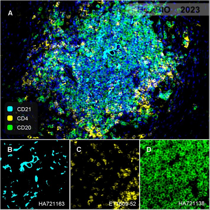
Fluorescence multiplex immunohistochemical analysis of tertiary lymphoid structures in human prostate cancer (Formalin/PFA-fixed paraffin-embedded sections). Panel A: the merged image of anti-CD20 (HA721138, green), anti-CD21 (HA721163, cyan) and anti-CD4 (ET1609-52, yellow) on tertiary lymphoid structures. Panel B: anti- CD20 stained on B cells. Panel C: anti-CD21 stained on naive B-cell, memory B-cell and plasma cells. Panel D: anti-CD4 stained on helper T cells and Treg cells. HRP Conjugated UltraPolymer Goat Polyclonal Antibody HA1119/HA1120 was used as a secondary antibody. The immunostaining was performed with the Sequential Immuno-staining Kit (IRISKit™MH010101, www.luminiris.cn). The section was incubated in three rounds of staining: in the order of HA721138 (1/1,500 dilution), HA721163 (1/1,000 dilution), and ET1609-52 (1/1,000 dilution) for 20 mins at room temperature. Each round was followed by a separate fluorescent tyramide signal amplification system. Heat mediated antigen retrieval with Tris-EDTA buffer (pH 9.0) for 30 mins at 95℃. DAPI (blue) was used as a nuclear counter stain. Image acquisition was performed with Olympus VS200 Slide Scanner.
-

Fluorescence multiplex immunohistochemical analysis of Human tonsil (Formalin/PFA-fixed paraffin-embedded sections). Panel A: the merged image of anti-BCL6 (HA601083, Red), anti-HLA-DPB1 (ET1704-13, Green), anti-Tryptase (ET1610-64, White), anti-CD20 (HA721138, Magenta) and anti-CD45 (ET7111-03, Yellow) on tonsil. HRP Conjugated UltraPolymer Goat Polyclonal Antibody HA1119/HA1120 was used as a secondary antibody. The immunostaining was performed with the Sequential Immuno-staining Kit (IRISKit™MH010101, www.luminiris.cn). The section was incubated in five rounds of staining: in the order of HA601083 (1/200 dilution), ET1704-13 (1/2,000 dilution), ET1610-64 (1/5,000 dilution), HA721138 (1/2,000 dilution) and ET7111-03 (1/500 dilution) for 20 mins at room temperature. Each round was followed by a separate fluorescent tyramide signal amplification system. Heat mediated antigen retrieval with Tris-EDTA buffer (pH 9.0) for 30 mins at 95℃. DAPI (blue) was used as a nuclear counter stain. Image acquisition was performed with Olympus VS200 Slide Scanner.
-

Fluorescence multiplex immunohistochemical analysis of Human tonsil (Formalin/PFA-fixed paraffin-embedded sections). Panel A: the merged image of anti-CD68 (HA601115, Red), anti-CD38 (HA721268, Green), anti-CD23 (HA721139, White), anti-CD11C (ET1606-19, Cyan), anti-CD45 (ET7111-03, Magenta) and anti-CD20 (HA721138, Yellow) on tonsil. Panel B: anti-CD68 stained on Macrophage. Panel C: anti-CD38 stained on lymphocyte subsets. Panel D: anti-CD11C stained on dendritic cells. Panel E: CD45 stained on lymphocytes. Panel F: anti-CD20 stained on B cells. Panel G: anti-CD23 stained on follicular dendritic cells. HRP Conjugated UltraPolymer Goat Polyclonal Antibody HA1119/HA1120 was used as a secondary antibody. The immunostaining was performed with the Sequential Immuno-staining Kit (IRISKit™MH010101, www.luminiris.cn). The section was incubated in six rounds of staining: in the order of HA601115 (1/2,000 dilution), HA721268 (1/1,000 dilution), ET1606-19 (1/1,000 dilution), ET7111-03 (1/500 dilution), HA721138 (1/2,000 dilution) and HA721139 (1/800 dilution) for 20 mins at room temperature. Each round was followed by a separate fluorescent tyramide signal amplification system. Heat mediated antigen retrieval with Tris-EDTA buffer (pH 9.0) for 30 mins at 95℃. DAPI (blue) was used as a nuclear counter stain. Image acquisition was performed with Olympus VS200 Slide Scanner.
-

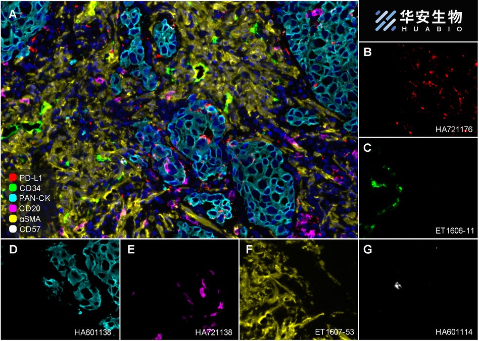
Fluorescence multiplex immunohistochemical analysis of Human non-small cell lung cancer (Formalin/PFA-fixed paraffin-embedded sections). Panel A: the merged image of anti-PD-L1 (HA721176, red), anti-CD34 (ET1606-11, green), anti-Pan-CK (HA601138, cyan), anti-CD20 (HA721138, magenta), anti-αSMA (ET1607-53, yellow) and anti-CD57 (HA601114, white) on NSCLC. Panel B: anti-PD-L1 stained on dendritic cells and macrophages cells. Panel C: anti- CD34 stained on endothelial cells. Panel D: anti-Pan-CK stained on cancer cells. Panel E: CD20 stained on B cells. Panel F: anti-αSMA stained on cancer-associated fibroblasts and smooth muscle cells. Panel G: anti-CD57 stained on NK cells and T cells. HRP Conjugated UltraPolymer Goat Polyclonal Antibody HA1119/HA1120 was used as a secondary antibody. The immunostaining was performed with the Sequential Immuno-staining Kit (IRISKit™MH010101, www.luminiris.cn). The section was incubated in six rounds of staining: in the order of HA721176 (1/1,000 dilution), ET1606-11 (1/1,000 dilution), HA601138 (1/3,000 dilution), HA721138 (1/2,000 dilution), ET1607-53 (1/3,000 dilution) and HA601114 (1/1,000 dilution) for 20 mins at room temperature. Each round was followed by a separate fluorescent tyramide signal amplification system. Heat mediated antigen retrieval with Tris-EDTA buffer (pH 9.0) for 30 mins at 95℃. DAPI (blue) was used as a nuclear counter stain. Image acquisition was performed with Olympus VS200 Slide Scanner.
-

Fluorescence multiplex immunohistochemical analysis of human tonsil (Formalin/PFA-fixed paraffin-embedded sections). Panel A: the merged image of anti-CD20 (HA721138, Cyan), anti-CD38 (HA721268, Violet) and anti-CD57 (HA601114, Yellow) on tonsil. HRP Conjugated UltraPolymer Goat Polyclonal Antibody HA1119/HA1120 was used as a secondary antibody. The immunostaining was performed with the Sequential Immuno-staining Kit (IRISKit™MH010101, www.luminiris.cn). The section was incubated in three rounds of staining: in the order of HA721138 (1/2,000 dilution), HA721268 (1/1,000 dilution) and HA601114 (1/1,000 dilution) for 20 mins at room temperature. Each round was followed by a separate fluorescent tyramide signal amplification system. Heat mediated antigen retrieval with Tris-EDTA buffer (pH 9.0) for 30 mins at 95℃. DAPI (blue) was used as a nuclear counter stain. Image acquisition was performed with Zeiss Observer 7 Inverted Fluorescence Microscope.
-

☑ Relative expression (RE)
Western blot analysis of CD20 on different lysates with Rabbit anti-CD20 antibody (HA721138) at 1/2,000 dilution.
Lane 1: Raji cell lysate
Lane 2: 293T cell lysate (negative)
Lane 3: Ramos cell lysate
Lane 4: HeLa cell lysate (negative)
Lane 5: Daudi cell lysate
Lysates/proteins at 20 µg/Lane.
Predicted band size: 33 kDa
Observed band size: 33 kDa
Exposure time: 4 seconds; ECL: K1801;
4-20% SDS-PAGE gel.
Proteins were transferred to a PVDF membrane and blocked with 5% NFDM/TBST for 1 hour at room temperature. The primary antibody (HA721138) at 1/2,000 dilution was used in 5% NFDM/TBST at 4℃ overnight. Goat Anti-Rabbit IgG - HRP Secondary Antibody (HA1001) at 1/50,000 dilution was used for 1 hour at room temperature. -

Immunohistochemical analysis of paraffin-embedded human tonsil tissue with Rabbit anti-CD20 antibody (HA721138) at 1/2,000 dilution.
The section was pre-treated using heat mediated antigen retrieval with Tris-EDTA buffer (pH 9.0) for 20 minutes. The tissues were blocked in 1% BSA for 20 minutes at room temperature, washed with ddH2O and PBS, and then probed with the primary antibody (HA721138) at 1/2,000 dilution for 1 hour at room temperature. The detection was performed using an HRP conjugated compact polymer system. DAB was used as the chromogen. Tissues were counterstained with hematoxylin and mounted with DPX. -

Immunohistochemical analysis of paraffin-embedded human lymph nodes tissue with Rabbit anti-CD20 antibody (HA721138) at 1/2,000 dilution.
The section was pre-treated using heat mediated antigen retrieval with Tris-EDTA buffer (pH 9.0) for 20 minutes. The tissues were blocked in 1% BSA for 20 minutes at room temperature, washed with ddH2O and PBS, and then probed with the primary antibody (HA721138) at 1/2,000 dilution for 1 hour at room temperature. The detection was performed using an HRP conjugated compact polymer system. DAB was used as the chromogen. Tissues were counterstained with hematoxylin and mounted with DPX. -

Immunohistochemical analysis of paraffin-embedded human spleen tissue with Rabbit anti-CD20 antibody (HA721138) at 1/1,000 dilution.
The section was pre-treated using heat mediated antigen retrieval with Tris-EDTA buffer (pH 9.0) for 20 minutes. The tissues were blocked in 1% BSA for 20 minutes at room temperature, washed with ddH2O and PBS, and then probed with the primary antibody (HA721138) at 1/1,000 dilution for 1 hour at room temperature. The detection was performed using an HRP conjugated compact polymer system. DAB was used as the chromogen. Tissues were counterstained with hematoxylin and mounted with DPX. -

Immunohistochemical analysis of paraffin-embedded mouse spleen tissue with Rabbit anti-CD20 antibody (HA721138) at 1/1,000 dilution.
The section was pre-treated using heat mediated antigen retrieval with Tris-EDTA buffer (pH 9.0) for 20 minutes. The tissues were blocked in 1% BSA for 20 minutes at room temperature, washed with ddH2O and PBS, and then probed with the primary antibody (HA721138) at 1/1,000 dilution for 1 hour at room temperature. The detection was performed using an HRP conjugated compact polymer system. DAB was used as the chromogen. Tissues were counterstained with hematoxylin and mounted with DPX. -

☑ Relative expression (RE)
Immunocytochemistry analysis of Raji (positive) and 293T (negative) labeling CD20 with Rabbit anti-CD20 antibody (HA721138) at 1/500 dilution.
Cells were fixed in 4% paraformaldehyde for 15 minutes at room temperature, permeabilized with 0.1% Triton X-100 in PBS for 15 minutes at room temperature, then blocked with 1% BSA in 10% negative goat serum for 1 hour at room temperature. Cells were then incubated with Rabbit anti-CD20 antibody (HA721138) at 1/500 dilution in 1% BSA in PBST overnight at 4 ℃. Goat Anti-Rabbit IgG H&L (iFluor™ 488, HA1121) was used as the secondary antibody at 1/1,000 dilution. PBS instead of the primary antibody was used as the secondary antibody only control. Nuclear DNA was labelled in blue with DAPI.
Beta tubulin (HA601187, red) was stained at 1/100 dilution overnight at +4℃. Goat Anti-Mouse IgG H&L (iFluor™ 594, HA1126) was used as the secondary antibody at 1/1,000 dilution. -

CD20 was immunoprecipitated from 0.2 mg Raji cell lysate with HA721138 at 2 µg/25 µl agarose. Western blot was performed from the immunoprecipitate using HA721138 at 1/2,000 dilution. Mouse anti Rabbit IgG heavy chain (Fc) secondary antibody (M1003-7) at 1/10,000 dilution was used for 1 hour at room temperature.
Lane 1: Raji cell lysate (input)
Lane 2: HA721138 IP in Raji cell lysate
Lane 3: Rabbit IgG instead of HA721138 in Raji cell lysate
Blocking/Dilution buffer: 5% NFDM/TBST
Exposure time: 59 seconds; ECL: K1801
Please note: All products are "FOR RESEARCH USE ONLY AND ARE NOT INTENDED FOR DIAGNOSTIC OR THERAPEUTIC USE"
引文
-
Single-cell chemokine receptor profiles delineate the immune contexture of tertiary lymphoid structures in head and neck squamous cell carcinoma
Author:
PMID: 36841416

应用: IF-tissue
反应种属: Human
发表时间: 2023 Apr
-
Citation
同靶点&同通路的产品
CD20 Recombinant Rabbit Monoclonal Antibody [SY12-01]
Application: WB,IHC-P,IF-Tissue,IF-Cell,FC,IP
Reactivity: Human,Monkey
Conjugate: unconjugated
CD20 Recombinant Rabbit Monoclonal Antibody [PSH08-12]
Application: WB,IF-Cell,IHC-P,FC,IP
Reactivity: Human
Conjugate: unconjugated
CD20 Recombinant Rabbit Monoclonal Antibody [PD00-02]
Application: mIHC
Reactivity: Human
Conjugate: unconjugated


