概述
产品名称
CD4 Recombinant Rabbit Monoclonal Antibody [ST0488]
抗体类型
Recombinant Rabbit monoclonal Antibody
免疫原
Recombinant protein within Human CD4 aa 196-416 / 458.
种属反应性
Human
验证应用
WB, IF-Cell, IF-Tissue, IHC-P, FC, mIHC
分子量
Predicted band size: 51 kDa
阳性对照
U937 cell lysate, THP-1 cell lysate, THP-1, human tonsil tissue, human spleen tissue, human lymph nodes tissue, human liver tissue, human prostate cancer, human cervical cancer.
偶联
unconjugated
克隆号
ST0488
RRID
产品特性
形态
Liquid
浓度
1ug/ul
存放说明
Store at +4℃ after thawing. Aliquot store at -20℃ or -80℃. Avoid repeated freeze / thaw cycles.
存储缓冲液
1*TBS (pH7.4), 0.05% BSA, 40% Glycerol. Preservative: 0.05% Sodium Azide.
亚型
IgG
纯化方式
Protein A affinity purified.
应用稀释度
-
WB
-
1:1,000-1:2,000
-
IF-Cell
-
1:50-1:200
-
IF-Tissue
-
1:50-1:200
-
IHC-P
-
1:400-1:800
-
FC
-
1:500-1:1,000
-
mIHC
-
1:800-1:1,000
发表文章中的应用
发表文章中的种属
| Human | See 4 publications below |
| human | See 2 publications below |
| Mouse | See 1 publications below |
靶点
功能
The T cell receptor (TCR) is a heterodimer composed of either α and β or γ and δ chains. CD3 chains and the CD4 or CD8 co-receptors are also required for efficient signal transduction through the TCR. The TCR is expressed on T helper and T cytotoxic cells that can be distinguished by their expression of CD4 and CD8; T helper cells express CD4 proteins and T cytotoxic cells display CD8. CD4 is also expressed on cortical cells, mature medullary thymocytes, microglial cells and dendritic cells. CD4 (also designated T4 and Leu 3), is a membrane glycoprotein that contains four extracellular immunoglobin-like domains. The TCR in association with CD4 can bind class II MHC molecules presented by the antigen-presenting cells. The CD4 protein functions by increasing the avidity of the interaction between the TCR and an antigen-class II MHC complex. An additional role of CD4 is to function as a receptor for HIV.
背景文献
1. Kim EJ et al. Costimulation blockade alters germinal center responses and prevents antibody-mediated rejection. Am J Transplant 14:59-69 (2014).
2. Liu XD et al. Resistance to Antiangiogenic Therapy Is Associated with an Immunosuppressive Tumor Microenvironment in Metastatic Renal Cell Carcinoma. Cancer Immunol Res 3:1017-29 (2015).
组织特异性
Highly expressed in T-helper cells. The presence of CD4 is a hallmark of T-helper cells which are specialized in the activation and growth of cytotoxic T-cells, regulation of B cells, or activation of phagocytes. CD4 is also present in other immune cells such as macrophages, dendritic cells or NK cells.
翻译后修饰
Palmitoylation and association with LCK contribute to the enrichment of CD4 in lipid rafts.; Phosphorylated by PKC; phosphorylation at Ser-433 plays an important role for CD4 internalization.
亚细胞定位
Cell membrane.
UNIPROT #
别名
CD 4 antibody
CD4 (L3T4) antibody
CD4 antibody
CD4 antigen (p55) antibody
CD4 antigen antibody
CD4 molecule antibody
CD4 receptor antibody
CD4+ Lymphocyte deficiency, included antibody
CD4_HUMAN antibody
CD4mut antibody
展开CD 4 antibody
CD4 (L3T4) antibody
CD4 antibody
CD4 antigen (p55) antibody
CD4 antigen antibody
CD4 molecule antibody
CD4 receptor antibody
CD4+ Lymphocyte deficiency, included antibody
CD4_HUMAN antibody
CD4mut antibody
L3T4 antibody
Leu3 antibody
Ly-4 antibody
Lymphocyte antigen CD4 antibody
MGC165891 antibody
OTTHUMP00000238897 antibody
p55 antibody
T cell antigen T4 antibody
T cell antigen T4/LEU3 antibody
T cell differentiation antigen L3T4 antibody
T cell OKT4 deficiency, included antibody
T cell surface antigen T4/Leu 3 antibody
T cell surface antigen T4/Leu3 antibody
T cell surface glycoprotein CD4 antibody
T-cell surface antigen T4/Leu-3 antibody
T-cell surface glycoprotein CD4 antibody
W3/25 antibody
W3/25 antigen antibody
折叠图片
-
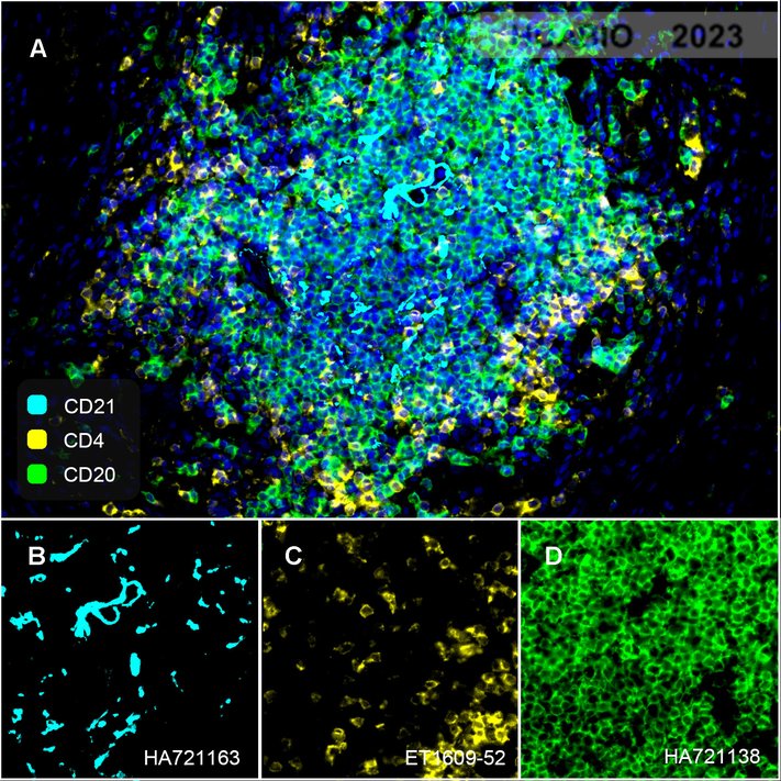
Fluorescence multiplex immunohistochemical analysis of tertiary lymphoid structures in human prostate cancer (Formalin/PFA-fixed paraffin-embedded sections). Panel A: the merged image of anti-CD20 (HA721138, green), anti-CD21 (HA721163, cyan) and anti-CD4 (ET1609-52, yellow) on tertiary lymphoid structures. Panel B: anti- CD20 stained on B cells. Panel C: anti-CD21 stained on naive B-cell, memory B-cell and plasma cells. Panel D: anti-CD4 stained on helper T cells and Treg cells. HRP Conjugated UltraPolymer Goat Polyclonal Antibody HA1119/HA1120 was used as a secondary antibody. The immunostaining was performed with the Sequential Immuno-staining Kit (IRISKit™MH010101, www.luminiris.cn). The section was incubated in three rounds of staining: in the order of HA721138 (1/1,500 dilution), HA721163 (1/1,000 dilution), and ET1609-52 (1/1,000 dilution) for 20 mins at room temperature. Each round was followed by a separate fluorescent tyramide signal amplification system. Heat mediated antigen retrieval with Tris-EDTA buffer (pH 9.0) for 30 mins at 95℃. DAPI (blue) was used as a nuclear counter stain. Image acquisition was performed with Olympus VS200 Slide Scanner.
-
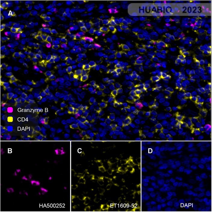
Fluorescence multiplex immunohistochemical analysis of tertiary lymphoid structures in human cervical cancer (Formalin/PFA-fixed paraffin-embedded sections). Panel A: the merged image of anti-Granzyme B (HA500252, magenta), anti-CD4 (ET1609-52, yellow) on tertiary lymphoid structures. Panel B: anti- Granzyme B stained on cytotoxic NK cells and dendritic cells. Panel C: anti-CD4 stained on helper T cells and Treg cells. HRP Conjugated UltraPolymer Goat Polyclonal Antibody HA1119/HA1120 was used as a secondary antibody. The immunostaining was performed with the Sequential Immuno-staining Kit (IRISKit™MH010101, www.luminiris.cn). The section was incubated in three rounds of staining: in the order of HA500252 (1/200 dilution), ET1609-52 (1/1,000 dilution) for 20 mins at room temperature. Each round was followed by a separate fluorescent tyramide signal amplification system. Heat mediated antigen retrieval with Tris-EDTA buffer (pH 9.0) for 30 mins at 95℃. DAPI (blue) was used as a nuclear counter stain. Image acquisition was performed with Olympus VS200 Slide Scanner.
-

Fluorescence multiplex immunohistochemical analysis of Human tonsil (Formalin/PFA-fixed paraffin-embedded sections). Panel A: the merged image of anti-CD14 (ET1610-85, Red), anti-CD4 (ET1609-52, Green), anti-CD57 (HA601114, White), anti-CD15 (HA721246, Cyan)and anti-Tryptase (ET1610-64, Magenta) on tonsil. Panel B: anti- CD14 stained on monocytes. Panel C: anti-CD4 stained on helper T cells and Treg cells. Panel D: anti-CD57 stained on NK cells and T cells. Panel E: CD15 stained on granulocytes and monocytes. Panel F: anti-Tryptase stained on Mast cells. HRP Conjugated UltraPolymer Goat Polyclonal Antibody HA1119/HA1120 was used as a secondary antibody. The immunostaining was performed with the Sequential Immuno-staining Kit (IRISKit™MH010101, www.luminiris.cn). The section was incubated in five rounds of staining: in the order of ET1610-85 (1/800 dilution), ET1609-52 (1/800 dilution), HA601114 (1/1,000 dilution), HA721246 (1/500 dilution), and ET1610-64 (1/3,000 dilution) for 20 mins at room temperature. Each round was followed by a separate fluorescent tyramide signal amplification system. Heat mediated antigen retrieval with Tris-EDTA buffer (pH 9.0) for 30 mins at 95℃. DAPI (blue) was used as a nuclear counter stain. Image acquisition was performed with Olympus VS200 Slide Scanner.
-

Fluorescence multiplex immunohistochemical analysis of human tonsil (Formalin/PFA-fixed paraffin-embedded sections). Panel A: the merged image of anti-CD68 (HA601115, Red), anti-BCL6 (HA601083, Yellow) and anti-CD4 (ET1609-52, Green) on tonsil. HRP Conjugated UltraPolymer Goat Polyclonal Antibody HA1119/HA1120 was used as a secondary antibody. The immunostaining was performed with the Sequential Immuno-staining Kit (IRISKit™MH010101, www.luminiris.cn). The section was incubated in three rounds of staining: in the order of HA601115 (1/2,000 dilution), HA601083 (1/200 dilution) and ET1609-52 (1/800 dilution) for 20 mins at room temperature. Each round was followed by a separate fluorescent tyramide signal amplification system. Heat mediated antigen retrieval with Tris-EDTA buffer (pH 9.0) for 30 mins at 95℃. DAPI (blue) was used as a nuclear counter stain. Image acquisition was performed with Zeiss Observer 7 Inverted Fluorescence Microscope.
-

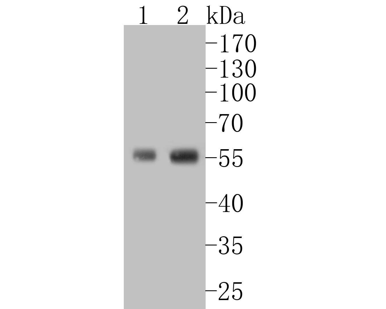
Western blot analysis of CD4 on different lysates with Rabbit anti-CD4 antibody (ET1609-52) at 1/1,000 dilution.
Lane 1: THP-1 cell lysate
Lane 2: U937 cell lysate
Lysates/proteins at 10 µg/Lane.
Predicted band size: 51 kDa
Observed band size: 55 kDa
Exposure time: 1 minute 30 seconds;
10% SDS-PAGE gel.
Proteins were transferred to a PVDF membrane and blocked with 5% NFDM/TBST for 1 hour at room temperature. The primary antibody (ET1609-52) at 1/1,000 dilution was used in 5% NFDM/TBST at room temperature for 2 hours. Goat Anti-Rabbit IgG - HRP Secondary Antibody (HA1001) at 1:100,000 dilution was used for 1 hour at room temperature. -

☑ Knockdown (KD)
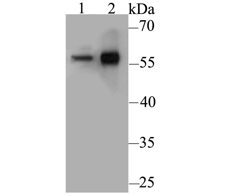
Western blot analysis of CD4 on different lysates with Rabbit anti-CD4 antibody (ET1609-52) at 1/2,000 dilution.
Lane 1: THP-1 WT cell lysate
Lane 2: THP-1 CD4 KD cell lysate
Lysates/proteins at 10 µg/Lane.
Predicted band size: 51 kDa
Observed band size: 55 kDa
Exposure time: 30 seconds;
ECL: Ori Supersensitive
4-20% SDS-PAGE gel.
ET1609-52 was shown to specifically react with CD4 in THP-1 WT cells. Weakened band was observed when THP-1 CD4 KD sample was tested. THP-1 WT and THP-1 CD4 KD samples were subjected to SDS-PAGE. Proteins were transferred to a PVDF membrane and blocked with 5% NFDM in TBST for 1 hour at room temperature. The primary antibody (ET1609-52, 1/2,000) and Loading control antibody (Rabbit anti-GAPDH, ET1601-4, 1/10,000) were used in 5% BSA at room temperature for 2 hours. Goat Anti-rabbit IgG-HRP Secondary Antibody (HA1001) at 1:50,000 dilution was used for 1 hour at room temperature. -

Immunocytochemistry analysis of THP-1 cells labeling CD4 with Rabbit anti-CD4 antibody (ET1609-52) at 1/50 dilution.
Cells were fixed in 4% paraformaldehyde for 10 minutes at 37 ℃, permeabilized with 0.05% Triton X-100 in PBS for 20 minutes, and then blocked with 2% negative goat serum for 30 minutes at room temperature. Cells were then incubated with Rabbit anti-CD4 antibody (ET1609-52) at 1/50 dilution in 2% negative goat serum overnight at 4 ℃. Goat Anti-Rabbit IgG H&L (iFluor™ 488, HA1121) was used as the secondary antibody at 1/1,000 dilution. Nuclear DNA was labelled in blue with DAPI.
Beta tubulin (M1305-2, red) was stained at 1/100 dilution overnight at +4℃. Goat Anti-Mouse IgG H&L (iFluor™ 594, HA1126) was used as the secondary antibody at 1/1,000 dilution. -

Immunohistochemical analysis of paraffin-embedded human tonsil tissue with Rabbit anti-CD4 antibody (ET1609-52) at 1/800 dilution.
The section was pre-treated using heat mediated antigen retrieval with Tris-EDTA buffer (pH 9.0) for 20 minutes. The tissues were blocked in 1% BSA for 20 minutes at room temperature, washed with ddH2O and PBS, and then probed with the primary antibody (ET1609-52) at 1/800 dilution for 1 hour at room temperature. The detection was performed using an HRP conjugated compact polymer system. DAB was used as the chromogen. Tissues were counterstained with hematoxylin and mounted with DPX. -

Immunohistochemical analysis of paraffin-embedded human spleen tissue with Rabbit anti-CD4 antibody (ET1609-52) at 1/400 dilution.
The section was pre-treated using heat mediated antigen retrieval with Tris-EDTA buffer (pH 9.0) for 20 minutes. The tissues were blocked in 1% BSA for 20 minutes at room temperature, washed with ddH2O and PBS, and then probed with the primary antibody (ET1609-52) at 1/400 dilution for 1 hour at room temperature. The detection was performed using an HRP conjugated compact polymer system. DAB was used as the chromogen. Tissues were counterstained with hematoxylin and mounted with DPX. -

Immunohistochemical analysis of paraffin-embedded human lymph nodes tissue with Rabbit anti-CD4 antibody (ET1609-52) at 1/400 dilution.
The section was pre-treated using heat mediated antigen retrieval with Tris-EDTA buffer (pH 9.0) for 20 minutes. The tissues were blocked in 1% BSA for 20 minutes at room temperature, washed with ddH2O and PBS, and then probed with the primary antibody (ET1609-52) at 1/400 dilution for 1 hour at room temperature. The detection was performed using an HRP conjugated compact polymer system. DAB was used as the chromogen. Tissues were counterstained with hematoxylin and mounted with DPX. -

Immunohistochemical analysis of paraffin-embedded human liver tissue with Rabbit anti-CD4 antibody (ET1609-52) at 1/800 dilution.
The section was pre-treated using heat mediated antigen retrieval with Tris-EDTA buffer (pH 9.0) for 20 minutes. The tissues were blocked in 1% BSA for 20 minutes at room temperature, washed with ddH2O and PBS, and then probed with the primary antibody (ET1609-52) at 1/800 dilution for 1 hour at room temperature. The detection was performed using an HRP conjugated compact polymer system. DAB was used as the chromogen. Tissues were counterstained with hematoxylin and mounted with DPX. -

Flow cytometric analysis of THP-1 cells labeling CD4.
Cells were washed twice with cold PBS and resuspend. Then stained with the primary antibody (ET1609-52, 1ug/ml) (red) compared with Rabbit IgG Isotype Control (green). After incubation of the primary antibody at +4℃ for an hour, the cells were stained with a iFluor™ 488 conjugate-Goat anti-Rabbit IgG Secondary antibody (HA1121) at 1/1,000 dilution for 30 minutes at +4℃. Unlabelled sample was used as a control (cells without incubation with primary antibody; black).
Please note: All products are "FOR RESEARCH USE ONLY AND ARE NOT INTENDED FOR DIAGNOSTIC OR THERAPEUTIC USE"
引文
-
Synergistic approach to combating triple-negative breast cancer: ddr1-targeted antibody-drug conjugate combined with pembrolizumab
Author: Shoubing Zhou,et al
PMID: NO PMID 20240930
应用: IF
反应种属: Human
发表时间: 2024 Sep
-
Citation
-
Lichenoid mucocutaneous reactions associated with sintilimab therapy in a non-small cell lung adenocarcinoma patient: case report and review
Author: Zhou Shuting,et al
PMID: 38161699
应用: IF
反应种属: Human
发表时间: 2024 Jan
-
Citation
-
FBXO38 mediates FGL1 ubiquitination and degradation to enhance cancer immunity and suppress inflammation
Author: Tian T, Xie X, Yi W, et al
PMID: 37938970
应用: IHC
反应种属: Mouse
发表时间: 2023 Nov
-
Citation
-
Chimeric antigen receptor T cells targeting cell surface GRP78 efficiently kill glioblastoma and cancer stem cells
Author:
PMID: 37481592
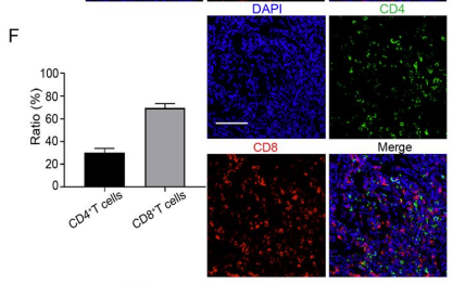
应用: IF
反应种属: Human
发表时间: 2023 Jul
-
Citation
-
The PD-L1 Expression and Tumor-Infiltrating Immune Cells Predict an Unfavorable Prognosis in Pancreatic Ductal Adenocarcinoma and Adenosquamous Carcinoma
Author: Zhang, Z., Xiong, Q., Xu, Y., Cai, X., Zhang, L., & Zhu, Q.
PMID: 36835933
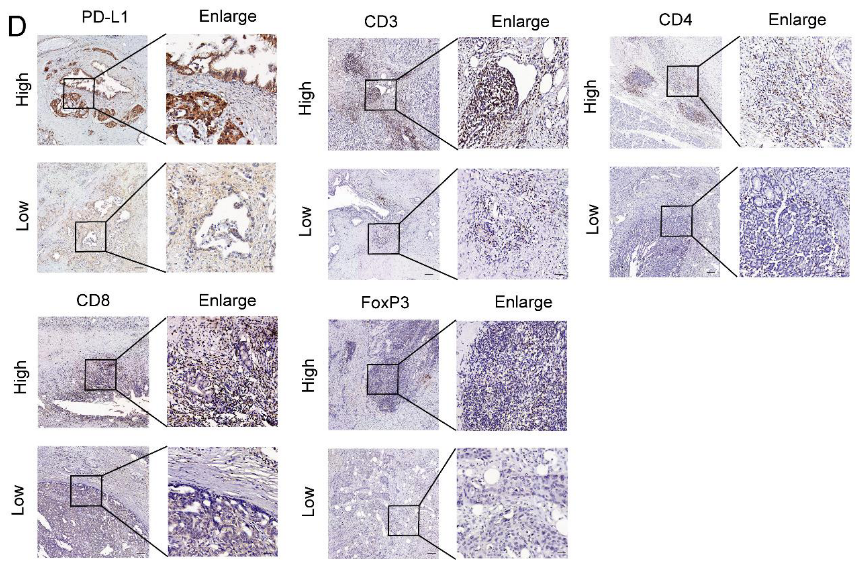
应用: IHC-P
反应种属: Human
发表时间: 2023 Feb
-
Citation
-
Proteomic Analyses Reveal Common Promiscuous Patterns of Cell Surface Proteins on Human Embryonic Stem Cells and Sperms
Author: Ming Zhang,Kangshou Yao
PMID: 21559292
应用: FC
反应种属: human
发表时间: 2011 May
-
Citation
-
Global Expression of Cell Surface Proteins in Embryonic Stem Cells
Author: Ming Zhang
PMID: 21209962

应用: WB,IHC
反应种属: human
发表时间: 2010 Dec
-
Citation
同靶点&同通路的产品
CD4 Rabbit Polyclonal Antibody
Application: WB,IF-Cell,FC
Reactivity: Human,Mouse,Rat
Conjugate: unconjugated
CD4 Recombinant Rabbit Monoclonal Antibody [JE56-36]
Application: WB,IHC-P
Reactivity: Human
Conjugate: unconjugated
CD4 Rabbit Polyclonal Antibody
Application: WB,IF-Cell,IHC-P,FC
Reactivity: Human
Conjugate: unconjugated
CD4
Application:
Reactivity:
Conjugate:
CD4 Rabbit Polyclonal Antibody
Application: WB,IF-Cell
Reactivity: Human
Conjugate: unconjugated
CD4 Rabbit Polyclonal Antibody
Application: WB,FC
Reactivity: Human
Conjugate: unconjugated


