概述
产品名称
CD34 Recombinant Rabbit Monoclonal Antibody [SI16-01]
抗体类型
Recombinant Rabbit monoclonal Antibody
免疫原
Synthetic peptide within Human CD34 aa 336-385 / 385.
种属反应性
Human, Mouse, Rat
验证应用
WB, IF-Cell, IF-Tissue, IHC-P, IP, mIHC, FC
分子量
Predicted band size: 41 kDa
阳性对照
Human non-small cell lung cancer, TF-1 cell lysate, TF-1, human tonsil tissue, human kidney tissue, mouse kidney tissue, rat kidney tissue, rat cerebellum tissue, rat cerebral cortex tissue, rat hippocampus tissue, mouse cerebellum tissue, mouse cerebral cortex tissue, mouse hippocampus tissue, mouse bladder tissue, human uterus tissue, mouse uterus tissue, rat uterus tissue.
偶联
unconjugated
克隆号
SI16-01
RRID
产品特性
形态
Liquid
浓度
1ug/ul
存放说明
Store at +4℃ after thawing. Aliquot store at -20℃ or -80℃. Avoid repeated freeze / thaw cycles.
存储缓冲液
1*TBS (pH7.4), 0.05% BSA, 40% Glycerol. Preservative: 0.05% Sodium Azide.
亚型
IgG
纯化方式
Protein A affinity purified.
应用稀释度
-
WB
-
1:2,000
-
IF-Cell
-
1:100
-
IF-Tissue
-
1:500-1:1,000
-
IHC-P
-
1:400-1:10,000
-
IP
-
1-2μg/sample
-
mIHC
-
1:1,000-1:2,000
-
FC
-
1:1,000
发表文章中的应用
发表文章中的种属
| Human | See 5 publications below |
| Mouse | See 4 publications below |
| mouse | See 2 publications below |
靶点
功能
CD34 is a heavily glycosylated, transmembrane glycoprotein that is expressed on the surface of lymphohematopoietic stem and progenitor cells, small-vessel endothelial cells, embryonic fibroblasts and some cells in fetal and adult nervous tissue. CD34 antigen expression is highest in the most primitive stem cells and is gradually lost as lineage committed progenitors differentiate. The CD34 antigen is also present on capillary endothelial cells and on bone marrow stromal cells. The CD34 cytoplasmic domain has an intracellular domain that contains consensus sites for activated protein kinase C (PKC) phosphorylation as well as serine, threonine and tyrosine phosphorylation consensus sites.
背景文献
1. Lin, SZ. et al. 2015. Emodin inhibits angiogenesis in pancreatic cancer by regulating the transforming growth factor-β/drosophila mothers against decapentaplegic pathway and angiogenesis-associated microRNAs. Molecular medicine reports. 12: 5865-71.
2. Corradi, LS. et al. 2013. Structural and ultrastructural evidence for telocytes in prostate stroma. J. Cell. Mol. Med. 17: 398-406.
序列相似性
Belongs to the CD34 family.
组织特异性
Selectively expressed on hematopoietic progenitor cells and the small vessel endothelium of a variety of tissues.
翻译后修饰
Highly glycosylated.; Phosphorylated on serine residues by PKC.
亚细胞定位
Membrane.
别名
CD34 antibody
CD34 antigen antibody
CD34 molecule antibody
CD34_HUMAN antibody
Cluster designation 34 antibody
Hematopoietic progenitor cell antigen CD34 antibody
HPCA1 antibody
Mucosialin antibody
OTTHUMP00000034733 antibody
OTTHUMP00000034734 antibody
图片
-
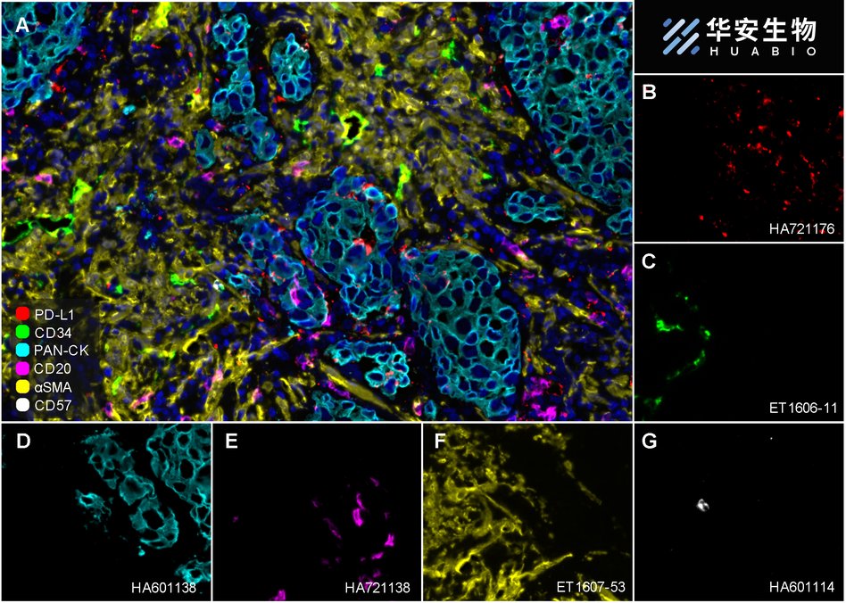
Fluorescence multiplex immunohistochemical analysis of Human non-small cell lung cancer (Formalin/PFA-fixed paraffin-embedded sections). Panel A: the merged image of anti-PD-L1 (HA721176, red), anti-CD34 (ET1606-11, green), anti-Pan-CK (HA601138, cyan), anti-CD20 (HA721138, magenta), anti-αSMA (ET1607-53, yellow) and anti-CD57 (HA601114, white) on NSCLC. Panel B: anti-PD-L1 stained on dendritic cells and macrophages cells. Panel C: anti- CD34 stained on endothelial cells. Panel D: anti-Pan-CK stained on cancer cells. Panel E: CD20 stained on B cells. Panel F: anti-αSMA stained on cancer-associated fibroblasts and smooth muscle cells. Panel G: anti-CD57 stained on NK cells and T cells. HRP Conjugated UltraPolymer Goat Polyclonal Antibody HA1119/HA1120 was used as a secondary antibody. The immunostaining was performed with the Sequential Immuno-staining Kit (IRISKit™MH010101, www.luminiris.cn). The section was incubated in six rounds of staining: in the order of HA721176 (1/1,000 dilution), ET1606-11 (1/1,000 dilution), HA601138 (1/3,000 dilution), HA721138 (1/2,000 dilution), ET1607-53 (1/3,000 dilution) and HA601114 (1/1,000 dilution) for 20 mins at room temperature. Each round was followed by a separate fluorescent tyramide signal amplification system. Heat mediated antigen retrieval with Tris-EDTA buffer (pH 9.0) for 30 mins at 95℃. DAPI (blue) was used as a nuclear counter stain. Image acquisition was performed with Olympus VS200 Slide Scanner.
-

Fluorescence multiplex immunohistochemical analysis of mouse brain (Formalin/PFA-fixed paraffin-embedded sections). Panel A: the merged image of anti-NeuN (ET1602-12, red), anti-Iba1 (ET1705-78, green), anti-GFAP (ET1601-23, gray), anti-Olig2 (ET1604-29, cyan), anti-MAP2 (HA500177, magenta) and anti-CD34 (ET1606-11, yellow) on mouse brain. HRP Conjugated UltraPolymer Goat Polyclonal Antibody HA1119/HA1120 was used as a secondary antibody. The immunostaining was performed with the Sequential Immuno-staining Kit (IRISKit™MH010101, www.luminiris.cn). The section was incubated in six rounds of staining: in the order of ET1602-12(1/5,000 dilution), ET1705-78 (1/2,000 dilution), ET1601-23 (1/5,000 dilution), ET1604-29 (1/1,000 dilution), HA500177 (1/5,000 dilution) and ET1606-11 (1/2,000 dilution) for 20 mins at room temperature. Each round was followed by a separate fluorescent tyramide signal amplification system. Heat mediated antigen retrieval with Tris-EDTA buffer (pH 9.0) for 30 mins at 95℃. DAPI (blue) was used as a nuclear counter stain. Image acquisition was performed with Olympus VS200 Slide Scanner.
-

Fluorescence multiplex immunohistochemical analysis of mouse brain (Formalin/PFA-fixed paraffin-embedded sections). Panel A: the merged image of anti-NeuN (ET1602-12, red), anti-PAX6 (ET1612-58, green), anti-CD34 (ET1606-11, gray), anti-MAP2 (HA500177, magenta) and anti-TBR1 (ET1702-97, yellow) on mouse brain. HRP Conjugated UltraPolymer Goat Polyclonal Antibody HA1119/HA1120 was used as a secondary antibody. The immunostaining was performed with the Sequential Immuno-staining Kit (IRISKit™MH010101, www.luminiris.cn). The section was incubated in five rounds of staining: in the order of ET1602-12 (1/5,000 dilution), ET1612-58 (1/1,000 dilution), ET1606-11 (1/2,000 dilution), HA500177 (1/5,000 dilution) and ET1702-97 (1/1,000 dilution) for 20 mins at room temperature. Each round was followed by a separate fluorescent tyramide signal amplification system. Heat mediated antigen retrieval with Tris-EDTA buffer (pH 9.0) for 30 mins at 95℃. DAPI (blue) was used as a nuclear counter stain. Image acquisition was performed with Olympus VS200 Slide Scanner.
-

Fluorescence multiplex immunohistochemical analysis of human non-small cell lung cancer (Formalin/PFA-fixed paraffin-embedded sections). Panel A: the merged image of anti-CD34 (ET1606-11, White), anti-PD-L1 (HA721176, Violet) and anti-pan Cytokeratin (HA601138, Yellow) on NSCLC. HRP Conjugated UltraPolymer Goat Polyclonal Antibody HA1119/HA1120 was used as a secondary antibody. The immunostaining was performed with the Sequential Immuno-staining Kit (IRISKit™MH010101, www.luminiris.cn). The section was incubated in three rounds of staining: in the order of ET1606-11 (1/2,000 dilution), HA721176 (1/1,000 dilution) and HA601138 (1/3,000 dilution) for 20 mins at room temperature. Each round was followed by a separate fluorescent tyramide signal amplification system. Heat mediated antigen retrieval with Tris-EDTA buffer (pH 9.0) for 30 mins at 95℃. DAPI (blue) was used as a nuclear counter stain. Image acquisition was performed with Zeiss Observer 7 Inverted Fluorescence Microscope.
-

Immunofluorescence analysis of paraffin-embedded human tonsil tissue labeling CD34 with Rabbit anti-CD34 antibody (ET1606-11) at 1/50 dilution.
The section was pre-treated using heat mediated antigen retrieval with Tris-EDTA buffer (pH 9.0) for 20 minutes. The tissues were blocked in 10% negative goat serum for 1 hour at room temperature, washed with PBS, and then probed with the primary antibody (ET1606-11, green) at 1/50 dilution overnight at 4 ℃, washed with PBS. Goat Anti-Rabbit IgG H&L (iFluor™ 488, HA1121) was used as the secondary antibody at 1/1,000 dilution. Nuclei were counterstained with DAPI (blue). -

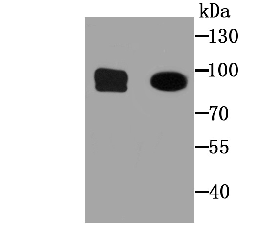
Western blot analysis of CD34 on TF-1 cell lysates with Rabbit anti-CD34 antibody (ET1606-11) at 1/2,000 dilution.
Lysates/proteins at 10 µg/Lane.
Predicted band size: 41 kDa
Observed band size: 100 kDa
Exposure time: 5 minutes;
4-20% SDS-PAGE gel.
Proteins were transferred to a PVDF membrane and blocked with 5% NFDM/TBST for 1 hour at room temperature. The primary antibody (ET1606-11) at 1/2,000 dilution was used in 5% NFDM/TBST at 4℃ overnight. Goat Anti-Rabbit IgG - HRP Secondary Antibody (HA1001) at 1:50,000 dilution was used for 1 hour at room temperature. -

Immunocytochemistry analysis of TF-1 cells labeling CD34 with Rabbit anti-CD34 antibody (ET1606-11) at 1/100 dilution.
Cells were fixed in 4% paraformaldehyde for 20 minutes at room temperature, permeabilized with 0.1% Triton X-100 in PBS for 5 minutes at room temperature, then blocked with 1% BSA in 10% negative goat serum for 1 hour at room temperature. Cells were then incubated with Rabbit anti-CD34 antibody (ET1606-11) at 1/100 dilution in 1% BSA in PBST overnight at 4 ℃. Goat Anti-Rabbit IgG H&L (iFluor™ 488, HA1121) was used as the secondary antibody at 1/1,000 dilution. PBS instead of the primary antibody was used as the secondary antibody only control. Nuclear DNA was labelled in blue with DAPI.
Beta tubulin (M1305-2, red) was stained at 1/100 dilution overnight at +4℃. Goat Anti-Mouse IgG H&L (iFluor™ 594, HA1126) was used as the secondary antibody at 1/1,000 dilution. -

Immunohistochemical analysis of paraffin-embedded human tonsil tissue with Rabbit anti-CD34 antibody (ET1606-11) at 1/1,000 dilution.
The section was pre-treated using heat mediated antigen retrieval with Tris-EDTA buffer (pH 9.0) for 20 minutes. The tissues were blocked in 1% BSA for 20 minutes at room temperature, washed with ddH2O and PBS, and then probed with the primary antibody (ET1606-11) at 1/1,000 dilution for 1 hour at room temperature. The detection was performed using an HRP conjugated compact polymer system. DAB was used as the chromogen. Tissues were counterstained with hematoxylin and mounted with DPX. -

Immunohistochemical analysis of paraffin-embedded human kidney tissue with Rabbit anti-CD34 antibody (ET1606-11) at 1/1,000 dilution.
The section was pre-treated using heat mediated antigen retrieval with Tris-EDTA buffer (pH 9.0) for 20 minutes. The tissues were blocked in 1% BSA for 20 minutes at room temperature, washed with ddH2O and PBS, and then probed with the primary antibody (ET1606-11) at 1/1,000 dilution for 1 hour at room temperature. The detection was performed using an HRP conjugated compact polymer system. DAB was used as the chromogen. Tissues were counterstained with hematoxylin and mounted with DPX. -

Immunohistochemical analysis of paraffin-embedded mouse kidney tissue with Rabbit anti-CD34 antibody (ET1606-11) at 1/1,000 dilution.
The section was pre-treated using heat mediated antigen retrieval with Tris-EDTA buffer (pH 9.0) for 20 minutes. The tissues were blocked in 1% BSA for 20 minutes at room temperature, washed with ddH2O and PBS, and then probed with the primary antibody (ET1606-11) at 1/1,000 dilution for 1 hour at room temperature. The detection was performed using an HRP conjugated compact polymer system. DAB was used as the chromogen. Tissues were counterstained with hematoxylin and mounted with DPX. -

Immunohistochemical analysis of paraffin-embedded human placenta tissue with Rabbit anti-CD34 antibody (ET1606-11) at 1/400 dilution.
The section was pre-treated using heat mediated antigen retrieval with Tris-EDTA buffer (pH 9.0) for 20 minutes. The tissues were blocked in 1% BSA for 20 minutes at room temperature, washed with ddH2O and PBS, and then probed with the primary antibody (ET1606-11) at 1/400 dilution for 1 hour at room temperature. The detection was performed using an HRP conjugated compact polymer system. DAB was used as the chromogen. Tissues were counterstained with hematoxylin and mounted with DPX. -

Immunohistochemical analysis of paraffin-embedded rat kidney tissue with Rabbit anti-CD34 antibody (ET1606-11) at 1/1,000 dilution.
The section was pre-treated using heat mediated antigen retrieval with Tris-EDTA buffer (pH 9.0) for 20 minutes. The tissues were blocked in 1% BSA for 20 minutes at room temperature, washed with ddH2O and PBS, and then probed with the primary antibody (ET1606-11) at 1/1,000 dilution for 1 hour at room temperature. The detection was performed using an HRP conjugated compact polymer system. DAB was used as the chromogen. Tissues were counterstained with hematoxylin and mounted with DPX. -

Immunohistochemical analysis of paraffin-embedded rat cerebellum tissue with Rabbit anti-CD34 antibody (ET1606-11) at 1/10,000 dilution.
The section was pre-treated using heat mediated antigen retrieval with Tris-EDTA buffer (pH 9.0) for 20 minutes. The tissues were blocked in 1% BSA for 20 minutes at room temperature, washed with ddH2O and PBS, and then probed with the primary antibody (ET1606-11) at 1/10,000 dilution for 1 hour at room temperature. The detection was performed using an HRP conjugated compact polymer system. DAB was used as the chromogen. Tissues were counterstained with hematoxylin and mounted with DPX. -

Immunohistochemical analysis of paraffin-embedded rat cerebral cortex tissue with Rabbit anti-CD34 antibody (ET1606-11) at 1/10,000 dilution.
The section was pre-treated using heat mediated antigen retrieval with Tris-EDTA buffer (pH 9.0) for 20 minutes. The tissues were blocked in 1% BSA for 20 minutes at room temperature, washed with ddH2O and PBS, and then probed with the primary antibody (ET1606-11) at 1/10,000 dilution for 1 hour at room temperature. The detection was performed using an HRP conjugated compact polymer system. DAB was used as the chromogen. Tissues were counterstained with hematoxylin and mounted with DPX. -

Immunohistochemical analysis of paraffin-embedded rat hippocampus tissue with Rabbit anti-CD34 antibody (ET1606-11) at 1/10,000 dilution.
The section was pre-treated using heat mediated antigen retrieval with Tris-EDTA buffer (pH 9.0) for 20 minutes. The tissues were blocked in 1% BSA for 20 minutes at room temperature, washed with ddH2O and PBS, and then probed with the primary antibody (ET1606-11) at 1/10,000 dilution for 1 hour at room temperature. The detection was performed using an HRP conjugated compact polymer system. DAB was used as the chromogen. Tissues were counterstained with hematoxylin and mounted with DPX. -

Immunohistochemical analysis of paraffin-embedded mouse cerebellum tissue with Rabbit anti-CD34 antibody (ET1606-11) at 1/10,000 dilution.
The section was pre-treated using heat mediated antigen retrieval with Tris-EDTA buffer (pH 9.0) for 20 minutes. The tissues were blocked in 1% BSA for 20 minutes at room temperature, washed with ddH2O and PBS, and then probed with the primary antibody (ET1606-11) at 1/10,000 dilution for 1 hour at room temperature. The detection was performed using an HRP conjugated compact polymer system. DAB was used as the chromogen. Tissues were counterstained with hematoxylin and mounted with DPX. -

Immunohistochemical analysis of paraffin-embedded mouse cerebral cortex tissue with Rabbit anti-CD34 antibody (ET1606-11) at 1/10,000 dilution.
The section was pre-treated using heat mediated antigen retrieval with Tris-EDTA buffer (pH 9.0) for 20 minutes. The tissues were blocked in 1% BSA for 20 minutes at room temperature, washed with ddH2O and PBS, and then probed with the primary antibody (ET1606-11) at 1/10,000 dilution for 1 hour at room temperature. The detection was performed using an HRP conjugated compact polymer system. DAB was used as the chromogen. Tissues were counterstained with hematoxylin and mounted with DPX. -

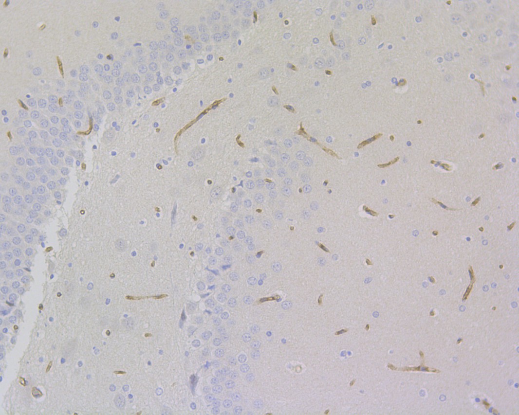
Immunohistochemical analysis of paraffin-embedded mouse hippocampus tissue with Rabbit anti-CD34 antibody (ET1606-11) at 1/10,000 dilution.
The section was pre-treated using heat mediated antigen retrieval with Tris-EDTA buffer (pH 9.0) for 20 minutes. The tissues were blocked in 1% BSA for 20 minutes at room temperature, washed with ddH2O and PBS, and then probed with the primary antibody (ET1606-11) at 1/10,000 dilution for 1 hour at room temperature. The detection was performed using an HRP conjugated compact polymer system. DAB was used as the chromogen. Tissues were counterstained with hematoxylin and mounted with DPX. -

Flow cytometric analysis of TF-1 cells labeling CD34.
Cells were fixed and permeabilized. Then stained with the primary antibody (ET1606-11, 1/1,000) (red) compared with Rabbit IgG Isotype Control (green). After incubation of the primary antibody at +4℃ for an hour, the cells were stained with a iFluor™ 488 conjugate-Goat anti-Rabbit IgG Secondary antibody (HA1121) at 1/1,000 dilution for 30 minutes at +4℃. Unlabelled sample was used as a control (cells without incubation with primary antibody; black). -

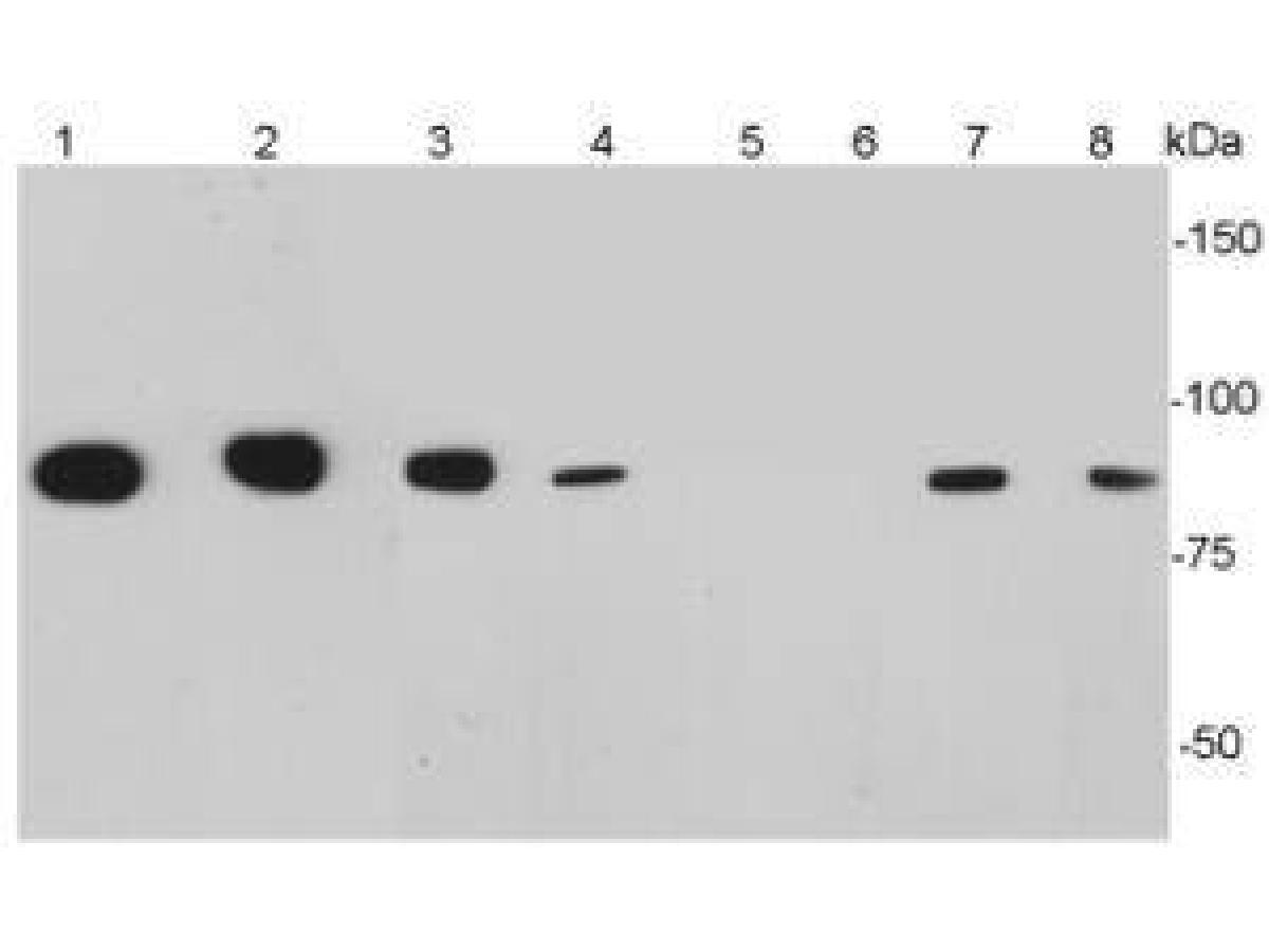
CD34 was immunoprecipitated from 0.2 mg TF-1 cell lysate with ET1606-11 at 2 µg/25 µl agarose. Western blot was performed from the immunoprecipitate using ET1606-11 at 1/2,000 dilution. Anti-Rabbit IgG for IP Nano-secondary antibody (NBI01H) at 1/5,000 dilution was used for 1 hour at room temperature.
Lane 1: TF-1 cell lysate (input)
Lane 2: ET1606-11 IP in TF-1 cell lysate
Lane 3: Rabbit IgG instead of ET1606-11 in TF-1 cell lysate
Blocking/Dilution buffer: 5% NFDM/TBST
Exposure time: 6 seconds; ECL: K1801 -

Immunofluorescence analysis of paraffin-embedded mouse bladder tissue labeling CD34 with Rabbit anti-CD34 antibody (ET1606-11) at 1/1,000 dilution.
The section was pre-treated using heat mediated antigen retrieval with Tris-EDTA buffer (pH 9.0) for 20 minutes. The tissues were blocked in 10% negative goat serum for 1 hour at room temperature, washed with PBS, and then probed with the primary antibody (ET1606-11, green) at 1/1,000 dilution overnight at 4 ℃, washed with PBS. Goat Anti-Rabbit IgG H&L (iFluor™ 488, HA1121) was used as the secondary antibody at 1/1,000 dilution. Nuclei were counterstained with DAPI (blue). -

Immunohistochemical analysis of paraffin-embedded human uterus tissue with Rabbit anti-CD34 antibody (ET1606-11) at 1/5,000 dilution.
The section was pre-treated using heat mediated antigen retrieval with Tris-EDTA buffer (pH 9.0) for 20 minutes. The tissues were blocked in 1% BSA for 20 minutes at room temperature, washed with ddH2O and PBS, and then probed with the primary antibody (ET1606-11) at 1/5,000 dilution for 1 hour at room temperature. The detection was performed using an HRP conjugated compact polymer system. DAB was used as the chromogen. Tissues were counterstained with hematoxylin and mounted with DPX. -

Immunohistochemical analysis of paraffin-embedded mouse uterus tissue with Rabbit anti-CD34 antibody (ET1606-11) at 1/5,000 dilution.
The section was pre-treated using heat mediated antigen retrieval with Tris-EDTA buffer (pH 9.0) for 20 minutes. The tissues were blocked in 1% BSA for 20 minutes at room temperature, washed with ddH2O and PBS, and then probed with the primary antibody (ET1606-11) at 1/5,000 dilution for 1 hour at room temperature. The detection was performed using an HRP conjugated compact polymer system. DAB was used as the chromogen. Tissues were counterstained with hematoxylin and mounted with DPX. -

Immunohistochemical analysis of paraffin-embedded rat uterus tissue with Rabbit anti-CD34 antibody (ET1606-11) at 1/5,000 dilution.
The section was pre-treated using heat mediated antigen retrieval with Tris-EDTA buffer (pH 9.0) for 20 minutes. The tissues were blocked in 1% BSA for 20 minutes at room temperature, washed with ddH2O and PBS, and then probed with the primary antibody (ET1606-11) at 1/5,000 dilution for 1 hour at room temperature. The detection was performed using an HRP conjugated compact polymer system. DAB was used as the chromogen. Tissues were counterstained with hematoxylin and mounted with DPX.
Please note: All products are "FOR RESEARCH USE ONLY AND ARE NOT INTENDED FOR DIAGNOSTIC OR THERAPEUTIC USE"
引文
-
Exosomal long non-coding RNA TRPM2-AS promotes angiogenesis in gallbladder cancer through interacting with PABPC1 to activate NOTCH1 signaling pathway
Author: Zhiqiang He , Yuhan Zhong , Parbatraj Regmi , et al
PMID: 38532427
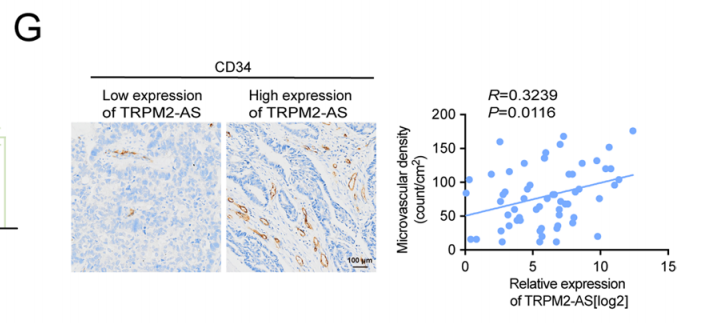
应用: IHC
反应种属: Human
发表时间: 2024 Mar
-
Citation
-
MSX1+ PDGFRAlow limb mesenchyme-like cells as an efficient stem cell source for human cartilage regeneration
Author: Liao Yuansong,et al
PMID: 38428414
应用: IF
反应种属: Mouse
发表时间: 2024 Mar
-
Citation
-
Human amniotic mesenchymal stem cell-islet organoids enhance the efficiency of islet engraftment in a mouse diabetes model
Author: Zhou Jiaxin,et al
PMID: 38862063
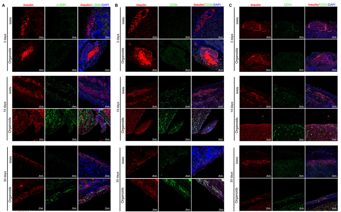
应用: IF
反应种属: Human
发表时间: 2024 Jun
-
Citation
-
Modulating cell stiffness for improved vascularization: leveraging the MIL-53(fe) for improved interaction of titanium implant and endothelial cell
Author: Wu Jie,et al
PMID: 39014416
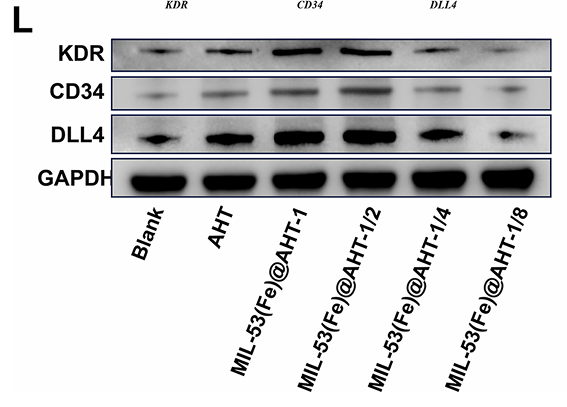
应用: WB,IF
反应种属: Human
发表时间: 2024 Jul
-
Citation
-
PP4R1 promotes glycolysis and gallbladder cancer progression through facilitating ERK1/2 mediated PKM2 nuclear translocation
Author: He Zhiqiang,et al
PMID: 38301910
应用: IHC
反应种属: Mouse
发表时间: 2024 Feb
-
Citation
-
Novel hKDR mouse model depicts the antiangiogenesis and apoptosis‐promoting effects of neutralizing antibodies targeting vascular endothelial growth factor receptor 2
Author:
PMID: 36114822

应用: IF-tissue
反应种属: mouse
发表时间: 2023 Jan
-
Citation
-
Impediment of Cerebrospinal Fluid Drainage Through Glymphatic System in Glioma
Author: Xu, D., Zhou, J., Mei, H., Li, H., Sun, W., & Xu, H.
PMID: 35083148

应用: IHC
反应种属: Human
发表时间: 2022 Jan
-
Citation
-
Dental niche cells directly contribute to tooth reconstitution and morphogenesis
Author: Hu, H., Duan, Y., Wang, K., Fu, H., Liao, Y., Wang, T., Zhang, Z., Kang, F., Zhang, B., Zhang, H., Huo, F., Yin, Y., Chen, G., Hu, H., Cai, H., Tian, W., & Li, Z.
PMID: 36476878

应用: IF
反应种属: Mouse
发表时间: 2022 Dec
-
Citation
-
Porcine Acellular Dermal Matrix Increases Fat Survival Rate after Fat Grafting in Nude Mice. Aesthetic plastic surgery, 10.1007/s00266-021-02299-z. Advance online publication.
Author: Zhu, M., Zhu, M., Wu, X., Xu, M., Fan, K., Wang, J., Zhang, L., Yin, M., Wu, J., Zhu, Z., & Yang, G.
PMID: 33959783
应用: IHC-P
反应种属: Pig
发表时间: 2021 Oct
-
Citation
-
<html>Suppression of Heterogeneous Nuclear Ribonucleoprotein C Inhibit Hepatocellular Carcinoma Proliferation, Migration, and Invasion <i>via</i> Ras/MAPK Signaling Pathway. Frontiers in oncology, 11, 659676.</html>
Author: Hu, J., Cai, D., Zhao, Z., Zhong, G. C., & Gong, J.
PMID: 33937074

应用: WB,IHC
反应种属: Human
发表时间: 2021 Apr
-
Citation
-
Interplay between cancer cells and M2 macrophages is necessary for miR-550a-3- 5p down-regulation-mediated HPV-positive OSCC progression
Author:
PMID: 32493454

应用: IHC
反应种属: Mouse
发表时间: 2020 Jun
-
Citation
-
Effect of cold stress on ovarian & uterine microcirculation in rats and the role of endothelin system
Author: Xiumei Cheng
PMID: 32290862
应用: IHC
反应种属: rat
发表时间: 2020 Apr
-
Citation
-
Toxicological Risk Assessments of Iron Oxide Nanocluster- and Gadolinium-Based T1MRI Contrast Agents in Renal Failure Rats. ACS nano, 13(6), 6801–6812.
Author: Weng, Q., Hu, X., Zheng, J., Xia, F., Wang, N., Liao, H., Liu, Y., Kim, D., Liu, J., Li, F., He, Q., Yang, B., Chen, C., Hyeon, T., & Ling, D.
PMID: 31141658

应用: ICC
反应种属: Rat
发表时间: 2019 Jun
-
Citation
-
Proteomic Analyses Reveal Common Promiscuous Patterns of Cell Surface Proteins on Human Embryonic Stem Cells and Sperms
Author: Ming Zhang,Kangshou Yao
PMID: 21559292
应用: ICC
反应种属: human,mouse
发表时间: 2011 May
-
Citation
-
Global Expression of Cell Surface Proteins in Embryonic Stem Cells
Author: Ming Zhang
PMID: 21209962

应用: WB,IHC
反应种属: human
发表时间: 2010 Dec
-
Citation
同靶点&同通路的产品
CD34 Mouse Monoclonal Antibody [15H1]
Application: IHC-P,FC
Reactivity: Human,Mouse,Rat
Conjugate: unconjugated
CD34 Rabbit Polyclonal Antibody
Application: WB,IHC-P
Reactivity: Human,Mouse
Conjugate: unconjugated
CD34 Rabbit Polyclonal Antibody
Application: WB,IF-Cell,IHC-P,FC
Reactivity: Human,Mouse,Rat,Zebrafish
Conjugate: unconjugated
CD34 Recombinant Mouse Monoclonal Antibody [PDM0-12]
Application: IHC-P
Reactivity: Human
Conjugate: unconjugated
CD34 Rabbit Polyclonal Antibody
Application: WB,IF-Cell,IHC-P
Reactivity: Human,Mouse,Zebrafish
Conjugate: unconjugated


