RMB: 900 特惠 1600 1600
产品规格
概述
产品名称
Histone H3 (acetyl K18) Mouse Monoclonal Antibody [A7B5]
抗体类型
Mouse Monoclonal Antibody
免疫原
Synthetic peptide within Human Histone H3 aa 1-50 / 136.
种属反应性
Human, Mouse, Rat
验证应用
WB, IHC-P, ChIP
分子量
Predicted band size: 15 kDa
阳性对照
NIH/3T3 cell lysate, NIH/3T3 treated with 400nM Calyculin A for 18 hours cell lysate, HeLa cell lysate, HeLa treated with 1μM Calyculin A for 18 hours cell lysate, C6 cell lysate, C6 treated with 1μM Calyculin A for 18 hours cell lysate, rat liver tissue, rat hippocampus tissue, rat kidney tissue, rat myocardium tissue, human lung tissue, human thyroid tissue, human colon carcinoma tissue, human skin tissue, human spleen tissue, human breast carcinoma tissue, human esophagus tissue, human placenta tissue.
偶联
unconjugated
克隆号
A7B5
RRID
产品特性
形态
Liquid
存放说明
Shipped at 4℃. Store at +4℃ short term (1-2 weeks). It is recommended to aliquot into single-use upon delivery. Store at -20℃ long term.
存储缓冲液
PBS (pH7.4), 0.05% BSA, 40% Glycerol. Preservative: 0.05% Sodium Azide.
亚型
IgG1
纯化方式
Protein G affinity purified.
应用稀释度
-
WB
-
1:2,000
-
IHC-P
-
1:500
-
ChIP
-
Use 0.5~2 μg for 25 μg of chromatin.
靶点
功能
Histones are basic nuclear proteins that are responsible for the nucleosome structure of the chromosomal fiber in eukaryotes. Two molecules of each of the four core histones (H2A, H2B, H3, and H4) form an octamer, around which approximately 146 bp of DNA is wrapped in repeating units, called nucleosomes. The linker histone, H1, interacts with linker DNA between nucleosomes and functions in the compaction of chromatin into higher order structures. This gene is intronless and encodes a replication-dependent histone that is a member of the histone H3 family. Transcripts from this gene lack polyA tails but instead contain a palindromic termination element. This gene is found in the small histone gene cluster on chromosome 6p22-p21.3.
背景文献
1. Benard A et al. Nuclear expression of histone deacetylases and their histone modifications predicts clinical outcome in colorectal cancer. Histopathology 66:270-82 (2015).
2. Zhang J et al. SOX4 inhibits GBM cell growth and induces G0/G1 cell cycle arrest through Akt-p53 axis. BMC Neurol 14:207 (2014).
亚细胞定位
Nucleus, Chromosome.
UNIPROT
别名
H3.3A antibody
HIST1 cluster, H3E antibody
H3 histone family, member A antibody
H3.1 antibody
H3/l antibody
H3F3 antibody
H3FF antibody
H3FJ antibody
H3FL antibody
Histone gene cluster 1, H3 histone family, member E antibody
展开H3.3A antibody
HIST1 cluster, H3E antibody
H3 histone family, member A antibody
H3.1 antibody
H3/l antibody
H3F3 antibody
H3FF antibody
H3FJ antibody
H3FL antibody
Histone gene cluster 1, H3 histone family, member E antibody
histone H3.1t antibody
Histone H3/o antibody
FLJ92264 antibody
H 3 antibody
H3 antibody
H3 histone family, member B antibody
H3 histone family, member C antibody
H3 histone family, member D antibody
H3 histone family, member F antibody
H3 histone family, member H antibody
H3 histone family, member I antibody
H3 histone family, member J antibody
H3 histone family, member K antibody
H3 histone family, member L antibody
H3 histone family, member T antibody
H3 histone, family 3A antibody
H3/A antibody
H3/b antibody
H3/c antibody
H3/d antibody
h3/f antibody
H3/h antibody
H3/i antibody
H3/j antibody
H3/k antibody
H3/t antibody
H31_HUMAN antibody
H3F1K antibody
H3F3A antibody
H3FA antibody
H3FB antibody
H3FC antibody
H3FD antibody
H3FH antibody
H3FI antibody
H3FK antibody
HIST1 cluster, H3A antibody
HIST1 cluster, H3B antibody
HIST1 cluster, H3C antibody
HIST1 cluster, H3D antibody
HIST1 cluster, H3F antibody
HIST1 cluster, H3G antibody
HIST1 cluster, H3H antibody
HIST1 cluster, H3I antibody
HIST1 cluster, H3J antibody
HIST1H3A antibody
HIST1H3B antibody
HIST1H3C antibody
HIST1H3D antibody
HIST1H3E antibody
HIST1H3F antibody
HIST1H3G antibody
HIST1H3H antibody
HIST1H3I antibody
HIST1H3J antibody
HIST3H3 antibody
Histone 1, H3a antibody
Histone 1, H3b antibody
Histone 1, H3c antibody
Histone 1, H3d antibody
Histone 1, H3e antibody
Histone 1, H3f antibody
Histone 1, H3g antibody
Histone 1, H3h antibody
Histone 1, H3i antibody
Histone 3, H3 antibody
histone cluster 1 H3 family member a antibody
histone cluster 1 H3 family member b antibody
histone cluster 1 H3 family member c antibody
histone cluster 1 H3 family member d antibody
histone cluster 1 H3 family member e antibody
histone cluster 1 H3 family member f antibody
histone cluster 1 H3 family member g antibody
histone cluster 1 H3 family member h antibody
histone cluster 1 H3 family member i antibody
histone cluster 1 H3 family member j antibody
Histone cluster 1, H3a antibody
Histone cluster 1, H3b antibody
Histone cluster 1, H3c antibody
Histone cluster 1, H3d antibody
Histone cluster 1, H3e antibody
Histone cluster 1, H3f antibody
Histone cluster 1, H3g antibody
Histone cluster 1, H3i antibody
Histone cluster 1, H3j antibody
Histone gene cluster 1, H3 histone family, member A antibody
Histone gene cluster 1, H3 histone family, member B antibody
Histone gene cluster 1, H3 histone family, member C antibody
Histone gene cluster 1, H3 histone family, member D antibody
Histone gene cluster 1, H3 histone family, member F antibody
Histone gene cluster 1, H3 histone family, member G antibody
Histone gene cluster 1, H3 histone family, member H antibody
Histone gene cluster 1, H3 histone family, member I antibody
Histone gene cluster 1, H3 histone family, member J antibody
Histone gene cluster 1, H3A antibody
Histone gene cluster 1, H3B antibody
Histone gene cluster 1, H3C antibody
Histone gene cluster 1, H3D antibody
Histone gene cluster 1, H3E antibody
Histone gene cluster 1, H3F antibody
Histone gene cluster 1, H3G antibody
Histone gene cluster 1, H3H antibody
Histone gene cluster 1, H3I antibody
Histone gene cluster 1, H3J antibody
Histone H 3 antibody
Histone H3.1 antibody
Histone H3.2 antibody
Histone H3.3 antibody
Histone H3/a antibody
Histone H3/b antibody
Histone H3/c antibody
Histone H3/d antibody
Histone H3/f antibody
Histone H3/h antibody
Histone H3/i antibody
Histone H3/j antibody
Histone H3/k antibody
Histone H3/l antibody
Histone H3/m antibody
H3K18ac antibody
折叠图片
-

☑ Cell treatment (CT)
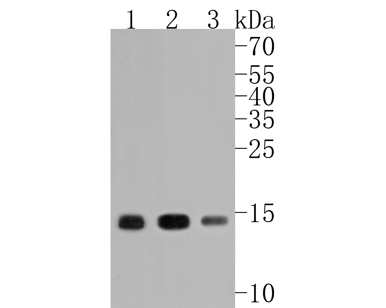
Western blot analysis of Histone H3 (acetyl K18) on different lysates with Mouse anti-Histone H3 (acetyl K18) antibody (HA600090) at 1/2,000 dilution.
Lane 1: NIH/3T3 cell lysate
Lane 2: NIH/3T3 treated with 400nM Calyculin A for 18 hours cell lysate
Lane 3: HeLa cell lysate
Lane 4: HeLa treated with 1μM Calyculin A for 18 hours cell lysate
Lane 5: C6 cell lysate
Lane 6: C6 treated with 1μM Calyculin A for 18 hours cell lysate
Lysates/proteins at 20 µg/Lane.
Predicted band size: 15 kDa
Observed band size: 15 kDa
Exposure time: 2 minutes; ECL: K1801;
4-20% SDS-PAGE gel.
Proteins were transferred to a PVDF membrane and blocked with 5% NFDM/TBST for 1 hour at room temperature. The primary antibody (HA600090) at 1/2,000 dilution was used in 5% NFDM/TBST at 4℃ overnight. Goat Anti-Mouse IgG - HRP Secondary Antibody (HA1006) at 1/50,000 dilution was used for 1 hour at room temperature. -

Immunohistochemical analysis of paraffin-embedded rat liver tissue with Mouse anti-Histone H3 (acetyl K18) antibody (HA600090) at 1/500 dilution.
The section was pre-treated using heat mediated antigen retrieval with sodium citrate buffer (pH 6.0) (high pressure) for 2 minutes. The tissues were blocked in 1% BSA for 20 minutes at room temperature, washed with ddH2O and PBS, and then probed with the primary antibody (HA600090) at 1/500 dilution for 1 hour at room temperature. The detection was performed using an HRP conjugated compact polymer system. DAB was used as the chromogen. Tissues were counterstained with hematoxylin and mounted with DPX. -

Immunohistochemical analysis of paraffin-embedded rat hippocampus tissue with Mouse anti-Histone H3 (acetyl K18) antibody (HA600090) at 1/500 dilution.
The section was pre-treated using heat mediated antigen retrieval with sodium citrate buffer (pH 6.0) (high pressure) for 2 minutes. The tissues were blocked in 1% BSA for 20 minutes at room temperature, washed with ddH2O and PBS, and then probed with the primary antibody (HA600090) at 1/500 dilution for 1 hour at room temperature. The detection was performed using an HRP conjugated compact polymer system. DAB was used as the chromogen. Tissues were counterstained with hematoxylin and mounted with DPX. -

Immunohistochemical analysis of paraffin-embedded rat kidney tissue with Mouse anti-Histone H3 (acetyl K18) antibody (HA600090) at 1/500 dilution.
The section was pre-treated using heat mediated antigen retrieval with sodium citrate buffer (pH 6.0) (high pressure) for 2 minutes. The tissues were blocked in 1% BSA for 20 minutes at room temperature, washed with ddH2O and PBS, and then probed with the primary antibody (HA600090) at 1/500 dilution for 1 hour at room temperature. The detection was performed using an HRP conjugated compact polymer system. DAB was used as the chromogen. Tissues were counterstained with hematoxylin and mounted with DPX. -

Immunohistochemical analysis of paraffin-embedded rat myocardium tissue with Mouse anti-Histone H3 (acetyl K18) antibody (HA600090) at 1/500 dilution.
The section was pre-treated using heat mediated antigen retrieval with sodium citrate buffer (pH 6.0) (high pressure) for 2 minutes. The tissues were blocked in 1% BSA for 20 minutes at room temperature, washed with ddH2O and PBS, and then probed with the primary antibody (HA600090) at 1/500 dilution for 1 hour at room temperature. The detection was performed using an HRP conjugated compact polymer system. DAB was used as the chromogen. Tissues were counterstained with hematoxylin and mounted with DPX. -

Immunohistochemical analysis of paraffin-embedded human lung tissue with Mouse anti-Histone H3 (acetyl K18) antibody (HA600090) at 1/500 dilution.
The section was pre-treated using heat mediated antigen retrieval with sodium citrate buffer (pH 6.0) (high pressure) for 2 minutes. The tissues were blocked in 1% BSA for 20 minutes at room temperature, washed with ddH2O and PBS, and then probed with the primary antibody (HA600090) at 1/500 dilution for 1 hour at room temperature. The detection was performed using an HRP conjugated compact polymer system. DAB was used as the chromogen. Tissues were counterstained with hematoxylin and mounted with DPX. -

Immunohistochemical analysis of paraffin-embedded human thyroid tissue with Mouse anti-Histone H3 (acetyl K18) antibody (HA600090) at 1/500 dilution.
The section was pre-treated using heat mediated antigen retrieval with sodium citrate buffer (pH 6.0) (high pressure) for 2 minutes. The tissues were blocked in 1% BSA for 20 minutes at room temperature, washed with ddH2O and PBS, and then probed with the primary antibody (HA600090) at 1/500 dilution for 1 hour at room temperature. The detection was performed using an HRP conjugated compact polymer system. DAB was used as the chromogen. Tissues were counterstained with hematoxylin and mounted with DPX. -

Immunohistochemical analysis of paraffin-embedded human colon carcinoma tissue with Mouse anti-Histone H3 (acetyl K18) antibody (HA600090) at 1/500 dilution.
The section was pre-treated using heat mediated antigen retrieval with sodium citrate buffer (pH 6.0) (high pressure) for 2 minutes. The tissues were blocked in 1% BSA for 20 minutes at room temperature, washed with ddH2O and PBS, and then probed with the primary antibody (HA600090) at 1/500 dilution for 1 hour at room temperature. The detection was performed using an HRP conjugated compact polymer system. DAB was used as the chromogen. Tissues were counterstained with hematoxylin and mounted with DPX. -

Immunohistochemical analysis of paraffin-embedded human skin tissue with Mouse anti-Histone H3 (acetyl K18) antibody (HA600090) at 1/500 dilution.
The section was pre-treated using heat mediated antigen retrieval with sodium citrate buffer (pH 6.0) (high pressure) for 2 minutes. The tissues were blocked in 1% BSA for 20 minutes at room temperature, washed with ddH2O and PBS, and then probed with the primary antibody (HA600090) at 1/500 dilution for 1 hour at room temperature. The detection was performed using an HRP conjugated compact polymer system. DAB was used as the chromogen. Tissues were counterstained with hematoxylin and mounted with DPX. -

Immunohistochemical analysis of paraffin-embedded human spleen tissue with Mouse anti-Histone H3 (acetyl K18) antibody (HA600090) at 1/500 dilution.
The section was pre-treated using heat mediated antigen retrieval with sodium citrate buffer (pH 6.0) (high pressure) for 2 minutes. The tissues were blocked in 1% BSA for 20 minutes at room temperature, washed with ddH2O and PBS, and then probed with the primary antibody (HA600090) at 1/500 dilution for 1 hour at room temperature. The detection was performed using an HRP conjugated compact polymer system. DAB was used as the chromogen. Tissues were counterstained with hematoxylin and mounted with DPX. -

Immunohistochemical analysis of paraffin-embedded human breast carcinoma tissue with Mouse anti-Histone H3 (acetyl K18) antibody (HA600090) at 1/500 dilution.
The section was pre-treated using heat mediated antigen retrieval with sodium citrate buffer (pH 6.0) (high pressure) for 2 minutes. The tissues were blocked in 1% BSA for 20 minutes at room temperature, washed with ddH2O and PBS, and then probed with the primary antibody (HA600090) at 1/500 dilution for 1 hour at room temperature. The detection was performed using an HRP conjugated compact polymer system. DAB was used as the chromogen. Tissues were counterstained with hematoxylin and mounted with DPX. -

Immunohistochemical analysis of paraffin-embedded human esophagus tissue with Mouse anti-Histone H3 (acetyl K18) antibody (HA600090) at 1/500 dilution.
The section was pre-treated using heat mediated antigen retrieval with sodium citrate buffer (pH 6.0) (high pressure) for 2 minutes. The tissues were blocked in 1% BSA for 20 minutes at room temperature, washed with ddH2O and PBS, and then probed with the primary antibody (HA600090) at 1/500 dilution for 1 hour at room temperature. The detection was performed using an HRP conjugated compact polymer system. DAB was used as the chromogen. Tissues were counterstained with hematoxylin and mounted with DPX. -

Immunohistochemical analysis of paraffin-embedded human placenta tissue with Mouse anti-Histone H3 (acetyl K18) antibody (HA600090) at 1/500 dilution.
The section was pre-treated using heat mediated antigen retrieval with sodium citrate buffer (pH 6.0) (high pressure) for 2 minutes. The tissues were blocked in 1% BSA for 20 minutes at room temperature, washed with ddH2O and PBS, and then probed with the primary antibody (HA600090) at 1/500 dilution for 1 hour at room temperature. The detection was performed using an HRP conjugated compact polymer system. DAB was used as the chromogen. Tissues were counterstained with hematoxylin and mounted with DPX. -

Chromatin immunoprecipitations were performed with cross-linked chromatin from HeLa cells with Histone H3 (acetyl K18) (HA600090) or Normal Mouse IgG according to the ChIP protocol. The enriched DNA was quantified by real-time PCR using indicated primers. The amount of immunoprecipitated DNA in each sample is represented as signal relative to the total amount of input chromatin, which is equivalent to one.
请注意: All products are "FOR RESEARCH USE ONLY AND ARE NOT INTENDED FOR DIAGNOSTIC OR THERAPEUTIC USE"
同靶点 & 同通路的产品
Histone H3 (mono methyl K36) Recombinant Rabbit Monoclonal Antibody [SR04-20]
Application: WB,IF-Cell,IF-Tissue,ChIP
Reactivity: Human,Mouse
Conjugate: unconjugated
Histone H3 Mouse Monoclonal Antibody [6-A7]
Application: WB,IHC-P,IF-Tissue,ChIP
Reactivity: Human,Mouse,Rat
Conjugate: unconjugated
Histone H3 (acetyl K14) Recombinant Rabbit Monoclonal Antibody [JU43-26]
Application: WB,IF-Cell,IF-Tissue,IHC-P,IP,SNAP-ChIP,CUT&Tag-seq
Reactivity: Human,Mouse,Rat
Conjugate: unconjugated
Histone H3 (acetyl K18) Rabbit Polyclonal Antibody
Application: WB,IHC-P,IF-Cell
Reactivity: Human,Mouse,Rat
Conjugate: unconjugated
Histone H3 (tri methyl K36) Recombinant Rabbit Monoclonal Antibody [PSH15-01]
Application: WB,IHC-P,IF-Cell,IP,Dot Blot,ChIP
Reactivity: Human,Mouse,Rat,Monkey
Conjugate: unconjugated
Histone H3 (acetyl K23) Recombinant Rabbit Monoclonal Antibody [JE46-39]
Application: WB,IF-Cell
Reactivity: Human,Mouse
Conjugate: unconjugated
Phospho-Histone H3 (S10) Recombinant Rabbit Monoclonal Antibody [SA31-01] - BSA and Azide free
Application: WB,IF-Cell,IHC-P,IP,FC,ChIP
Reactivity: Human,Mouse,Rat
Conjugate: unconjugated
Histone H3 (acetyl K56) Recombinant Rabbit Monoclonal Antibody [SU30-10]
Application: WB,IF-Cell,IF-Tissue,IHC-P,ChIP,CUT&Tag-seq
Reactivity: Human,Mouse,Rat
Conjugate: unconjugated
Histone H3 (acetyl K9) Recombinant Rabbit Monoclonal Antibody [PSH04-47] - BSA and Azide free
Application: WB,IF-Cell,IHC-P,IF-Tissue,FC,ChIP,Dot Blot,IP
Reactivity: Human,Mouse,Rat
Conjugate: unconjugated
Histone H3 Rabbit Polyclonal Antibody
Application: WB
Reactivity: Zebrafish,Human,Mouse
Conjugate: unconjugated
Histone H3 (acetyl K27) Recombinant Rabbit Monoclonal Antibody [PSH13-93] - BSA and Azide free
Application: WB,IHC-P,IF-Cell
Reactivity: Human,Mouse,Rat
Conjugate: unconjugated
Histone H3 (mono+di+tri methyl K79) Recombinant Rabbit Monoclonal Antibody [SR42-06] - BSA and Azide free
Application: WB,IHC-P,ChIP
Reactivity: Human,Mouse,Rat
Conjugate: unconjugated
Histone H3 Mouse Monoclonal Antibody [A11-D7]
Application: WB,IF-Cell,IHC-P,IF-Tissue
Reactivity: Human,Mouse,Rat,Zebrafish
Conjugate: unconjugated
Histone H3 (tri methyl K9) Mouse Monoclonal Antibody [1-6]
Application: WB,IF-Cell,IHC-P,IF-Tissue
Reactivity: Human,Mouse,Rat
Conjugate: unconjugated
Histone H3 (acetyl K27) Recombinant Rabbit Monoclonal Antibody [PSH13-93]
Application: WB,IHC-P,IF-Cell
Reactivity: Human,Mouse,Rat
Conjugate: unconjugated
Histone H3 Rabbit Polyclonal Antibody
Application: WB
Reactivity: Human,Mouse,Rat
Conjugate: unconjugated
Histone H3 (mono+di+tri methyl K79) Recombinant Rabbit Monoclonal Antibody [PSH09-10] - BSA and Azide free
Application: WB,IHC-P,IF-Cell,ChIP
Reactivity: Human,Mouse,Rat,Monkey
Conjugate: unconjugated
Histone H3 (di methyl K4) Rabbit Polyclonal Antibody
Application: WB,IF-Cell,IHC-P,FC,Dot Blot
Reactivity: Human,Mouse,Rat
Conjugate: unconjugated
Histone H3 (mono methyl K18) Recombinant Rabbit Monoclonal Antibody [SA42-07]
Application: WB,IF-Cell,IF-Tissue,IHC-P
Reactivity: Human,Mouse,Rat
Conjugate: unconjugated
Histone H3 (acetyl K27) Mouse Monoclonal Antibody [A6D6]
Application: WB,IF-Cell,IHC-P,FC,ChIP
Reactivity: Human,Mouse,Rat
Conjugate: unconjugated
Phospho-Histone H3 (T3) Recombinant Rabbit Monoclonal Antibody [JE42-48]
Application: WB,IHC-P,IF-Cell,FC
Reactivity: Human,Mouse,Rat
Conjugate: unconjugated
Histone H3 (mono methyl R2) Recombinant Rabbit Monoclonal Antibody [ST0427]
Application: WB,IF-Cell,IF-Tissue,IHC-P,ChIP
Reactivity: Human,Mouse,Rat
Conjugate: unconjugated
Histone H3 (acetyl K9) Recombinant Rabbit Monoclonal Antibody [PSH04-47] - ChIP Grade
Application: WB,IF-Cell,IHC-P,IF-Tissue,FC,ChIP,Dot Blot,IP
Reactivity: Human,Mouse,Rat
Conjugate: unconjugated
Histone H3 Recombinant Mouse Monoclonal Antibody [6-A7-R] - Loading control
Application: WB,IHC-P,IF-Tissue,ChIP
Reactivity: Human,Mouse,Rat
Conjugate: unconjugated
Histone H3 (mono methyl K14) Recombinant Rabbit Monoclonal Antibody [JE43-29]
Application: WB,IF-Cell,ChIP,Dot Blot
Reactivity: Human,Mouse,Rat
Conjugate: unconjugated
Histone H3 (tri methyl K27) Recombinant Rabbit Monoclonal Antibody [PSH05-15]
Application: WB,IF-Cell,IHC-P,IF-Tissue,ChIP
Reactivity: Human,Mouse,Rat
Conjugate: unconjugated
Histone H3 (acetyl K36) Recombinant Rabbit Monoclonal Antibody [JE77-32]
Application: WB,IHC-P,IF-Cell,ChIP
Reactivity: Human,Mouse,Rat
Conjugate: unconjugated
Histone H3 (mono+di+tri methyl K36) Recombinant Rabbit Monoclonal Antibody [PSH07-14]
Application: WB,IHC-P,IF-Cell,FC,ChIP
Reactivity: Human,Mouse,Rat
Conjugate: unconjugated
Histone H3 (mono methyl K18) Recombinant Rabbit Monoclonal Antibody [SA42-07] - BSA and Azide free
Application: WB,IF-Cell,IF-Tissue,IHC-P
Reactivity: Human,Mouse,Rat
Conjugate: unconjugated
Histone H3 (acetyl K56) Recombinant Rabbit Monoclonal Antibody [SU30-10] - BSA and Azide free
Application: WB,IF-Cell,IF-Tissue,IHC-P,ChIP,CUT&Tag-seq
Reactivity: Human,Mouse,Rat
Conjugate: unconjugated
Histone H3 (mono methyl R2) Recombinant Rabbit Monoclonal Antibody [ST0427] - BSA and Azide free
Application: WB,IF-Cell,IF-Tissue,IHC-P,ChIP
Reactivity: Human,Mouse,Rat
Conjugate: unconjugated
Histone H3 Mouse Monoclonal Antibody [3-C4]
Application: WB,IHC-P,mIHC,IF-Tissue,ChIP
Reactivity: Human,Mouse,Rat
Conjugate: unconjugated
Histone H3 (di methyl K9) Recombinant Rabbit Monoclonal Antibody [SN07-30] - BSA and Azide free
Application: WB,IF-Cell,IF-Tissue,IHC-P
Reactivity: Human,Mouse,Rat
Conjugate: unconjugated
Histone H3 Recombinant Rabbit Monoclonal Antibody [JJ090-07] - BSA and Azide free
Application: WB,IF-Cell,IF-Tissue,IHC-P,ChIP,IP
Reactivity: Human,Mouse,Rat
Conjugate: unconjugated
Histone H3 (mono methyl K9) Recombinant Rabbit Monoclonal Antibody [JE43-27]
Application: WB,IF-Cell,IHC-P,FC,ChIP,IF-Tissue,Dot Blot
Reactivity: Human,Mouse,Rat,Monkey
Conjugate: unconjugated
Histone H3 (acetyl K14) Recombinant Rabbit Monoclonal Antibody [JU43-26] - BSA and Azide free
Application: WB,IF-Cell,IF-Tissue,IHC-P,IP,SNAP-ChIP,CUT&Tag-seq
Reactivity: Human,Mouse,Rat
Conjugate: unconjugated
Histone H3 (acetyl K18) Mouse Monoclonal Antibody [A7B6]
Application: WB,IF-Cell
Reactivity: Human
Conjugate: unconjugated
Histone H3 Recombinant Mouse Monoclonal Antibody [6-A7-R] - BSA and Azide free
Application: WB,IHC-P,IF-Tissue,ChIP
Reactivity: Human,Mouse,Rat
Conjugate: unconjugated
Histone H3 (mono methyl K4) Recombinant Rabbit Monoclonal Antibody [PSH07-15]
Application: WB,IF-Cell,ChIP,Dot Blot
Reactivity: Human,Mouse,Rat
Conjugate: unconjugated
Histone H3 (tri methyl K27) Recombinant Rabbit Monoclonal Antibody [PSH05-15] - BSA and Azide free
Application: WB,IF-Cell,IHC-P,IF-Tissue,ChIP
Reactivity: Human,Mouse,Rat
Conjugate: unconjugated
Histone H3 (mono methyl K27) Recombinant Rabbit Monoclonal Antibody [PSH05-67] - BSA and Azide free
Application: WB,IF-Cell,FC,ChIP
Reactivity: Human,Mouse,Rat
Conjugate: unconjugated
Histone H3 (acetyl K4) Recombinant Rabbit Monoclonal Antibody [PSH03-71] - BSA and Azide free
Application: WB,IF-Cell,ChIP,Dot Blot
Reactivity: Human,Mouse,Rat
Conjugate: unconjugated
Histone H3 (di methyl K27) Recombinant Rabbit Monoclonal Antibody [PSH05-88] - BSA and Azide free
Application: WB,IF-Cell,ChIP
Reactivity: Human,Mouse,Rat
Conjugate: unconjugated
Histone H3 (mono+di+tri methyl K4) Recombinant Rabbit Monoclonal Antibody [PSH06-30] - BSA and Azide free
Application: WB,IHC-P,IF-Cell,FC,ChIP
Reactivity: Human,Mouse,Rat
Conjugate: unconjugated
Histone H3 (mono+di+tri methyl K36) Recombinant Rabbit Monoclonal Antibody [PSH07-14] - BSA and Azide free
Application: WB,IHC-P,IF-Cell,FC,ChIP
Reactivity: Human,Mouse,Rat
Conjugate: unconjugated
Histone H3 (mono methyl K4) Recombinant Rabbit Monoclonal Antibody [PSH07-15] - BSA and Azide free
Application: WB,IF-Cell,ChIP,Dot Blot
Reactivity: Human,Mouse,Rat
Conjugate: unconjugated
Histone H3 (mono methyl K79) Recombinant Rabbit Monoclonal Antibody [PSH07-66] - BSA and Azide free
Application: WB,IF-Cell,IHC-P,FC,ChIP
Reactivity: Human,Mouse,Rat,Monkey
Conjugate: unconjugated
Histone H3 Recombinant Mouse Monoclonal Antibody [A11-D7-R] - BSA and Azide free
Application: WB,IF-Cell,IHC-P
Reactivity: Human,Mouse,Rat
Conjugate: unconjugated
Phospho-Histone H3 (S10) Recombinant Rabbit Monoclonal Antibody [SA31-01]
Application: WB,IF-Cell,IHC-P,IP,FC,ChIP
Reactivity: Human,Mouse,Rat
Conjugate: unconjugated
Histone H3 (mono methyl K4) Rabbit Polyclonal Antibody
Application: WB,IF-Cell,IHC-P,FC,Dot Blot
Reactivity: Human,Mouse,Rat
Conjugate: unconjugated
Histone H3 (mono methyl K27) Recombinant Rabbit Monoclonal Antibody [PSH05-67]
Application: WB,IF-Cell,FC,ChIP
Reactivity: Human,Mouse,Rat
Conjugate: unconjugated
Histone H3 (mono+di+tri methyl K4) Recombinant Rabbit Monoclonal Antibody [PSH06-30]
Application: WB,IHC-P,IF-Cell,FC,ChIP
Reactivity: Human,Mouse,Rat
Conjugate: unconjugated
Histone H3 (acetyl K4) Recombinant Rabbit Monoclonal Antibody [PSH03-71]
Application: WB,IF-Cell,ChIP,Dot Blot
Reactivity: Human,Mouse,Rat
Conjugate: unconjugated
Histone H3 (mono methyl K79) Recombinant Rabbit Monoclonal Antibody [PSH07-66]
Application: WB,IF-Cell,IHC-P,FC,ChIP
Reactivity: Human,Mouse,Rat,Monkey
Conjugate: unconjugated
Histone H3 (di methyl K9) Recombinant Rabbit Monoclonal Antibody [SN07-30]
Application: WB,IF-Cell,IF-Tissue,IHC-P
Reactivity: Human,Mouse,Rat
Conjugate: unconjugated
Histone H3 (acetyl K27) Rabbit Polyclonal Antibody
Application: WB,IHC-P,ChIP
Reactivity: Human,Mouse,Rat
Conjugate: unconjugated
Histone H3 (mono+di+tri methyl K79) Recombinant Rabbit Monoclonal Antibody [SR42-06]
Application: WB,IHC-P,ChIP
Reactivity: Human,Mouse,Rat
Conjugate: unconjugated
Histone H3 (tri methyl K9) Mouse Monoclonal Antibody [2G1]
Application: WB,IF
Reactivity: Human
Conjugate: unconjugated
Histone H3 Recombinant Mouse Monoclonal Antibody [A11-D7-R]
Application: WB,IF-Cell,IHC-P
Reactivity: Human,Mouse,Rat
Conjugate: unconjugated
Histone H3 Recombinant Rabbit Monoclonal Antibody [JJ090-07]
Application: WB,IF-Cell,IF-Tissue,IHC-P,ChIP,IP
Reactivity: Human,Mouse,Rat
Conjugate: unconjugated
Histone H3 (di methyl K4) Recombinant Rabbit Monoclonal Antibody [JE00-98]
Application: WB,IF-Cell,ChIP,Dot Blot,IHC-P
Reactivity: Human,Mouse,Rat,Monkey
Conjugate: unconjugated
Phospho-Histone H3 (S28) Recombinant Rabbit Monoclonal Antibody [JE56-06]
Application: WB,IF-Cell,IHC-P,FC,ChIP
Reactivity: Human,Rat,Mouse
Conjugate: unconjugated
Histone H3 (di methyl K27) Recombinant Rabbit Monoclonal Antibody [PSH05-88]
Application: WB,IF-Cell,ChIP
Reactivity: Human,Mouse,Rat
Conjugate: unconjugated
Histone H3 (acetyl K27) Mouse Monoclonal Antibody [A6D7]
Application: WB,IF-Cell,IHC-P,ChIP,FC(Intra)
Reactivity: Human,Mouse,Rat
Conjugate: unconjugated
Histone H3 Recombinant Rabbit Monoclonal Antibody [JJ092-08]
Application: WB,IF-Tissue,IHC-P,ChIP
Reactivity: Human,Mouse,Rat
Conjugate: unconjugated
Histone H3 (mono+di+tri methyl K79) Recombinant Rabbit Monoclonal Antibody [PSH09-10]
Application: WB,IHC-P,IF-Cell,ChIP
Reactivity: Human,Mouse,Rat,Monkey
Conjugate: unconjugated











 浙公网安备 33019202000643号