GAPDH Rabbit Polyclonal Antibody
Catalog# R1210-1
GAPDH Rabbit Polyclonal Antibody
-
WB
-
IF-Cell
-
IHC-P
-
FC
-
Human
-
Mouse
-
Rat
-
Oryza sativa
-
unconjugated
概述
产品名称
GAPDH Rabbit Polyclonal Antibody
抗体类型
Rabbit Polyclonal Antibody
免疫原
This antibody is produced by immunizing rabbits with full length recombinant protein of GAPDH.
种属反应性
Human, Mouse, Rat, Oryza sativa
验证应用
WB, IF-Cell, IHC-P, FC
分子量
Predicted band size: 36 kDa
阳性对照
HeLa cell lysate, A431 cell lysate, HEK-293 cell lysate, Jurkat cell lysate, NIH/3T3 cell lysate, RAW264.7 cell lysate, PC-12 cell lysate, C6 cell lysate, mouse brain tissue lysate, rat brain tissue lysate, A549, LOVO, MCF-7, rat kidney tissue, human colon cancer tissue, human spleen tissue, mouse testis tissue, Hela.
偶联
unconjugated
RRID
产品特性
形态
Liquid
存放说明
Shipped at 4℃. Store at +4℃ short term (1-2 weeks). It is recommended to aliquot into single-use upon delivery. Store at -20℃ long term.
存储缓冲液
1*PBS (pH7.4), 0.2% BSA, 40% Glycerol. Preservative: 0.05% Sodium Azide.
亚型
IgG
纯化方式
Immunogen affinity purified.
应用稀释度
-
WB
-
1:5,000-1:20,000
-
IF-Cell
-
1:100-1:200
-
IHC-P
-
1:50-1:200
-
FC
-
1:50-1:100
靶点
功能
Glyceraldehyde-3-phosphate dehydrogenase (GAPDH) catalyzes the phosphorylation of glyceraldehyde-3-phosphate during glycolysis. It participates in nuclear events including transcription, RNA transport, DNA replication and apoptosis. GAPDH is thought to be a constitutively expressed housekeeping protein. For this reason, GAPDH mRNA and protein levels are often measured as controls in experiments quantifying specific changes in expression of other targets.
背景文献
1. Allen R.W et al. Identification of the 37-kDa protein displaying a variable interaction with the erythroid cell membrane as glyceraldehyde-3-phosphate dehydrogenase. J Biol Chem 262:649-653 (1987).
2. Meyer-Siegler K et al. A human nuclear uracil DNA glycosylase is the 37-kDa subunit of glyceraldehyde-3-phosphate dehydrogenase. Proc Natl Acad Sci USA 88:8460-8464 (1991).
序列相似性
Belongs to the glyceraldehyde-3-phosphate dehydrogenase family.
翻译后修饰
S-nitrosylation of Cys-152 leads to interaction with SIAH1, followed by translocation to the nucleus (By similarity). S-nitrosylation of Cys-247 is induced by interferon-gamma and LDL(ox) implicating the iNOS-S100A8/9 transnitrosylase complex and seems to prevent interaction with phosphorylated RPL13A and to interfere with GAIT complex activity.; ISGylated.; Sulfhydration at Cys-152 increases catalytic activity.; Oxidative stress can promote the formation of high molecular weight disulfide-linked GAPDH aggregates, through a process called nucleocytoplasmic coagulation. Such aggregates can be observed in vivo in the affected tissues of patients with Alzheimer disease or alcoholic liver cirrhosis, or in cell cultures during necrosis. Oxidation at Met-46 may play a pivotal role in the formation of these insoluble structures. This modification has been detected in vitro following treatment with free radical donor (+/-)-(E)-4-ethyl-2-[(E)-hydroxyimino]-5-nitro-3-hexenamide. It has been proposed to destabilize nearby residues, increasing the likelihood of secondary oxidative damages, including oxidation of Tyr-45 and Met-105. This cascade of oxidations may augment GAPDH misfolding, leading to intermolecular disulfide cross-linking and aggregation.; Succination of Cys-152 and Cys-247 by the Krebs cycle intermediate fumarate, which leads to S-(2-succinyl)cysteine residues, inhibits glyceraldehyde-3-phosphate dehydrogenase activity. Fumarate concentration as well as succination of cysteine residues in GAPDH is significantly increased in muscle of diabetic mammals. It was proposed that the S-(2-succinyl)cysteine chemical modification may be a useful biomarker of mitochondrial and oxidative stress in diabetes and that succination of GAPDH and other thiol proteins by fumarate may contribute to the metabolic changes underlying the development of diabetes complications.
亚细胞定位
Cytoplasm, Nucleus.
别名
38 kDa BFA-dependent ADP-ribosylation substrate antibody
aging associated gene 9 protein antibody
Aging-associated gene 9 protein antibody
BARS-38 antibody
cb609 antibody
EC 1.2.1.12 antibody
Epididymis secretory sperm binding protein Li 162eP antibody
G3P_HUMAN antibody
G3PD antibody
G3PDH antibody
展开38 kDa BFA-dependent ADP-ribosylation substrate antibody
aging associated gene 9 protein antibody
Aging-associated gene 9 protein antibody
BARS-38 antibody
cb609 antibody
EC 1.2.1.12 antibody
Epididymis secretory sperm binding protein Li 162eP antibody
G3P_HUMAN antibody
G3PD antibody
G3PDH antibody
GAPD antibody
GAPDH antibody
Glyceraldehyde 3 phosphate dehydrogenase antibody
Glyceraldehyde-3-phosphate dehydrogenase antibody
HEL-S-162eP antibody
KNC-NDS6 antibody
MGC102544 antibody
MGC102546 antibody
MGC103190 antibody
MGC103191 antibody
MGC105239 antibody
MGC127711 antibody
MGC88685 antibody
OCAS, p38 component antibody
OCT1 coactivator in S phase, 38-KD component antibody
peptidyl cysteine S nitrosylase GAPDH antibody
Peptidyl-cysteine S-nitrosylase GAPDH antibody
wu:fb33a10 antibody
折叠图片
-

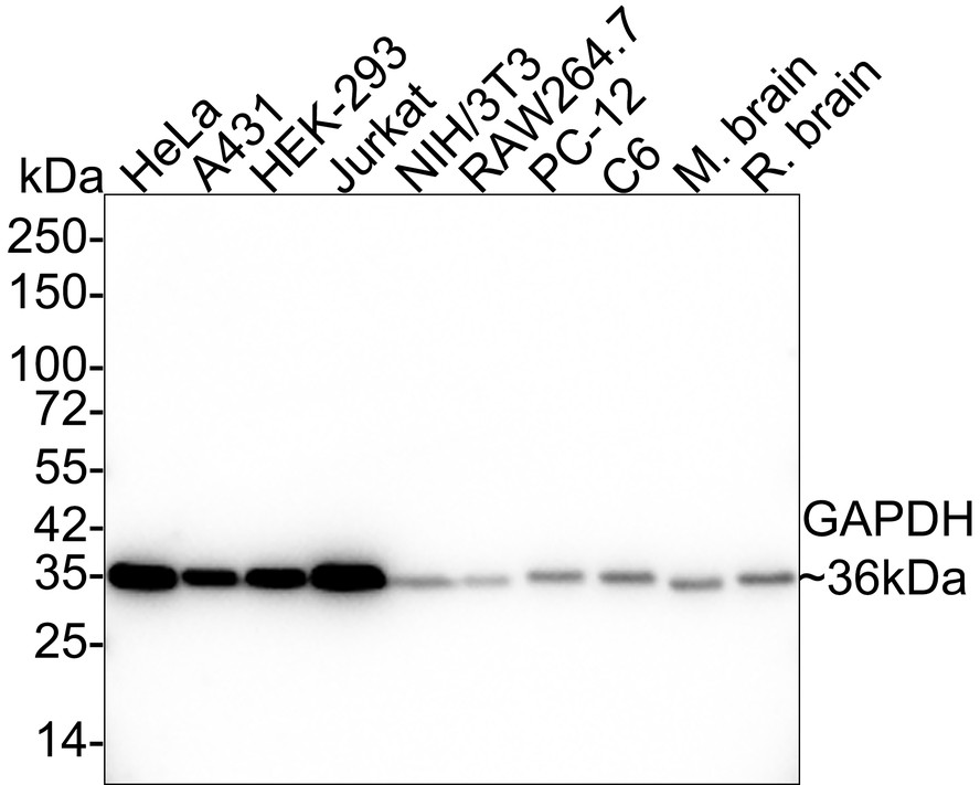
Western blot analysis of GAPDH on different lysates with Rabbit anti-GAPDH antibody (R1210-1) at 1/20,000 dilution.
Lane 1: HeLa cell lysate
Lane 2: A431 cell lysate
Lane 3: HEK-293 cell lysate
Lane 4: Jurkat cell lysate
Lane 5: NIH/3T3 cell lysate
Lane 6: RAW264.7 cell lysate
Lane 7: PC-12 cell lysate
Lane 8: C6 cell lysate
Lane 9: Mouse brain tissue lysate
Lane 10: Rat brain tissue lysate
Lysates/proteins at 20 µg/Lane.
Predicted band size: 36 kDa
Observed band size: 36 kDa
Exposure time: 20 seconds;
4-20% SDS-PAGE gel.
Proteins were transferred to a PVDF membrane and blocked with 5% NFDM/TBST for 1 hour at room temperature. The primary antibody (R1210-1) at 1/20,000 dilution was used in 5% NFDM/TBST at 4℃ overnight. Goat Anti-Rabbit IgG - HRP Secondary Antibody (HA1001) at 1:50,000 dilution was used for 1 hour at room temperature. -

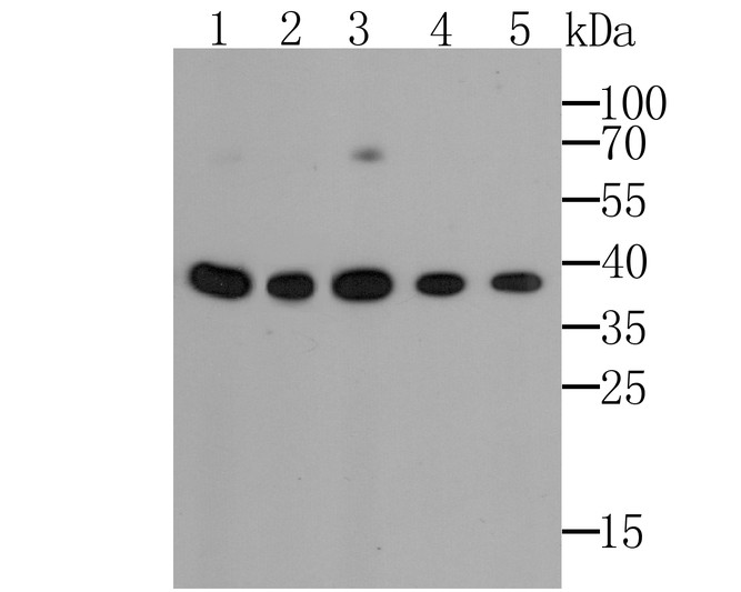
Western blot analysis of GAPDH on Oryza sativa lysates with Rabbit anti-GAPDH antibody (R1210-1) at 1/5,000 dilution.
Lysates/proteins at 15 µg/Lane.
Predicted band size: 36 kDa
Observed band size: 36 kDa
Exposure time: 42 seconds; ECL: K1801;
4-20% SDS-PAGE gel.
Proteins were transferred to a PVDF membrane and blocked with 5% NFDM/TBST for 1 hour at room temperature. The primary antibody (R1210-1) at 1/5,000 dilution was used in 5% NFDM/TBST at 4℃ overnight. Goat Anti-Rabbit IgG - HRP Secondary Antibody (HA1001) at 1/50,000 dilution was used for 1 hour at room temperature. -

Western blot analysis of GAPDH on COS-1 cell/tissue lysates with Rabbit anti-GAPDH antibody (R1210-1) at 1/20,000 dilution.
Lysates/proteins at 10 µg/Lane.
Predicted band size: 36 kDa
Observed band size: 36 kDa
Exposure time: 2 minutes;
4-20% SDS-PAGE gel.
Proteins were transferred to a PVDF membrane and blocked with 5% NFDM/TBST for 1 hour at room temperature. The primary antibody (R1210-1) at 1/20,000 dilution was used in 5% NFDM/TBST at 4℃ overnight. Goat Anti-Rabbit IgG - HRP Secondary Antibody (HA1001) at 1/50,000 dilution was used for 1 hour at room temperature. -

Immunocytochemistry analysis of HeLa cells labeling GAPDH with Rabbit anti-GAPDH antibody (R1210-1) at 1/100 dilution.
Cells were fixed in 4% paraformaldehyde for 20 minutes at room temperature, permeabilized with 0.1% Triton X-100 in PBS for 5 minutes at room temperature, then blocked with 1% BSA in 10% negative goat serum for 1 hour at room temperature. Cells were then incubated with Rabbit anti-GAPDH antibody (R1210-1) at 1/100 dilution in 1% BSA in PBST overnight at 4 ℃. Goat Anti-Rabbit IgG H&L (iFluor™ 488, HA1121) was used as the secondary antibody at 1/1,000 dilution. PBS instead of the primary antibody was used as the secondary antibody only control. Nuclear DNA was labelled in blue with DAPI.
Beta tubulin (M1305-2, red) was stained at 1/100 dilution overnight at +4℃. Goat Anti-Mouse IgG H&L (iFluor™ 594, HA1126) was used as the secondary antibody at 1/1,000 dilution. -

Immunocytochemistry analysis of NIH/3T3 cells labeling GAPDH with Rabbit anti-GAPDH antibody (R1210-1) at 1/100 dilution.
Cells were fixed in 4% paraformaldehyde for 20 minutes at room temperature, permeabilized with 0.1% Triton X-100 in PBS for 5 minutes at room temperature, then blocked with 1% BSA in 10% negative goat serum for 1 hour at room temperature. Cells were then incubated with Rabbit anti-GAPDH antibody (R1210-1) at 1/100 dilution in 1% BSA in PBST overnight at 4 ℃. Goat Anti-Rabbit IgG H&L (iFluor™ 488, HA1121) was used as the secondary antibody at 1/1,000 dilution. PBS instead of the primary antibody was used as the secondary antibody only control. Nuclear DNA was labelled in blue with DAPI.
Beta tubulin (M1305-2, red) was stained at 1/100 dilution overnight at +4℃. Goat Anti-Mouse IgG H&L (iFluor™ 594, HA1126) was used as the secondary antibody at 1/1,000 dilution. -

Immunocytochemistry analysis of C6 cells labeling GAPDH with Rabbit anti-GAPDH antibody (R1210-1) at 1/100 dilution.
Cells were fixed in 4% paraformaldehyde for 20 minutes at room temperature, permeabilized with 0.1% Triton X-100 in PBS for 5 minutes at room temperature, then blocked with 1% BSA in 10% negative goat serum for 1 hour at room temperature. Cells were then incubated with Rabbit anti-GAPDH antibody (R1210-1) at 1/100 dilution in 1% BSA in PBST overnight at 4 ℃. Goat Anti-Rabbit IgG H&L (iFluor™ 488, HA1121) was used as the secondary antibody at 1/1,000 dilution. PBS instead of the primary antibody was used as the secondary antibody only control. Nuclear DNA was labelled in blue with DAPI.
Beta tubulin (M1305-2, red) was stained at 1/100 dilution overnight at +4℃. Goat Anti-Mouse IgG H&L (iFluor™ 594, HA1126) was used as the secondary antibody at 1/1,000 dilution. -

Immunohistochemical analysis of paraffin-embedded rat kidney tissue using anti-GAPDH antibody. The section was pre-treated using heat mediated antigen retrieval with sodium citrate buffer (pH 6.0) (high pressure) for 2 minutes. The tissues were blocked in 5% BSA for 30 minutes at room temperature, washed with ddH2O and PBS, and then probed with the antibody (R1210-1) at 1/100 dilution, for 30 minutes at room temperature and detected using an HRP conjugated compact polymer system. DAB was used as the chrogen. Counter stained with hematoxylin and mounted with DPX.
-

Immunohistochemical analysis of paraffin-embedded human colon cancer tissue using anti-GAPDH antibody. The section was pre-treated using heat mediated antigen retrieval with Tris-EDTA buffer (pH 8.0-8.4) for 20 minutes.The tissues were blocked in 5% BSA for 30 minutes at room temperature, washed with ddH2O and PBS, and then probed with the antibody (R1210-1) at 1/100 dilution, for 30 minutes at room temperature and detected using an HRP conjugated compact polymer system. DAB was used as the chrogen. Counter stained with hematoxylin and mounted with DPX.
-

Immunohistochemical analysis of paraffin-embedded human spleen tissue using anti-GAPDH antibody. The section was pre-treated using heat mediated antigen retrieval with sodium citrate buffer (pH 6.0) (high pressure) for 2 minutes. The tissues were blocked in 5% BSA for 30 minutes at room temperature, washed with ddH2O and PBS, and then probed with the antibody (R1210-1) at 1/100 dilution, for 30 minutes at room temperature and detected using an HRP conjugated compact polymer system. DAB was used as the chrogen. Counter stained with hematoxylin and mounted with DPX.
-

Immunohistochemical analysis of paraffin-embedded mouse testis tissue using anti-GAPDH antibody. The section was pre-treated using heat mediated antigen retrieval with Tris-EDTA buffer (pH 8.0-8.4) for 20 minutes.The tissues were blocked in 5% BSA for 30 minutes at room temperature, washed with ddH2O and PBS, and then probed with the antibody (R1210-1) at 1/100 dilution, for 30 minutes at room temperature and detected using an HRP conjugated compact polymer system. DAB was used as the chrogen. Counter stained with hematoxylin and mounted with DPX.
-

Flow cytometric analysis of HeLa cells labeling GAPDH.
Cells were fixed and permeabilized. Then stained with the primary antibody (R1210-1, 1μg/mL) (red) compared with Rabbit IgG Isotype Control (green). After incubation of the primary antibody at +4℃ for an hour, the cells were stained with a iFluor™ 488 conjugate-Goat anti-Rabbit IgG Secondary antibody (HA1121) at 1/1,000 dilution for 30 minutes at +4℃. Unlabelled sample was used as a control (cells without incubation with primary antibody; black).
请注意: All products are "FOR RESEARCH USE ONLY AND ARE NOT INTENDED FOR DIAGNOSTIC OR THERAPEUTIC USE"
引文
-
Leucine‑rich repeat‑containing G protein‑coupled receptor 4 promotes proliferation, invasion and migration, and inhibits apoptosis in non‑small cell lung cancer cells
期刊: Oncology Letters
DOI: 10.3892/ol.2025.15304
IF: 2.2
应用: WB
反应种属: Human
发表时间: 2025 Sept
-
PRMT5 encourages cell migration and metastasis of tongue squamous cell carcinoma through methylating ΔNp63α
期刊: Cell Death & Differentiation
DOI: 10.1038/s41418-025-01575-8
IF: 15.4
应用: WB
反应种属: Human
发表时间: 2025 Sept
-
Chrysin inhibits hypertrophic scar formation through TGF-β/Smad signaling pathways
期刊: Journal Of Molecular Histology
DOI: 10.1007/s10735-025-10576-3
IF: 2.2
应用: WB
反应种属: Human
发表时间: 2025 Sept
-
Development of a prognostic model based on seven mitochondrial autophagy- and ferroptosis-related genes in lung adenocarcinoma
期刊: BMC Medical Genomics
DOI: 10.1186/s12920-025-02216-2
IF: 2
应用: WB
反应种属: Human
发表时间: 2025 Oct
-
Activation of the MEK1-CHK2 axis in macrophages by Staphylococcus aureus promotes mitophagy, resulting in a reduction in bactericidal efficacy
期刊: Molecular Medicine
DOI: 10.1186/s10020-025-01274-7
IF: 6
应用: WB
反应种属: Mouse
发表时间: 2025 May
-
Transcriptome and proteome profile analysis of the regulation of chicken ovarian development
期刊: Poultry Science
DOI: 10.1016/j.psj.2025.105384
IF: 3.8
应用: WB
反应种属: Chicken
发表时间: 2025 May
-
Activation of HTR2B Suppresses Osteosarcoma Progression through the STAT1-NLRP3 Inflammasome Pathway and Promotes OASL1+ Macrophage Production to Enhance Antitumor Immunity
期刊: Advanced Science
DOI: 10.1002/advs.202415276
IF: 14.3
应用: WB
反应种属: Mouse
发表时间: 2025 May
-
Kaempferol Induces DNA Damage in Colorectal Cancer Cells by Regulating the MiR-195/miR-497-PFKFB4-Mediated Nonoxidative Pentose Phosphate Pathway
期刊: Journal Of Agricultural And Food Chemistry
DOI: 10.1021/acs.jafc.4c13123
IF: 5.7
应用: WB
反应种属: Human
发表时间: 2025 Mar
-
Adeno-associated virus-mediated inhibition of ROCK2 promotes synaptogenesis and neurogenesis in rats after ischemic stroke
期刊: Neural Regeneration Research
DOI: 10.4103/NRR.NRR-D-24-01474
IF: 6.7
应用: WB
反应种属: Rat
发表时间: 2025 Jun
-
Obacunone Alleviates Thalamic Pain via Promoting LCN2-Mediated Phagocytosis of Astrocytes in Mice
期刊: ACS Chemical Neuroscience
DOI: 10.1021/acschemneuro.5c00371
IF: 3.9
应用: WB
反应种属: Mouse
发表时间: 2025 Jun
-
Identification of lipid metabolism-associated biomarkers in lupus nephritis by SVM model and therapeutic potential of Alisol B 23-acetate
期刊: Gene
DOI: 10.1016/j.gene.2025.149628
IF: 2.4
应用: WB
反应种属: Human
发表时间: 2025 Jun
-
CD11b Blockade Ameliorates Myocardial Ischemia/Reperfusion Injury by Reducing Neutrophil and Monocyte Infiltration
期刊: Journal Of The American Heart Association
DOI: 10.1161/JAHA.124.038142
IF: 5.3
应用: WB
反应种属: Mouse
发表时间: 2025 Jun
-
Omega-3 Polyunsaturated Fatty Acids Prevent Sevoflurane-induced Cognitive and Fine Motor Dysfunctions in Neonatal Mice by Enhancing Phosphorylated Tau Glymphatic System Clearance Pathway
期刊: Molecular Neurobiology
DOI: 10.1007/s12035-025-05363-w
IF: 4.3
应用: WB
反应种属: Mouse
发表时间: 2025 Dec
-
Injectable Gypsogenin-Based Composite Hydrogel Enhances Osteoporotic Bone Regeneration by Alleviating Oxidative Injury via Promoting AMPKα Phosphorylation
期刊: Advanced Functional Materials
DOI: 10.1002/adfm.202424326
IF: 18.5
应用: WB
反应种属: Human
发表时间: 2025 Apr
-
Exploring the key target molecules of angiogenesis in diabetic cardiomyopathy based on bioinformatics analysis
期刊: Frontiers In Endocrinology
DOI: 10.3389/fendo.2025.1561142
IF: 3.9
应用: WB
反应种属: Human
发表时间: 2025 Apr
-
Evaluated NSUN3 in reticulocytes from HbH-CS disease that reflects cellular stress in erythroblasts
期刊: Annals of Hematology
DOI: 10.1007/s00277-025-06359-1
IF: 3
应用: WB
反应种属: Human
发表时间: 2025 Apr
-
A pH-Responsive Polyetheretherketone Implant Modified with a Core–Shell Metal–Organic Framework to Promote Antibacterial and Osseointegration Abilities
期刊: Biomaterials Research
DOI: 10.34133/bmr.0188
IF: 8.1
应用: WB
反应种属: Rat
发表时间: 2025 Apr
-
Zebrafish FKBP5 facilitates apoptosis and SVCV propagation by suppressing NF-κB signaling pathway
期刊: Fish & Shellfish Immunology
DOI:
IF: 4.1
应用: WB
反应种属: Zebrafish
发表时间: 2024 Nov
-
P2RX1-blocked neutrophils induce CD8+ T cell dysfunction and affect the immune escape of gastric cancer cells
期刊: Cellular Immunology
DOI:
IF: 3.7
应用: WB
反应种属: Human
发表时间: 2024 Dec
-
GCRV-II major outer capsid protein VP4 promotes cell apoptosis by VDAC2-mediated calcium pathway facilitation
期刊: International Journal Of Biological Macromolecules
DOI:
IF: 7.7
应用: WB
反应种属: Ctenopharyngodon
发表时间: 2024 Dec
-
Altered Iron-Mediated Metabolic Homeostasis Governs the Efficacy and Toxicity of Tripterygium Glycosides Tablets Against Rheumatoid Arthritis
期刊: Engineering
DOI: 10.1016/j.eng.2024.04.003
IF: 10.1
应用: WB
反应种属: Human
发表时间: 2024 Apr
-
JMJD5 inhibits lung cancer progression by facilitating EGFR proteasomal degradation
期刊: Cell Death & Disease
DOI:
IF: 9.0
应用: WB,IP
反应种属: Human,Mouse
发表时间: 2023 Oct
-
Chronic Pain Accelerates Cognitive Impairment by Reducing Hippocampal Neurogenesis may via CCL2/CCR2 Signaling in APP/PS1 Mice
期刊: Brain Research Bulletin
DOI:
IF: 3.8
应用: WB
反应种属: Mouse
发表时间: 2023 Nov
-
Aminoprocalcitonin protects against hippocampal neuronal death via preserving oxidative phosphorylation in refractory status epilepticus
期刊: Cell Death Discovery
DOI:
IF: 7.109
应用: WB
反应种属: Rat
发表时间: 2023 May
-
SARS-CoV-2 ORF3a positively regulates NF-κB activity by enhancing IKKβ-NEMO interaction
期刊: Virus Research
DOI:
IF: 6.286
应用: WB
反应种属: Human
发表时间: 2023 Mar
-
A defect in mitochondrial protein translation influences mitonuclear communication in the heart
期刊: Nature Communications
DOI:
IF: 17.694
应用: WB
反应种属: Mouse
发表时间: 2023 Mar
-
TRIM21 restricts influenza A virus replication by ubiquitination-dependent degradation of M1
期刊: PLoS Pathogens
DOI:
IF: 6.7
应用: WB
反应种属: Human
发表时间: 2023 Jun
-
ZNF32 prevents the activation of cancer‐associated fibroblasts through negative regulation of TGFB1 transcription in breast cancer
期刊: FASEB Journal
DOI:
IF: 4.8
应用: WB
反应种属: Mouse
发表时间: 2023 Apr
-
PHGDH promotes the proliferation and differentiation of primary chicken myoblasts
期刊: British Poultry Science
DOI:
IF: 2
应用: WB
反应种属: Human
发表时间: 2022 Oct
-
Inhibition of ferroptosis and iron accumulation alleviates pulmonary fibrosis in a bleomycin model
期刊: Redox Biology
DOI:
IF: 10.787
应用: WB
反应种属: Mouse
发表时间: 2022 Oct
-
Interleukin-17 promotes osteoclastogenesis and periodontal damage via autophagy in vitro and in vivo
期刊: International Immunopharmacology
DOI:
IF: 5.6
应用: WB
反应种属: Mouse
发表时间: 2022 Jun
-
TNF antagonist sensitizes synovial fibroblasts to ferroptotic cell death in collagen-induced arthritis mouse models
期刊: Nature Communications
DOI:
IF: 14.919
应用: WB
反应种属: Mouse
发表时间: 2022 Feb
-
Vitamin C deficiency induces hypoglycemia and cognitive disorder through S-nitrosylation-mediated activation of glycogen synthase kinase 3β
期刊: Redox Biology
DOI:
IF: 10.787
应用: WB
反应种属: Mouse
发表时间: 2022 Aug
-
Loss of Numb promotes hepatic progenitor expansion and intrahepatic cholangiocarcinoma by enhancing Notch signaling
期刊: Cell Death & Disease
DOI:
IF: 8.469
应用: WB
反应种属: Mouse
发表时间: 2021 Oct
-
CALR-TLR4 Complex Inhibits Non-Small Cell Lung Cancer Progression by Regulating the Migration and Maturation of Dendritic Cells
期刊: Frontiers In Oncology
DOI:
IF: 6.243
应用: WB
反应种属: Mouse
发表时间: 2021 Oct
-
Circular RNA hsa_circ_0000700 promotes cell proliferation and migration in Esophageal Squamous Cell Carcinoma by sponging miR-1229. Journal of Cancer, 12(9), 2610–2623.
期刊:
DOI:
IF:
应用: WB
反应种属: Human
发表时间: 2021 Mar
-
Interleukin-17 induces pyroptosis in osteoblasts through the NLRP3 inflammasome pathway in vitro. International immunopharmacology, 96, 107781. Advance online publication.
期刊: International Immunopharmacology
DOI:
IF: 3.943
应用:
反应种属: Mouse
发表时间: 2021 Jul
-
Novel optimized drug delivery systems for enhancing spinal cord injury repair in rats
期刊: Drug Delivery
DOI:
IF: 6.411
应用: WB
反应种属: Rat
发表时间: 2021 Dec
-
Delivery of siRNA Using Functionalized Gold Nanorods Enhances Anti-Osteosarcoma Efficacy
期刊: Frontiers In Pharmacology
DOI:
IF: 5.811
应用: WB
反应种属: Species independent
发表时间: 2021 Dec
-
Blockage of Extracellular Signal-Regulated Kinase Exerts an Antitumor Effect via Regulating Energy Metabolism and Enhances the Efficacy of Autophagy Inhibitors by Regulating Transcription Factor EB Nuclear Translocation in Osteosarcoma
期刊: Frontiers In Cell And Developmental Biology
DOI:
IF: 6.681
应用: WB
反应种属: Mouse
发表时间: 2021 Aug
-
STAT1-induced upregulation of lncRNA RHPN1-AS1 predicts a poor prognosis of hepatocellular carcinoma and contributes to tumor progression via the miR-485/CDCA5 axis
期刊: Journal Of Cellular Biochemistry
DOI: 10.1002/jcb.29689
IF: 4.237
应用:
反应种属:
发表时间: 2020 Feb
-
Bioinformatics-based analysis of the lncRNA–miRNA–mRNA and TF regulatory networks reveals functional genes in esophageal squamous cell carcinoma
期刊: Bioscience Reports
DOI:
IF: 2.942
应用: WB
反应种属: Human
发表时间: 2020 Aug
-
Dendrobiumpolysaccharides attenuate cognitive impairment in senescence-accelerated mouse prone 8 mice via modulation of microglial activation.
期刊: Brain Research
DOI:
IF: 3.125
应用: WB
反应种属: Mouse
发表时间: 2019 Feb
-
Combination of CALR and PDIA3 is a potential prognostic biomarker for non-small cell lung cancer
期刊: Oncotarget
DOI:
IF: 5.168
应用: WB
反应种属: Human
发表时间: 2017 Jun
-
Rabies virus matrix protein induces apoptosis by targeting mitochondria
期刊: Experimental Cell Research
DOI:
IF: 3.3
应用: WB,IF
反应种属: Mouse
发表时间: 2016 Sep
-
Knockdown of zinc transporter ZIP5 by RNA interference inhibits esophageal cancer growth in vivo
期刊: Oncology Research
DOI:
IF: 3.1
应用: WB
反应种属: Human
发表时间: 2016
Alternative Products
同靶点 & 同通路的产品
GAPDH Rabbit Polyclonal Antibody
Application: WB,IHC-P,IF-Cell,FC
Reactivity: Human,Mouse,Rat
Conjugate: unconjugated
HRP Conjugated GAPDH Recombinant Rabbit Monoclonal Antibody [JF81-04]
Application: WB
Reactivity: Human,Mouse,Rat,Monkey,Chicken,Fish
Conjugate: HRP
GAPDH Recombinant Rabbit Monoclonal Antibody [SA30-01] - Loading control
Application: WB,IF-Cell,IF-Tissue,IHC-P,FC,IP
Reactivity: Human,Mouse,Rat,Chicken,Drosophila melanogaster,Zebrafish
Conjugate: unconjugated
GAPDH Mouse Monoclonal Antibody [12D6]
Application: WB,IF-Cell,IHC-P
Reactivity: Human,Mouse,Rat,Zebrafish,Escherichia coli
Conjugate: unconjugated
GAPDH Recombinant Rabbit Monoclonal Antibody [SA30-01] - BSA and Azide free
Application: WB,IF-Cell,IF-Tissue,IHC-P,FC,IP
Reactivity: Human,Mouse,Rat,Chicken,Drosophila melanogaster,Zebrafish
Conjugate: unconjugated
GAPDH Recombinant Rabbit Monoclonal Antibody [PD00-07] - BSA and Azide free
Application: WB,IHC-P,IF-Cell,FC
Reactivity: Human,Mouse,Rat,Escherichia coli,Zebrafish,Drosophila melanogaster
Conjugate: unconjugated
GAPDH Mouse Monoclonal Antibody [12D7]
Application: WB,IHC-P,FC
Reactivity: Human,Mouse,Rat,Zebrafish
Conjugate: unconjugated
GAPDH Mouse Monoclonal Antibody [5-E10]
Application: WB,IF-Cell,IHC-P
Reactivity: Human,Mouse,Rat,Zebrafish,Rabbit
Conjugate: unconjugated
GAPDH Rabbit Polyclonal Antibody
Application: WB
Reactivity: Magnaporthe oryzae,Zebrafish
Conjugate: unconjugated
GAPDH Recombinant Rabbit Monoclonal Antibody [PD00-07]
Application: WB,IHC-P,IF-Cell,FC
Reactivity: Human,Mouse,Rat,Escherichia coli,Zebrafish,Drosophila melanogaster
Conjugate: unconjugated
HRP Conjugated GAPDH Recombinant Rabbit Monoclonal Antibody [SA30-01]
Application: WB,IHC-P
Reactivity: Human,Mouse,Rat
Conjugate: HRP
GAPDH Rabbit Polyclonal Antibody
Application: WB,IF-Cell,IHC-P,FC
Reactivity: Human,Mouse,Rat
Conjugate: unconjugated


 浙公网安备 33019202000643号