概述
产品名称
Histone H3 (acetyl K23) Recombinant Rabbit Monoclonal Antibody [JE46-39]
抗体类型
Recombinant Rabbit monoclonal Antibody
免疫原
Synthetic peptide corresponding to residues surrounding acetyl-Lys23 of human histone H3 protein.
种属反应性
Human, Mouse
验证应用
WB, IF-Cell
分子量
Predicted band size: 15 kDa
阳性对照
HeLa treated with 500 ng/mL TSA for 4 hours, HeLa treated with 500 ng/mL TSA for 4 hours cell lysate, NIH/3T3 treated with 500 ng/mL TSA for 4 hours cell lysate.
偶联
unconjugated
克隆号
JE46-39
RRID
产品特性
形态
Liquid
浓度
1ug/ul
存放说明
Store at +4℃ after thawing. Aliquot store at -20℃. Avoid repeated freeze / thaw cycles.
存储缓冲液
1*TBS (pH7.4), 0.05% BSA, 40% Glycerol. Preservative: 0.05% Sodium Azide.
亚型
IgG
纯化方式
Protein A affinity purified.
应用稀释度
-
WB
-
1:1,000
-
IF-Cell
-
1:100
靶点
功能
Core component of nucleosome. Nucleosomes wrap and compact DNA into chromatin, limiting DNA accessibility to the cellular machineries which require DNA as a template. Histones thereby play a central role in transcription regulation, DNA repair, DNA replication and chromosomal stability. DNA accessibility is regulated via a complex set of post-translational modifications of histones, also called histone code, and nucleosome remodeling.
背景文献
1. Mandal P et al. H3 clipping activity of glutamate dehydrogenase is regulated by stefin B and chromatin structure. FEBS J 281:5292-308 (2014).
2. Anderson L et al. Histone deacetylase inhibition modulates histone acetylation at gene promoter regions and affects genome-wide gene transcription in Schistosoma mansoni. PLoS Negl Trop Dis 11:e0005539 (2017).
序列相似性
Belongs to the histone H3 family.
翻译后修饰
Acetylation is generally linked to gene activation. Acetylation on Lys-10 (H3K9ac) impairs methylation at Arg-9 (H3R8me2s). Acetylation on Lys-19 (H3K18ac) and Lys-24 (H3K24ac) favors methylation at Arg-18 (H3R17me). Acetylation at Lys-123 (H3K122ac) by EP300/p300 plays a central role in chromatin structure: localizes at the surface of the histone octamer and stimulates transcription, possibly by promoting nucleosome instability.; Citrullination at Arg-9 (H3R8ci) and/or Arg-18 (H3R17ci) by PADI4 impairs methylation and represses transcription.; Asymmetric dimethylation at Arg-18 (H3R17me2a) by CARM1 is linked to gene activation. Symmetric dimethylation at Arg-9 (H3R8me2s) by PRMT5 is linked to gene repression. Asymmetric dimethylation at Arg-3 (H3R2me2a) by PRMT6 is linked to gene repression and is mutually exclusive with H3 Lys-5 methylation (H3K4me2 and H3K4me3). H3R2me2a is present at the 3' of genes regardless of their transcription state and is enriched on inactive promoters, while it is absent on active promoters.; Methylation at Lys-5 (H3K4me), Lys-37 (H3K36me) and Lys-80 (H3K79me) are linked to gene activation. Methylation at Lys-5 (H3K4me) facilitates subsequent acetylation of H3 and H4. Methylation at Lys-80 (H3K79me) is associated with DNA double-strand break (DSB) responses and is a specific target for TP53BP1. Methylation at Lys-10 (H3K9me) and Lys-28 (H3K27me) are linked to gene repression. Methylation at Lys-10 (H3K9me) is a specific target for HP1 proteins (CBX1, CBX3 and CBX5) and prevents subsequent phosphorylation at Ser-11 (H3S10ph) and acetylation of H3 and H4. Methylation at Lys-5 (H3K4me) and Lys-80 (H3K79me) require preliminary monoubiquitination of H2B at 'Lys-120'. Methylation at Lys-10 (H3K9me) and Lys-28 (H3K27me) are enriched in inactive X chromosome chromatin. Monomethylation at Lys-57 (H3K56me1) by EHMT2/G9A in G1 phase promotes interaction with PCNA and is required for DNA replication.; Phosphorylated at Thr-4 (H3T3ph) by HASPIN during prophase and dephosphorylated during anaphase. Phosphorylation at Ser-11 (H3S10ph) by AURKB is crucial for chromosome condensation and cell-cycle progression during mitosis and meiosis. In addition phosphorylation at Ser-11 (H3S10ph) by RPS6KA4 and RPS6KA5 is important during interphase because it enables the transcription of genes following external stimulation, like mitogens, stress, growth factors or UV irradiation and result in the activation of genes, such as c-fos and c-jun. Phosphorylation at Ser-11 (H3S10ph), which is linked to gene activation, prevents methylation at Lys-10 (H3K9me) but facilitates acetylation of H3 and H4. Phosphorylation at Ser-11 (H3S10ph) by AURKB mediates the dissociation of HP1 proteins (CBX1, CBX3 and CBX5) from heterochromatin. Phosphorylation at Ser-11 (H3S10ph) is also an essential regulatory mechanism for neoplastic cell transformation. Phosphorylated at Ser-29 (H3S28ph) by MAP3K20 isoform 1, RPS6KA5 or AURKB during mitosis or upon ultraviolet B irradiation. Phosphorylation at Thr-7 (H3T6ph) by PRKCB is a specific tag for epigenetic transcriptional activation that prevents demethylation of Lys-5 (H3K4me) by LSD1/KDM1A. At centromeres, specifically phosphorylated at Thr-12 (H3T11ph) from prophase to early anaphase, by DAPK3 and PKN1. Phosphorylation at Thr-12 (H3T11ph) by PKN1 is a specific tag for epigenetic transcriptional activation that promotes demethylation of Lys-10 (H3K9me) by KDM4C/JMJD2C. Phosphorylation at Thr-12 (H3T11ph) by chromatin-associated CHEK1 regulates the transcription of cell cycle regulatory genes by modulating acetylation of Lys-10 (H3K9ac). Phosphorylation at Tyr-42 (H3Y41ph) by JAK2 promotes exclusion of CBX5 (HP1 alpha) from chromatin.; Monoubiquitinated by RAG1 in lymphoid cells, monoubiquitination is required for V(D)J recombination (By similarity). Ubiquitinated by the CUL4-DDB-RBX1 complex in response to ultraviolet irradiation. This may weaken the interaction between histones and DNA and facilitate DNA accessibility to repair proteins.; Lysine deamination at Lys-5 (H3K4all) to form allysine is mediated by LOXL2. Allysine formation by LOXL2 only takes place on H3K4me3 and results in gene repression.; Crotonylation (Kcr) is specifically present in male germ cells and marks testis-specific genes in post-meiotic cells, including X-linked genes that escape sex chromosome inactivation in haploid cells. Crotonylation marks active promoters and enhancers and confers resistance to transcriptional repressors. It is also associated with post-meiotically activated genes on autosomes.; Butyrylation of histones marks active promoters and competes with histone acetylation. It is present during late spermatogenesis.; Succinylation at Lys-80 (H3K79succ) by KAT2A takes place with a maximum frequency around the transcription start sites of genes. It gives a specific tag for epigenetic transcription activation. Desuccinylation at Lys-123 (H3K122succ) by SIRT7 in response to DNA damage promotes chromatin condensation and double-strand breaks (DSBs) repair.; Serine ADP-ribosylation constitutes the primary form of ADP-ribosylation of proteins in response to DNA damage. Serine ADP-ribosylation at Ser-11 (H3S10ADPr) is mutually exclusive with phosphorylation at Ser-11 (H3S10ph) and impairs acetylation at Lys-10 (H3K9ac).
亚细胞定位
Nucleus. Chromosome.
别名
H3 histone family member E pseudogene antibody
H3 histone family, member A antibody
H3/A antibody
H31_HUMAN antibody
H3F3 antibody
H3FA antibody
Hist1h3a antibody
HIST1H3B antibody
HIST1H3C antibody
HIST1H3D antibody
展开H3 histone family member E pseudogene antibody
H3 histone family, member A antibody
H3/A antibody
H31_HUMAN antibody
H3F3 antibody
H3FA antibody
Hist1h3a antibody
HIST1H3B antibody
HIST1H3C antibody
HIST1H3D antibody
HIST1H3E antibody
HIST1H3F antibody
HIST1H3G antibody
HIST1H3H antibody
HIST1H3I antibody
HIST1H3J antibody
HIST3H3 antibody
histone 1, H3a antibody
Histone cluster 1, H3a antibody
Histone H3 3 pseudogene antibody
Histone H3.1 antibody
Histone H3/a antibody
Histone H3/b antibody
Histone H3/c antibody
Histone H3/d antibody
Histone H3/f antibody
Histone H3/h antibody
Histone H3/i antibody
Histone H3/j antibody
Histone H3/k antibody
Histone H3/l antibody
H3K23ac antibody
折叠图片
-

☑ Cell treatment (CT)
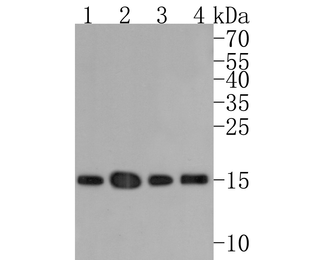
Western blot analysis of Histone H3 (acetyl K23) on different lysates with Rabbit anti-Histone H3 (acetyl K23) antibody (HA721840) at 1/1,000 dilution.
Lane 1: HeLa cell lysate
Lane 2: HeLa treated with 500 ng/mL TSA for 4 hours cell lysate
Lane 3: NIH/3T3 cell lysate
Lane 4: NIH/3T3 treated with 500 ng/mL TSA for 4 hours cell lysate
Lysates/proteins at 40 µg/Lane.
Predicted band size: 15 kDa
Observed band size: 15/14 kDa
Exposure time: 1 minute;
4-20% SDS-PAGE gel.
Proteins were transferred to a PVDF membrane and blocked with 5% NFDM/TBST for 1 hour at room temperature. The primary antibody (HA721840) at 1/1,000 dilution was used in 5% NFDM/TBST at 4℃ overnight. Goat Anti-Rabbit IgG - HRP Secondary Antibody (HA1001) at 1/50,000 dilution was used for 1 hour at room temperature. -

☑ Cell treatment (CT)
Immunocytochemistry analysis of HeLa cells treated with or without 500 ng/mL TSA for 4 hours labeling Histone H3 (acetyl K23) with Rabbit anti-Histone H3 (acetyl K23) antibody (HA721840) at 1/100 dilution.
Cells were fixed in 4% paraformaldehyde for 10 minutes at 37 ℃, permeabilized with 0.05% Triton X-100 in PBS for 20 minutes, and then blocked with 2% negative goat serum for 30 minutes at room temperature. Cells were then incubated with Rabbit anti-Histone H3 (acetyl K23) antibody (HA721840) at 1/100 dilution in 2% negative goat serum overnight at 4 ℃. Goat Anti-Rabbit IgG H&L (iFluor™ 488, HA1121) was used as the secondary antibody at 1/1,000 dilution. Nuclear DNA was labelled in blue with DAPI.
Beta tubulin (M1305-2, red) was stained at 1/100 dilution overnight at +4℃. Goat Anti-Mouse IgG H&L (iFluor™ 594, HA1126) was used as the secondary antibody at 1/1,000 dilution.
Please note: All products are "FOR RESEARCH USE ONLY AND ARE NOT INTENDED FOR DIAGNOSTIC OR THERAPEUTIC USE"
同靶点&同通路的产品
Phospho-Histone H3 (S28) Recombinant Rabbit Monoclonal Antibody [JE56-06]
Application: WB,IF-Cell,IHC-P,FC,ChIP
Reactivity: Human,Rat,Mouse
Conjugate: unconjugated
Histone H3 (mono methyl K36) Recombinant Rabbit Monoclonal Antibody [SR04-20]
Application: WB,IF-Cell,IF-Tissue,ChIP
Reactivity: Human,Mouse
Conjugate: unconjugated
Histone H3 (mono methyl R2) Recombinant Rabbit Monoclonal Antibody [ST0427]
Application: WB,IF-Cell,IF-Tissue,IHC-P,ChIP
Reactivity: Human,Mouse,Rat
Conjugate: unconjugated
Histone H3 (acetyl K56) Recombinant Rabbit Monoclonal Antibody [SU30-10]
Application: WB,IF-Cell,IF-Tissue,IHC-P,ChIP,CUT&Tag-seq
Reactivity: Human,Mouse,Rat
Conjugate: unconjugated
Histone H3 (tri methyl K9) Mouse Monoclonal Antibody [1-6]
Application: WB,IF-Cell,IHC-P,IF-Tissue
Reactivity: Human,Mouse,Rat
Conjugate: unconjugated
Histone H3 Mouse Monoclonal Antibody [A11-D7]
Application: WB,IF-Cell,IHC-P,IF-Tissue
Reactivity: Human,Mouse,Rat,Zebrafish
Conjugate: unconjugated
Histone H3 (tri methyl K27) Recombinant Rabbit Monoclonal Antibody [PSH05-15]
Application: WB,IF-Cell,IHC-P,IF-Tissue,ChIP
Reactivity: Human,Mouse,Rat
Conjugate: unconjugated
Histone H3 (di methyl K4) Rabbit Polyclonal Antibody
Application: WB,IF-Cell,IHC-P,FC,Dot Blot
Reactivity: Human,Mouse,Rat
Conjugate: unconjugated
Histone H3 (mono methyl K9) Recombinant Rabbit Monoclonal Antibody [JE43-27]
Application: WB,IF-Cell,IHC-P,FC,ChIP,IF-Tissue,Dot Blot
Reactivity: Human,Mouse,Rat,Monkey
Conjugate: unconjugated
Histone H3 (mono methyl K27) Recombinant Rabbit Monoclonal Antibody [PSH05-67]
Application: WB,IF-Cell,FC,ChIP
Reactivity: Human,Mouse,Rat
Conjugate: unconjugated
Histone H3 (acetyl K36) Recombinant Rabbit Monoclonal Antibody [JE77-32]
Application: WB,IHC-P,IF-Cell,ChIP
Reactivity: Human,Mouse,Rat
Conjugate: unconjugated
Histone H3 (mono+di+tri methyl K4) Recombinant Rabbit Monoclonal Antibody [PSH06-30]
Application: WB,IHC-P,IF-Cell,FC,ChIP
Reactivity: Human,Mouse,Rat
Conjugate: unconjugated
Histone H3 (di methyl K27) Recombinant Rabbit Monoclonal Antibody [PSH05-88]
Application: WB,IF-Cell,ChIP
Reactivity: Human,Mouse,Rat
Conjugate: unconjugated
Histone H3 (mono methyl K4) Recombinant Rabbit Monoclonal Antibody [PSH07-15]
Application: WB,IF-Cell,ChIP,Dot Blot
Reactivity: Human,Mouse,Rat
Conjugate: unconjugated
Histone H3 (mono+di+tri methyl K36) Recombinant Rabbit Monoclonal Antibody [PSH07-14]
Application: WB,IHC-P,IF-Cell,FC,ChIP
Reactivity: Human,Mouse,Rat
Conjugate: unconjugated
Histone H3 Mouse Monoclonal Antibody [3-C4]
Application: WB,IHC-P,mIHC,IF-Tissue,ChIP
Reactivity: Human,Mouse,Rat
Conjugate: unconjugated
Histone H3 (mono methyl K18) Recombinant Rabbit Monoclonal Antibody [SA42-07]
Application: WB,IF-Cell,IF-Tissue,IHC-P
Reactivity: Human,Mouse,Rat
Conjugate: unconjugated
Phospho-Histone H3 (S10) Recombinant Rabbit Monoclonal Antibody [SA31-01]
Application: WB,IF-Cell,IF-Tissue,IHC-P,IP,FC,ChIP
Reactivity: Human,Mouse,Rat
Conjugate: unconjugated
Histone H3 (di methyl K9) Recombinant Rabbit Monoclonal Antibody [SN07-30]
Application: WB,IF-Cell,IF-Tissue,IHC-P
Reactivity: Human,Mouse,Rat
Conjugate: unconjugated
Histone H3 Recombinant Rabbit Monoclonal Antibody [JJ092-08]
Application: WB,IF-Tissue,IHC-P,ChIP
Reactivity: Human,Mouse,Rat
Conjugate: unconjugated
Histone H3 (acetyl K4) Recombinant Rabbit Monoclonal Antibody [PSH03-71]
Application: WB,IF-Cell,ChIP,Dot Blot
Reactivity: Human,Mouse,Rat
Conjugate: unconjugated
Histone H3 (acetyl K14) Recombinant Rabbit Monoclonal Antibody [JU43-26]
Application: WB,IF-Cell,IF-Tissue,IHC-P,IP,SNAP-ChIP,CUT&Tag-seq
Reactivity: Human,Mouse,Rat
Conjugate: unconjugated
Histone H3 (tri methyl K9) Mouse Monoclonal Antibody [2G1]
Application: WB,IF
Reactivity: Human
Conjugate: unconjugated
Histone H3 (acetyl K27) Rabbit Polyclonal Antibody
Application: WB,IHC-P,ChIP
Reactivity: Human,Mouse
Conjugate: unconjugated
Histone H3 (acetyl K18) Rabbit Polyclonal Antibody
Application: WB,IHC-P,IF-Cell
Reactivity: Human,Mouse
Conjugate: unconjugated
Histone H3 Rabbit Polyclonal Antibody
Application: WB
Reactivity: Zebrafish,Human,Mouse
Conjugate: unconjugated
Phospho-Histone H3 (T3) Recombinant Rabbit Monoclonal Antibody [JE42-48]
Application: WB,IHC-P
Reactivity: Human,Mouse,Rat
Conjugate: unconjugated
Histone H3 Mouse Monoclonal Antibody [6-A7]
Application: WB,IHC-P,IF-Tissue,ChIP
Reactivity: Human,Mouse,Rat
Conjugate: unconjugated
Histone H3 Rabbit Polyclonal Antibody
Application: WB
Reactivity: Human,Mouse,Rat
Conjugate: unconjugated
Histone H3 (mono methyl K4) Rabbit Polyclonal Antibody
Application: WB,IF-Cell,IHC-P,FC,Dot Blot
Reactivity: Human,Mouse,Rat
Conjugate: unconjugated
Histone H3 (acetyl K27) Mouse Monoclonal Antibody [A6D7]
Application: WB,IF-Cell,IHC-P,FC,ChIP
Reactivity: Human,Mouse,Rat
Conjugate: unconjugated
Histone H3 (acetyl K27) Mouse Monoclonal Antibody [A6D6]
Application: WB,IF-Cell,IHC-P,FC,ChIP
Reactivity: Human,Mouse,Rat
Conjugate: unconjugated
Histone H3 (acetyl K18) Mouse Monoclonal Antibody [A7B5]
Application: WB,IHC-P,ChIP
Reactivity: Human,Rat
Conjugate: unconjugated
Histone H3 Recombinant Mouse Monoclonal Antibody [A11-D7-R]
Application: WB,IF-Cell,IHC-P
Reactivity: Human,Mouse,Rat
Conjugate: unconjugated
Histone H3 Recombinant Mouse Monoclonal Antibody [6-A7-R] - Loading control
Application: WB,IHC-P,IF-Tissue,ChIP
Reactivity: Human,Mouse,Rat
Conjugate: unconjugated
Histone H3 Recombinant Rabbit Monoclonal Antibody [JJ090-07]
Application: WB,IF-Cell,IF-Tissue,IHC-P,ChIP,IP
Reactivity: Human,Mouse,Rat
Conjugate: unconjugated
Histone H3 (acetyl K9) Recombinant Rabbit Monoclonal Antibody [PSH04-47] - ChIP Grade
Application: WB,IF-Cell,IHC-P,IF-Tissue,FC,ChIP,Dot Blot,IP
Reactivity: Human,Mouse,Rat
Conjugate: unconjugated
Histone H3 (mono methyl K14) Recombinant Rabbit Monoclonal Antibody [JE43-29]
Application: WB,IF-Cell,ChIP,Dot Blot
Reactivity: Human,Mouse,Rat
Conjugate: unconjugated
Histone H3 (mono+di+tri methyl K79) Recombinant Rabbit Monoclonal Antibody [SR42-06]
Application: WB,IHC-P,ChIP
Reactivity: Human,Mouse,Rat
Conjugate: unconjugated
Histone H3 (acetyl K18) Mouse Monoclonal Antibody [A7B6]
Application: WB,IF-Cell
Reactivity: Human
Conjugate: unconjugated
Histone H3 (di methyl K4) Recombinant Rabbit Monoclonal Antibody [JE00-98]
Application: WB,IF-Cell,ChIP,Dot Blot,IHC-P
Reactivity: Human,Mouse,Rat,Monkey
Conjugate: unconjugated


