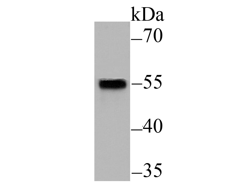
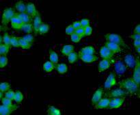
概述
产品名称
Smad2 Recombinant Rabbit Monoclonal Antibody [SP06-05]
抗体类型
Recombinant Rabbit monoclonal Antibody
免疫原
Synthetic peptide within human Smad2 aa 220-270.
种属反应性
Human, Mouse, Rat
验证应用
WB, IF-Cell, IHC-P, IP, FC
分子量
Predicted band size: 52 kDa
阳性对照
HeLa cell lysate, HT-29 cell lysate, Jurkat cell lysate, HL-60 cell lysate, C2C12 cell lysate, mouse lung tissue lysate, mouse placenta tissue lysate, human cerebellum tissue, mouse cerebellum tissue, rat cerebellum tissue, HepG2, NIH/3T3.
偶联
unconjugated
克隆号
SP06-05
RRID
产品特性
形态
Liquid
浓度
1ug/ul
存放说明
Store at +4℃ after thawing. Aliquot store at -20℃ or -80℃. Avoid repeated freeze / thaw cycles.
存储缓冲液
1*TBS (pH7.4), 0.05% BSA, 40% Glycerol. Preservative: 0.05% Sodium Azide.
亚型
IgG
纯化方式
Protein A affinity purified.
应用稀释度
-
WB
-
1:1,000-1:5,000
-
IF-Cell
-
1:100
-
IHC-P
-
1:200-1:1,000
-
FC
-
1:1,000
-
IP
-
Use at an assay dependent concentration.
发表文章中的应用
发表文章中的种属
| Mouse | See 6 publications below |
| Rat | See 2 publications below |
| Human | See 2 publications below |
靶点
功能
Smad proteins, the mammalian homologs of the Drosophila mothers against decapentaplegic (Mad), have been implicated as downstream effectors of TGFβ/BMP signaling. Smad1 (also designated Madr1 or JV4-1) and Smad5 are effectors of BMP-2 and BMP-4 function, while Smad2 (also designated Madr2 or JV18-1) and Smad3 are involved in TGFβ and Activin-mediated growth modulation. Smad4 (also designated DPC4) has been shown to mediate all of the above activities through interaction with various Smad family members. Smad6 and Smad7 regulate the response to Activin/TGFβ signaling by interfering with TGFβ-mediated phosphorylation of other Smad proteins.
背景文献
1. Ungefroren H et al. Rac1b negatively regulates TGF-1-induced cell motility in pancreatic ductal epithelial cells by suppressing Smad signalling. Oncotarget 5:277-90 (2014).
2. Harazono Y et al. miR-655 Is an EMT-suppressive MicroRNA targeting ZEB1 and TGFBR2. PLoS One 8:e62757 (2013).
序列相似性
Belongs to the dwarfin/SMAD family.
组织特异性
Expressed at high levels in skeletal muscle, endothelial cells, heart and placenta.
翻译后修饰
Phosphorylated on one or several of Thr-220, Ser-245, Ser-250, and Ser-255. In response to TGF-beta, phosphorylated on Ser-465/467 by TGF-beta and activin type 1 receptor kinases. TGF-beta-induced Ser-465/467 phosphorylation declines progressively in a KMT5A-dependent manner. Able to interact with SMURF2 when phosphorylated on Ser-465/467, recruiting other proteins, such as SNON, for degradation. In response to decorin, the naturally occurring inhibitor of TGF-beta signaling, phosphorylated on Ser-240 by CaMK2. Phosphorylated by MAPK3 upon EGF stimulation; which increases transcriptional activity and stability, and is blocked by calmodulin. Phosphorylated by PDPK1.; In response to TGF-beta, ubiquitinated by NEDD4L; which promotes its degradation. Monoubiquitinated, leading to prevent DNA-binding (By similarity). Deubiquitination by USP15 alleviates inhibition and promotes activation of TGF-beta target genes. Ubiquitinated by RNF111, leading to its degradation: only SMAD2 proteins that are 'in use' are targeted by RNF111, RNF111 playing a key role in activating SMAD2 and regulating its turnover (By similarity).; Acetylated on Lys-19 by coactivators in response to TGF-beta signaling, which increases transcriptional activity. Isoform short: Acetylation increases DNA binding activity in vitro and enhances its association with target promoters in vivo. Acetylation in the nucleus by EP300 is enhanced by TGF-beta.
亚细胞定位
Cytoplasm, Nucleus.
别名
Drosophila, homolog of, MADR2 antibody
hMAD-2 antibody
HsMAD2 antibody
JV18 antibody
JV18-1 antibody
JV181 antibody
MAD antibody
MAD homolog 2 antibody
MAD Related Protein 2 antibody
Mad-related protein 2 antibody
展开Drosophila, homolog of, MADR2 antibody
hMAD-2 antibody
HsMAD2 antibody
JV18 antibody
JV18-1 antibody
JV181 antibody
MAD antibody
MAD homolog 2 antibody
MAD Related Protein 2 antibody
Mad-related protein 2 antibody
MADH2 antibody
MADR2 antibody
MGC22139 antibody
MGC34440 antibody
Mother against DPP homolog 2 antibody
Mothers against decapentaplegic homolog 2 antibody
Mothers against decapentaplegic, Drosophila, homolog of, 2 antibody
Mothers against DPP homolog 2 antibody
OTTHUMP00000163489 antibody
Sma and Mad related protein 2 antibody
Sma- and Mad-related protein 2 MAD antibody
SMAD 2 antibody
SMAD family member 2 antibody
SMAD, mothers against DPP homolog 2 antibody
SMAD2 antibody
SMAD2_HUMAN antibody
折叠图片
-
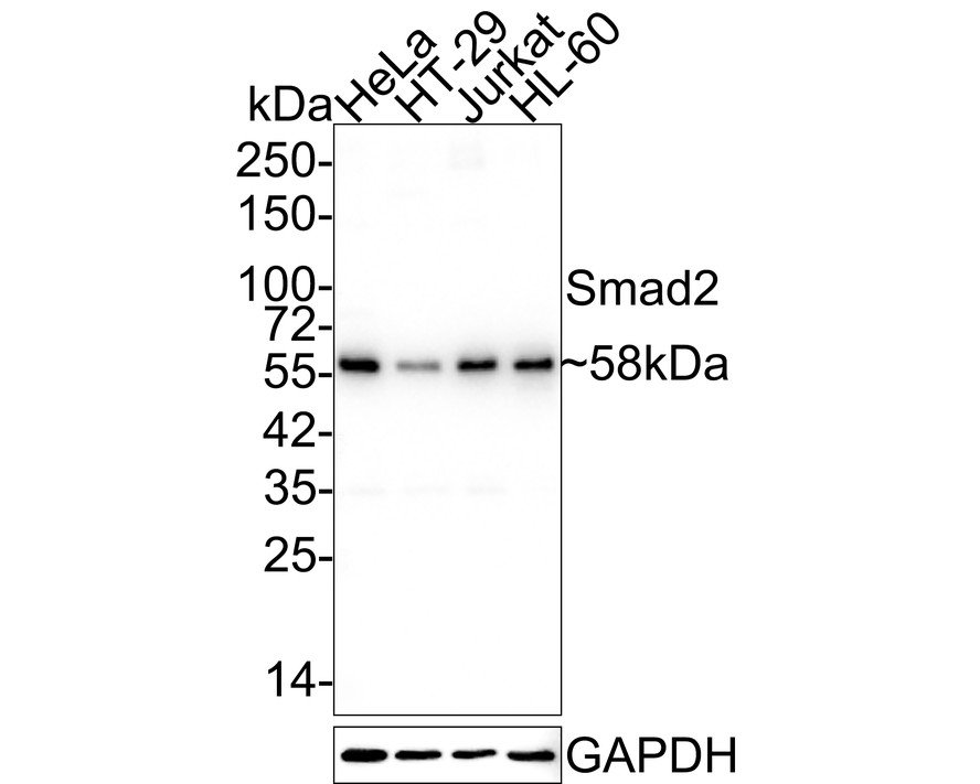
Western blot analysis of Smad2 on different lysates with Rabbit anti-Smad2 antibody (ET1604-22) at 1/5,000 dilution.
Lane 1: HeLa cell lysate
Lane 2: HT-29 cell lysate
Lane 3: Jurkat cell lysate
Lane 4: HL-60 cell lysate
Lysates/proteins at 15 µg/Lane.
Predicted band size: 52 kDa
Observed band size: 58 kDa
Exposure time: 1 minute 20 seconds;
4-20% SDS-PAGE gel.
Proteins were transferred to a PVDF membrane and blocked with 5% NFDM/TBST for 1 hour at room temperature. The primary antibody (ET1604-22) at 1/5,000 dilution was used in 5% NFDM/TBST at room temperature for 2 hours. Goat Anti-Rabbit IgG - HRP Secondary Antibody (HA1001) at 1:50,000 dilution was used for 1 hour at room temperature. -

Western blot analysis of Smad2 on different lysates with Rabbit anti-Smad2 antibody (ET1604-22) at 1/1,000 dilution.
Lane 1: Jurkat cell lysate (15 µg/Lane)
Lane 2: C2C12 cell lysate (15 µg/Lane)
Lane 3: Mouse lung tissue lysate (30 µg/Lane)
Lane 4: Mouse placenta tissue lysate (30 µg/Lane)
Predicted band size: 52 kDa
Observed band size: 58 kDa
Exposure time: 1 minute 21 seconds;
4-20% SDS-PAGE gel.
Proteins were transferred to a PVDF membrane and blocked with 5% NFDM/TBST for 1 hour at room temperature. The primary antibody (ET1604-22) at 1/1,000 dilution was used in 5% NFDM/TBST at 4℃ overnight. Goat Anti-Rabbit IgG - HRP Secondary Antibody (HA1001) at 1/50,000 dilution was used for 1 hour at room temperature. -

☑ Knockout (KO)
Western blot analysis of Smad2 with anti-Smad2 antibody (ET1604-22) at 1:500 dilution.
Lane 1: Wild-type HaCaT whole cell lysate (15 µg).
Lane 2: Smad2 knockout HaCaT whole cell lysate (15 µg).
ET1604-22 was shown to specifically react with Smad2 in wild-type HaCaT cells. NO band was observed when Smad2 knockout sample was tested. Wild-type and Smad2 knockout samples were subjected to SDS-PAGE. Proteins were transferred to a PVDF membrane and blocked with 5% NFDM in TBST for 1 hour at room temperature. The primary antibody (ET1604-22, 1:500) was used in 5% BSA at room temperature for 2 hours. Goat anti-Rabbit IgG-HRP antibody at 1:10,000 dilution was used for 1 hour at room temperature. -
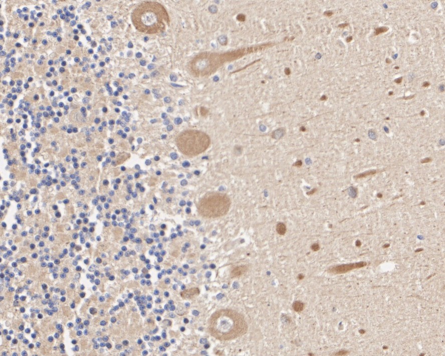
Immunohistochemical analysis of paraffin-embedded human cerebellum tissue with Rabbit anti-Smad2 antibody (ET1604-22) at 1/200 dilution.
The section was pre-treated using heat mediated antigen retrieval with sodium citrate buffer (pH 6.0) for 2 minutes. The tissues were blocked in 1% BSA for 20 minutes at room temperature, washed with ddH2O and PBS, and then probed with the primary antibody (ET1604-22) at 1/200 dilution for 1 hour at room temperature. The detection was performed using an HRP conjugated compact polymer system. DAB was used as the chromogen. Tissues were counterstained with hematoxylin and mounted with DPX. -

Immunohistochemical analysis of paraffin-embedded mouse cerebellum tissue with Rabbit anti-Smad2 antibody (ET1604-22) at 1/1,000 dilution.
The section was pre-treated using heat mediated antigen retrieval with sodium citrate buffer (pH 6.0) for 2 minutes. The tissues were blocked in 1% BSA for 20 minutes at room temperature, washed with ddH2O and PBS, and then probed with the primary antibody (ET1604-22) at 1/1,000 dilution for 1 hour at room temperature. The detection was performed using an HRP conjugated compact polymer system. DAB was used as the chromogen. Tissues were counterstained with hematoxylin and mounted with DPX. -

Immunohistochemical analysis of paraffin-embedded rat cerebellum tissue with Rabbit anti-Smad2 antibody (ET1604-22) at 1/200 dilution.
The section was pre-treated using heat mediated antigen retrieval with sodium citrate buffer (pH 6.0) for 2 minutes. The tissues were blocked in 1% BSA for 20 minutes at room temperature, washed with ddH2O and PBS, and then probed with the primary antibody (ET1604-22) at 1/200 dilution for 1 hour at room temperature. The detection was performed using an HRP conjugated compact polymer system. DAB was used as the chromogen. Tissues were counterstained with hematoxylin and mounted with DPX. -
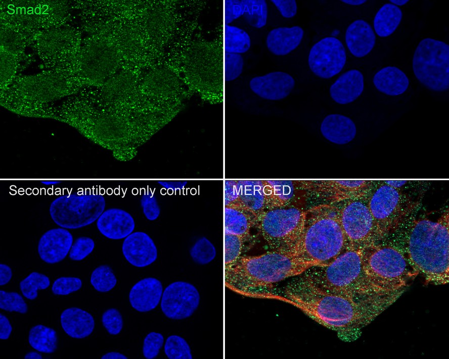
Immunocytochemistry analysis of HepG2 cells labeling Smad2 with Rabbit anti-Smad2 antibody (ET1604-22) at 1/100 dilution.
Cells were fixed in 4% paraformaldehyde for 20 minutes at room temperature, permeabilized with 0.1% Triton X-100 in PBS for 5 minutes at room temperature, then blocked with 1% BSA in 10% negative goat serum for 1 hour at room temperature. Cells were then incubated with Rabbit anti-Smad2 antibody (ET1604-22) at 1/100 dilution in 1% BSA in PBST overnight at 4 ℃. Goat Anti-Rabbit IgG H&L (iFluor™ 488, HA1121) was used as the secondary antibody at 1/1,000 dilution. PBS instead of the primary antibody was used as the secondary antibody only control. Nuclear DNA was labelled in blue with DAPI. Beta tubulin (M1305-2, red) was stained at 1/100 dilution overnight at +4℃. Goat Anti-Mouse IgG H&L (iFluor™ 594, HA1126) was used as the secondary antibody at 1/1,000 dilution. -
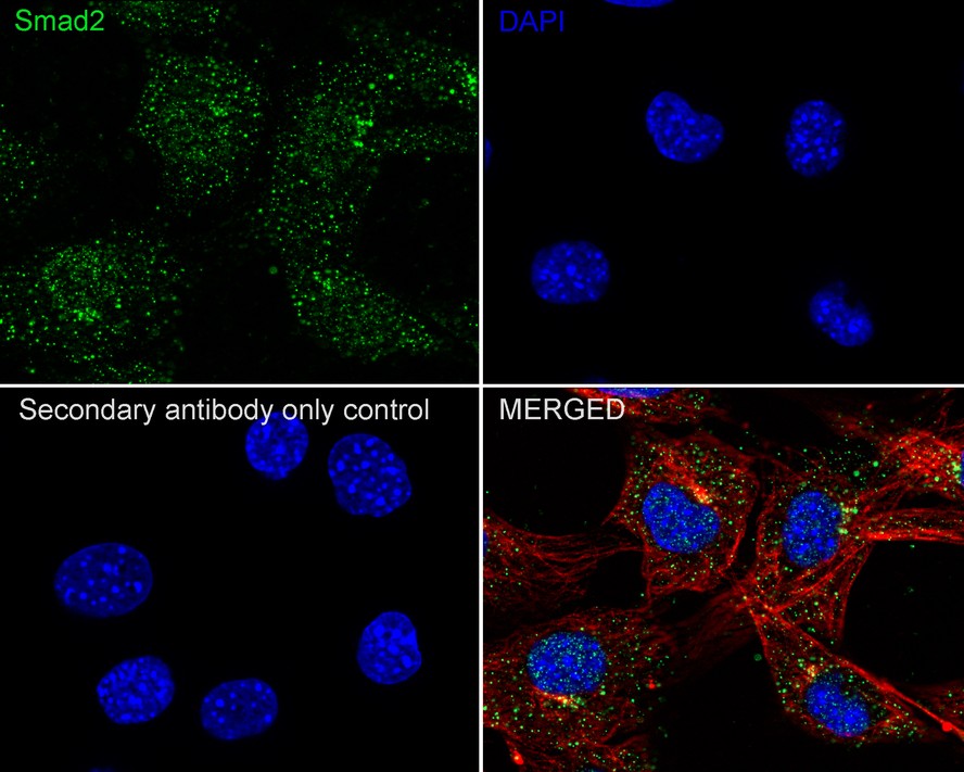
Immunocytochemistry analysis of NIH/3T3 cells labeling Smad2 with Rabbit anti-Smad2 antibody (ET1604-22) at 1/100 dilution.
Cells were fixed in 4% paraformaldehyde for 20 minutes at room temperature, permeabilized with 0.1% Triton X-100 in PBS for 5 minutes at room temperature, then blocked with 1% BSA in 10% negative goat serum for 1 hour at room temperature. Cells were then incubated with Rabbit anti-Smad2 antibody (ET1604-22) at 1/100 dilution in 1% BSA in PBST overnight at 4 ℃. Goat Anti-Rabbit IgG H&L (iFluor™ 488, HA1121) was used as the secondary antibody at 1/1,000 dilution. PBS instead of the primary antibody was used as the secondary antibody only control. Nuclear DNA was labelled in blue with DAPI. Beta tubulin (M1305-2, red) was stained at 1/100 dilution overnight at +4℃. Goat Anti-Mouse IgG H&L (iFluor™ 594, HA1126) was used as the secondary antibody at 1/1,000 dilution. -
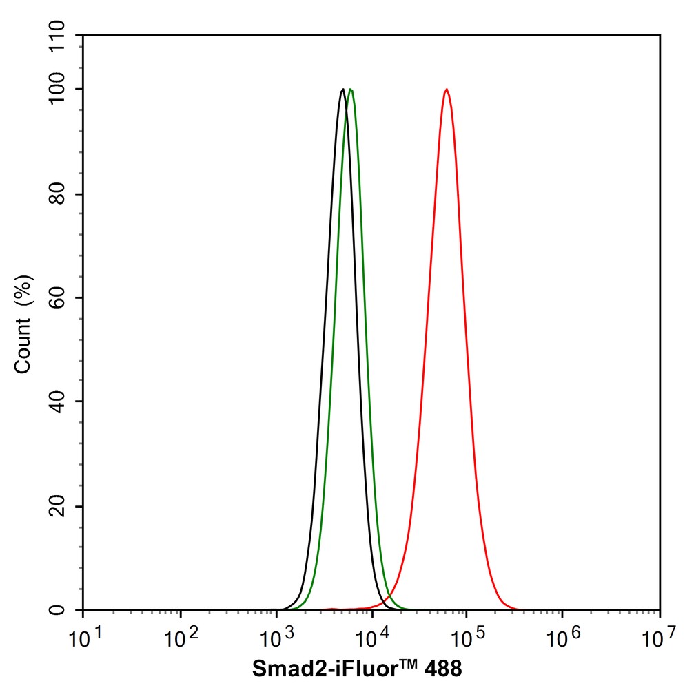
Flow cytometric analysis of HepG2 cells labeling Smad2.
Cells were fixed and permeabilized. Then stained with the primary antibody (ET1604-22, 1/1,000) (red) compared with Rabbit IgG Isotype Control (green). After incubation of the primary antibody at +4℃ for an hour, the cells were stained with a iFluor™ 488 conjugate-Goat anti-Rabbit IgG Secondary antibody (HA1121) at 1/1,000 dilution for 30 minutes at +4℃. Unlabelled sample was used as a control (cells without incubation with primary antibody; black). -
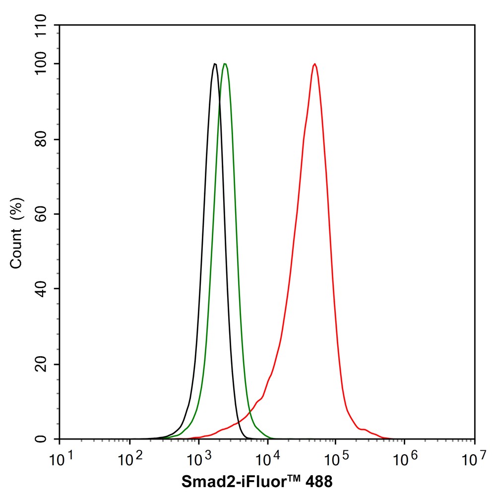
Flow cytometric analysis of NIH/3T3 cells labeling Smad2.
Cells were fixed and permeabilized. Then stained with the primary antibody (ET1604-22, 1/1,000) (red) compared with Rabbit IgG Isotype Control (green). After incubation of the primary antibody at +4℃ for an hour, the cells were stained with a iFluor™ 488 conjugate-Goat anti-Rabbit IgG Secondary antibody (HA1121) at 1/1,000 dilution for 30 minutes at +4℃. Unlabelled sample was used as a control (cells without incubation with primary antibody; black).
Please note: All products are "FOR RESEARCH USE ONLY AND ARE NOT INTENDED FOR DIAGNOSTIC OR THERAPEUTIC USE"
引文
-
PD-1 Inhibitor Aggravate Irradiation-Induced Myocardial Fibrosis by Regulating TGF-β1/Smads Signaling Pathway via GSDMD-Mediated Pyroptosis
Author: Wu Bibo,et al
PMID: 38773023
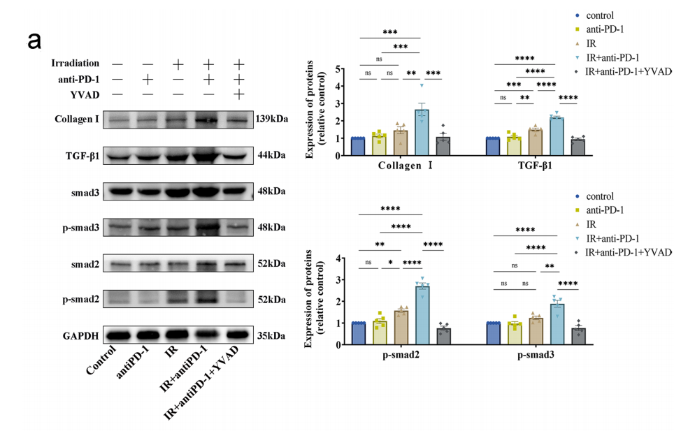
应用: WB
反应种属: Mouse
发表时间: 2024 May
-
Citation
-
RRM2 Regulates Hepatocellular Carcinoma Progression Through Activation of TGF-β/Smad Signaling and Hepatitis B Virus Transcription
Author: Dandan Wu,et al
PMID: NOPMID24122003
应用: WB
反应种属: Mouse
发表时间: 2024 Dec
-
Citation
-
The combination of paeoniflorin and metformin synergistically inhibits the progression of liver fibrosis in mice
Author: Meng Lingjie,et al
PMID: 39154824
应用: WB
反应种属: Mouse
发表时间: 2024 Aug
-
Citation
-
CD44 mediates hyaluronan to promote the differentiation of human amniotic mesenchymal stem cells into chondrocytes
Author:
PMID: 36680638

应用: WB
反应种属: Human
发表时间: 2023 Mar
-
Citation
-
TNFSF14/LIGHT promotes cardiac fibrosis and atrial fibrillation vulnerability via PI3Kγ/SGK1 pathway-dependent M2 macrophage polarisation
Author: Yirong Wu, Siyao Zhan, Lian Chen, Mingrui Sun, Miaofu Li, Xuanting Mou, Zhen Zhang, Linhao Xu, Yizhou Xu
PMID: 37580750

应用: WB
反应种属: Mouse
发表时间: 2023 Aug
-
Citation
-
Low intensity pulsed ultrasound ameliorates Adriamycin-induced chronic renal injury by inhibiting ferroptosis
Author: Ouyang, Z. Q., Shao, L. S., Wang, W. P., Ke, T. F., Chen, D., Zheng, G. R., Duan, X. R., Chu, J. X., Zhu, Y., Yang, L., Shan, H. Y., Huang, L., & Liao, C. D.
PMID: 37652897

应用: WB
反应种属: Rat
发表时间: 2023 Aug
-
Citation
-
TGF-β-Containing Small Extracellular Vesicles From PM2.5-Activated Macrophages Induces Cardiotoxicity
Author: Hu, X., Chen, M., Cao, X., Yuan, X., Zhang, F., & Ding, W.
PMID: 35872905

应用: WB
反应种属: Mouse
发表时间: 2022 Jul
-
Citation
-
TBX20 Contributes to Balancing the Differentiation of Perivascular Adipose-Derived Stem Cells to Vascular Lineages and Neointimal Hyperplasia
Author:
PMID: 34150759

应用: WB
反应种属: Mouse
发表时间: 2021 Jun
-
Citation
-
7,8-Dihydroxyflavone modulates bone formation and resorption and ameliorates ovariectomy-induced osteoporosis. eLife, 10, e64872.
Author: Xue, F., Zhao, Z., Gu, Y., Han, J., Ye, K., & Zhang, Y.
PMID: 34227467
应用: WB
反应种属: Rat
发表时间: 2021 Jul
-
Citation
-
Profilin 2 promotes growth, metastasis, and angiogenesis of small cell lung cancer through cancer-derived exosomes
Author:
PMID: 33234737

应用: WB
反应种属: Human
发表时间: 2020 Nov
-
Citation
同靶点&同通路的产品
Smad2
Application:
Reactivity:
Conjugate:
Phospho-Smad2 (S255) Recombinant Rabbit Monoclonal Antibody [JF0882]
Application: WB,IHC-P,IP
Reactivity: Human,Mouse,Rat
Conjugate: unconjugated
Smad2 Mouse Monoclonal Antibody [C9-B0]
Application: WB,IHC-P,IF-Cell
Reactivity: Human,Mouse,Rat
Conjugate: unconjugated
Phospho-Smad2 (S250) Recombinant Rabbit Monoclonal Antibody [SD207-1]
Application: WB,FC,IP
Reactivity: Human,Mouse,Rat
Conjugate: unconjugated
Smad2 Rabbit Polyclonal Antibody
Application: IF-Cell,IHC-P
Reactivity: Human,Mouse,Rat
Conjugate: unconjugated


