Hematopoietic Stem Cell Marker Antibody Kit
General Overview
Kit Components
| Product Includes | Specification | Application | Reactivity | Mw |
|---|---|---|---|---|
| CD34[ET1606-11] | 20µl | WB,IF-Cell,IF-Tissue,IHC-P,IP,mIHC,FC | Human,Mouse,Rat | Predicted band size: 41 kDa |
| CD38[HA721268] | 20µl | WB,IHC-P,IF-Cell,FC,mIHC,IF-Tissue | Human | Predicted band size: 34 kDa |
| CD59[ET1703-28] | 20µl | WB,IP | Human | Predicted band size: 14 kDa |
| CD90 / THY1[ET1702-92] | 20µl | WB,IF-Cell,IF-Tissue,IHC-P,IHC-Fr | Human,Mouse,Rat | Predicted band size: 18 kDa |
| c-Kit[HA721154] | 20µl | IHC-P,WB,mIHC | Human | Predicted band size: 110 kDa |
| Goat Anti-Rabbit IgG (H+L)[HA1001] | 100µl | WB,ELISA,IHC-P | Rabbit |
Product Description
Hematopoietic Stem Cell Marker Antibody Sampler Kit contains multiple trial-sized versions of anti-human antibody clones against CD34, CD38,CD59, CD90 / Thy1, c-Kit, specifically selected for high performance in various applications. This panel contains 5 recombinant rabbit monoclonal antibodies against human CD34, CD59, CD90 / Thy1, CD38, c-Kit. They are provided as a sampler panel to allow you to easily evaluate each in your required applications.
Product Features
Storage Buffer
1*TBS (pH7.4), 0.05% BSA, 40% Glycerol. Preservative: 0.05% Sodium Azide.
Storage Instructions
Store at +4℃ after thawing. Aliquot store at -20℃. Avoid repeated freeze / thaw cycles.
Background
Hematopoietic stem cells (HSC) possess multipotentiality, enabling them to self-renew and also to produce mature blood cells, such as erythrocytes, leukocytes, platelets, and lymphocytes. </br>CD34 is a marker of human HSC, and all colony-forming activity of human bone marrow (BM) cells is found in the CD34+ fraction. Cyclic ADP-ribose hydrolase 1 (CD38) is a transmembrane protein involved in several important biological processes, including immune response, insulin secretion, and social behavior. Originally described as a glycosylated immune cell surface marker, additional research determined that CD38 is a multifunctional enzyme that catalyzes the synthesis and hydrolysis of cyclic ADP ribose (cADPR) from NAD.</br>CD59 is a GPI-anchored membrane protein that functions as an inhibitor of the complement membrane attack complex (MAC). The Thy1/CD90 cell surface antigen is a GPI-anchored, developmentally regulated protein involved in signaling cascades that mediate neurite outgrowth, T cell activation, tumor suppression, apoptosis, and fibrosis .c-Kit is a member of the subfamily of receptor tyrosine kinases that includes PDGF, CSF-1, and FLT3/flk-2 receptors . It plays a critical role in activation and growth in a number of cell types, including hematopoietic stem cells, mast cells, melanocytes, and germ cells .
Background References
1. Pei X. Who is hematopoietic stem cell: CD34+ or CD34-? Int J Hematol. 1999 Dec;70(4):213-5.
2. Malavasi F, Deaglio S, Funaro A, Ferrero E, Horenstein AL, Ortolan E, Vaisitti T, Aydin S. Evolution and function of the ADP ribosyl cyclase/CD38 gene family in physiology and pathology. Physiol Rev. 2008 Jul;88(3):841-86.
3. Sugita Y, Masuho Y. CD59: its role in complement regulation and potential for therapeutic use. Immunotechnology. 1995 Dec;1(3-4):157-68.
4. Rege TA, Hagood JS. Thy-1 as a regulator of cell-cell and cell-matrix interactions in axon regeneration, apoptosis, adhesion, migration, cancer, and fibrosis. FASEB J. 2006 Jun;20(8):1045-54.
5. Martin FH, Suggs SV, Langley KE, Lu HS, Ting J, Okino KH, Morris CF, McNiece IK, Jacobsen FW, Mendiaz EA, et al. Primary structure and functional expression of rat and human stem cell factor DNAs. Cell. 1990 Oct 5;63(1):203-11.
Images
-

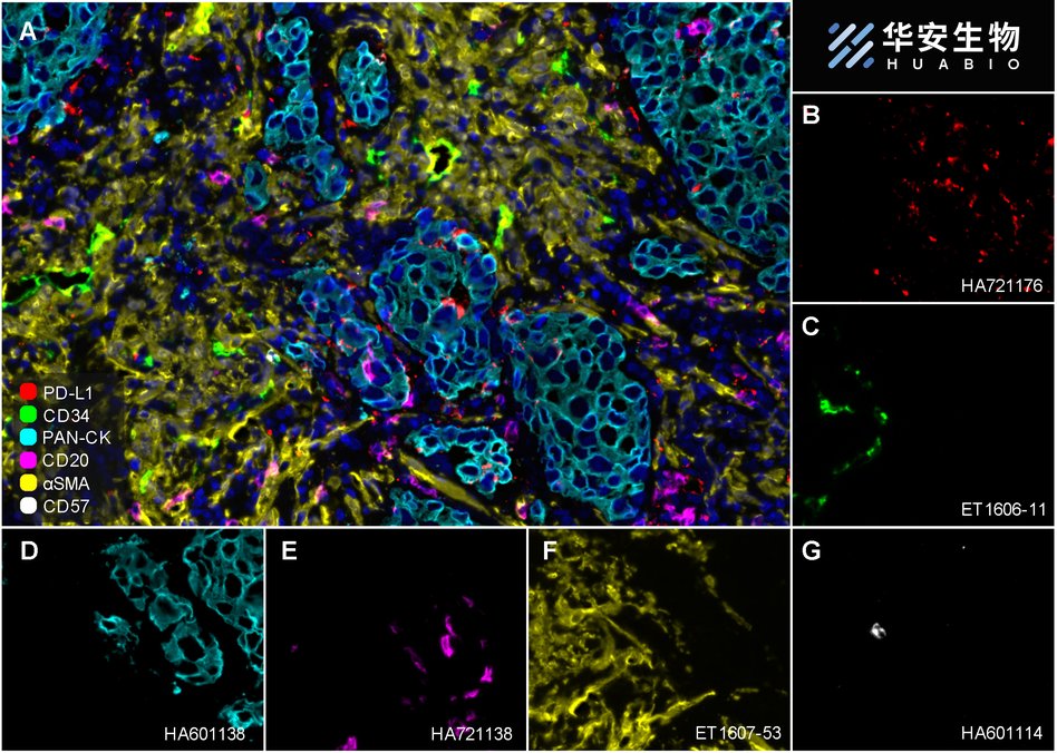
Fluorescence multiplex immunohistochemical analysis of Human non-small cell lung cancer (Formalin/PFA-fixed paraffin-embedded sections). Panel A: the merged image of anti-PD-L1 (HA721176, red), anti-CD34 (ET1606-11, green), anti-Pan-CK (HA601138, cyan), anti-CD20 (HA721138, magenta), anti-αSMA (ET1607-53, yellow) and anti-CD57 (HA601114, white) on NSCLC. Panel B: anti-PD-L1 stained on dendritic cells and macrophages cells. Panel C: anti- CD34 stained on endothelial cells. Panel D: anti-Pan-CK stained on cancer cells. Panel E: CD20 stained on B cells. Panel F: anti-αSMA stained on cancer-associated fibroblasts and smooth muscle cells. Panel G: anti-CD57 stained on NK cells and T cells. HRP Conjugated UltraPolymer Goat Polyclonal Antibody HA1119/HA1120 was used as a secondary antibody. The immunostaining was performed with the Sequential Immuno-staining Kit (IRISKit™MH010101, www.luminiris.cn). The section was incubated in six rounds of staining: in the order of HA721176 (1/1,000 dilution), ET1606-11 (1/1,000 dilution), HA601138 (1/3,000 dilution), HA721138 (1/2,000 dilution), ET1607-53 (1/3,000 dilution) and HA601114 (1/1,000 dilution) for 20 mins at room temperature. Each round was followed by a separate fluorescent tyramide signal amplification system. Heat mediated antigen retrieval with Tris-EDTA buffer (pH 9.0) for 30 mins at 95℃. DAPI (blue) was used as a nuclear counter stain. Image acquisition was performed with Olympus VS200 Slide Scanner.
-

Fluorescence multiplex immunohistochemical analysis of mouse brain (Formalin/PFA-fixed paraffin-embedded sections). Panel A: the merged image of anti-NeuN (ET1602-12, red), anti-PAX6 (ET1612-58, green), anti-CD34 (ET1606-11, gray), anti-MAP2 (HA500177, magenta) and anti-TBR1 (ET1702-97, yellow) on mouse brain. HRP Conjugated UltraPolymer Goat Polyclonal Antibody HA1119/HA1120 was used as a secondary antibody. The immunostaining was performed with the Sequential Immuno-staining Kit (IRISKit™MH010101, www.luminiris.cn). The section was incubated in five rounds of staining: in the order of ET1602-12 (1/5,000 dilution), ET1612-58 (1/1,000 dilution), ET1606-11 (1/2,000 dilution), HA500177 (1/5,000 dilution) and ET1702-97 (1/1,000 dilution) for 20 mins at room temperature. Each round was followed by a separate fluorescent tyramide signal amplification system. Heat mediated antigen retrieval with Tris-EDTA buffer (pH 9.0) for 30 mins at 95℃. DAPI (blue) was used as a nuclear counter stain. Image acquisition was performed with Olympus VS200 Slide Scanner.
-

Fluorescence multiplex immunohistochemical analysis of mouse brain (Formalin/PFA-fixed paraffin-embedded sections). Panel A: the merged image of anti-NeuN (ET1602-12, red), anti-Iba1 (ET1705-78, green), anti-GFAP (ET1601-23, gray), anti-Olig2 (ET1604-29, cyan), anti-MAP2 (HA500177, magenta) and anti-CD34 (ET1606-11, yellow) on mouse brain. HRP Conjugated UltraPolymer Goat Polyclonal Antibody HA1119/HA1120 was used as a secondary antibody. The immunostaining was performed with the Sequential Immuno-staining Kit (IRISKit™MH010101, www.luminiris.cn). The section was incubated in six rounds of staining: in the order of ET1602-12(1/5,000 dilution), ET1705-78 (1/2,000 dilution), ET1601-23 (1/5,000 dilution), ET1604-29 (1/1,000 dilution), HA500177 (1/5,000 dilution) and ET1606-11 (1/2,000 dilution) for 20 mins at room temperature. Each round was followed by a separate fluorescent tyramide signal amplification system. Heat mediated antigen retrieval with Tris-EDTA buffer (pH 9.0) for 30 mins at 95℃. DAPI (blue) was used as a nuclear counter stain. Image acquisition was performed with Olympus VS200 Slide Scanner.
-

Fluorescence multiplex immunohistochemical analysis of human non-small cell lung cancer (Formalin/PFA-fixed paraffin-embedded sections). Panel A: the merged image of anti-CD34 (ET1606-11, White), anti-PD-L1 (HA721176, Violet) and anti-pan Cytokeratin (HA601138, Yellow) on NSCLC. HRP Conjugated UltraPolymer Goat Polyclonal Antibody HA1119/HA1120 was used as a secondary antibody. The immunostaining was performed with the Sequential Immuno-staining Kit (IRISKit™MH010101, www.luminiris.cn). The section was incubated in three rounds of staining: in the order of ET1606-11 (1/2,000 dilution), HA721176 (1/1,000 dilution) and HA601138 (1/3,000 dilution) for 20 mins at room temperature. Each round was followed by a separate fluorescent tyramide signal amplification system. Heat mediated antigen retrieval with Tris-EDTA buffer (pH 9.0) for 30 mins at 95℃. DAPI (blue) was used as a nuclear counter stain. Image acquisition was performed with Zeiss Observer 7 Inverted Fluorescence Microscope.
-

Immunohistochemical analysis of paraffin-embedded human placenta tissue with Rabbit anti-CD34 antibody (ET1606-11) at 1/400 dilution.
The section was pre-treated using heat mediated antigen retrieval with Tris-EDTA buffer (pH 9.0) for 20 minutes. The tissues were blocked in 1% BSA for 20 minutes at room temperature, washed with ddH2O and PBS, and then probed with the primary antibody (ET1606-11) at 1/400 dilution for 1 hour at room temperature. The detection was performed using an HRP conjugated compact polymer system. DAB was used as the chromogen. Tissues were counterstained with hematoxylin and mounted with DPX. -

CD34 was immunoprecipitated from 0.2 mg TF-1 cell lysate with ET1606-11 at 2 µg/10 µl beads. Western blot was performed from the immunoprecipitate using ET1606-11 at 1/2,000 dilution. Anti-Rabbit IgG for IP Nano-secondary antibody (NBI01H) at 1/5,000 dilution was used for 1 hour at room temperature.
Lane 1: TF-1 cell lysate (input)
Lane 2: ET1606-11 IP in TF-1 cell lysate
Lane 3: Rabbit IgG instead of ET1606-11 in TF-1 cell lysate
Blocking/Dilution buffer: 5% NFDM/TBST
Exposure time: 6 seconds; ECL: K1801 -

Immunohistochemical analysis of paraffin-embedded human appendix tissue with Rabbit anti-CD38 antibody (HA721268) at 1/2,000 dilution.
The section was pre-treated using heat mediated antigen retrieval with Tris-EDTA buffer (pH 9.0) for 20 minutes. The tissues were blocked in 1% BSA for 20 minutes at room temperature, washed with ddH2O and PBS, and then probed with the primary antibody (HA721268) at 1/2,000 dilution for 1 hour at room temperature. The detection was performed using an HRP conjugated compact polymer system. DAB was used as the chromogen. Tissues were counterstained with hematoxylin and mounted with DPX. -

Fluorescence multiplex immunohistochemical analysis of Human tonsil (Formalin/PFA-fixed paraffin-embedded sections). Panel A: the merged image of anti-CD68 (HA601115, Red), anti-CD38 (HA721268, Green), anti-CD23 (HA721139, White), anti-CD11C (ET1606-19, Cyan), anti-CD45 (ET7111-03, Magenta) and anti-CD20 (HA721138, Yellow) on tonsil. Panel B: anti-CD68 stained on Macrophage. Panel C: anti-CD38 stained on lymphocyte subsets. Panel D: anti-CD11C stained on dendritic cells. Panel E: CD45 stained on lymphocytes. Panel F: anti-CD20 stained on B cells. Panel G: anti-CD23 stained on follicular dendritic cells. HRP Conjugated UltraPolymer Goat Polyclonal Antibody HA1119/HA1120 was used as a secondary antibody. The immunostaining was performed with the Sequential Immuno-staining Kit (IRISKit™MH010101, www.luminiris.cn). The section was incubated in six rounds of staining: in the order of HA601115 (1/2,000 dilution), HA721268 (1/1,000 dilution), ET1606-19 (1/1,000 dilution), ET7111-03 (1/500 dilution), HA721138 (1/2,000 dilution) and HA721139 (1/800 dilution) for 20 mins at room temperature. Each round was followed by a separate fluorescent tyramide signal amplification system. Heat mediated antigen retrieval with Tris-EDTA buffer (pH 9.0) for 30 mins at 95℃. DAPI (blue) was used as a nuclear counter stain. Image acquisition was performed with Olympus VS200 Slide Scanner.
-

Fluorescence multiplex immunohistochemical analysis of human tonsil (Formalin/PFA-fixed paraffin-embedded sections). Panel A: the merged image of anti-CD20 (HA721138, Cyan), anti-CD38 (HA721268, Violet) and anti-CD57 (HA601114, Yellow) on tonsil. HRP Conjugated UltraPolymer Goat Polyclonal Antibody HA1119/HA1120 was used as a secondary antibody. The immunostaining was performed with the Sequential Immuno-staining Kit (IRISKit™MH010101, www.luminiris.cn). The section was incubated in three rounds of staining: in the order of HA721138 (1/2,000 dilution), HA721268 (1/1,000 dilution) and HA601114 (1/1,000 dilution) for 20 mins at room temperature. Each round was followed by a separate fluorescent tyramide signal amplification system. Heat mediated antigen retrieval with Tris-EDTA buffer (pH 9.0) for 30 mins at 95℃. DAPI (blue) was used as a nuclear counter stain. Image acquisition was performed with Zeiss Observer 7 Inverted Fluorescence Microscope.
-

Immunohistochemical analysis of paraffin-embedded human thymus tissue with Rabbit anti-CD38 antibody (HA721268) at 1/2,000 dilution.
The section was pre-treated using heat mediated antigen retrieval with Tris-EDTA buffer (pH 9.0) for 20 minutes. The tissues were blocked in 1% BSA for 20 minutes at room temperature, washed with ddH2O and PBS, and then probed with the primary antibody (HA721268) at 1/2,000 dilution for 1 hour at room temperature. The detection was performed using an HRP conjugated compact polymer system. DAB was used as the chromogen. Tissues were counterstained with hematoxylin and mounted with DPX. -

Western blot analysis of CD59 on different lysates with Rabbit anti-CD59 antibody (ET1703-28) at 1/2,000 dilution.
Lane 1: Human placenta tissue lysate (20 µg/Lane)
Lane 2: HUVEC cell lysate (10 µg/Lane)
Lane 3: K562 cell lysate (10 µg/Lane)
Predicted band size: 14 kDa
Observed band size: 15 kDa
Exposure time: 2 minutes;
15% SDS-PAGE gel.
Proteins were transferred to a PVDF membrane and blocked with 5% NFDM/TBST for 1 hour at room temperature. The primary antibody (ET1703-28) at 1/2,000 dilution was used in 5% NFDM/TBST at room temperature for 2 hours. Goat Anti-Rabbit IgG - HRP Secondary Antibody (HA1001) at 1:300,000 dilution was used for 1 hour at room temperature. -

Immunofluorescence analysis of frozen mouse hippocampus tissue labeling CD90 / THY1 with Rabbit anti-CD90 / THY1 antibody (ET1702-92).
The tissues were blocked in 3% BSA for 30 minutes at room temperature, washed with PBS, and then probed with the primary antibody (ET1702-92, green) at 1/100 dilution overnight at 4℃, washed with PBS. Goat Anti-Rabbit IgG H&L (Alexa Fluor® 488) was used as the secondary antibody at 1/200 dilution. Nuclei were counterstained with DAPI (blue). Image acquisition was performed with KFBIO KF-FL-400 Scanner. -
![All lanes: Western blot analysis of THY1 with anti-CD90 / THY1 antibody[JF10-09] (<a href="/products/ET1702-92" style="font-weight: bold;text-decoration: underline;">ET1702-92</a>) at 1:500 dilution.<br />Lane 1: Wild-type Hela whole cell lysate (10 µg).<br />Lane 2: THY1 knockdown Hela whole cell lysate (10 µg).<br /><br /><a href="/products/ET1702-92" style="font-weight: bold;text-decoration: underline;">ET1702-92</a> was shown to specifically react with THY1 in wild-type Hela cells. Weakened bands were observed when THY1 knockdown samples were tested. Wild-type and THY1 knockdown samples were subjected to SDS-PAGE. Proteins were transferred to a PVDF membrane and blocked with 5% NFDM in TBST for 1 hour at room temperature. The primary antibody (<a href="/products/ET1702-92" style="font-weight: bold;text-decoration: underline;">ET1702-92</a>, 1/500) was used in 5% BSA at room temperature for 2 hours. Goat Anti-Rabbit IgG-HRP Secondary Antibody (<a href="/products/HA1001" style="font-weight: bold;text-decoration: underline;">HA1001</a>) at 1:100,000 dilution was used for 1 hour at room temperature.](https://storage.huabio.cn/huabio/productImg/HAK21017_13.jpg?v=20251212230501)
All lanes: Western blot analysis of THY1 with anti-CD90 / THY1 antibody[JF10-09] (ET1702-92) at 1:500 dilution.
Lane 1: Wild-type Hela whole cell lysate (10 µg).
Lane 2: THY1 knockdown Hela whole cell lysate (10 µg).
ET1702-92 was shown to specifically react with THY1 in wild-type Hela cells. Weakened bands were observed when THY1 knockdown samples were tested. Wild-type and THY1 knockdown samples were subjected to SDS-PAGE. Proteins were transferred to a PVDF membrane and blocked with 5% NFDM in TBST for 1 hour at room temperature. The primary antibody (ET1702-92, 1/500) was used in 5% BSA at room temperature for 2 hours. Goat Anti-Rabbit IgG-HRP Secondary Antibody (HA1001) at 1:100,000 dilution was used for 1 hour at room temperature. -

Immunohistochemical analysis of paraffin-embedded mouse hippocampus tissue using anti-CD90 / THY1 antibody. The section was pre-treated using heat mediated antigen retrieval with Tris-EDTA buffer (pH 9.0) for 20 minutes.The tissues were blocked in 1% BSA for 30 minutes at room temperature, washed with ddH2O and PBS, and then probed with the primary antibody (ET1702-92, 1/400) for 30 minutes at room temperature. The detection was performed using an HRP conjugated compact polymer system. DAB was used as the chromogen. Tissues were counterstained with hematoxylin and mounted with DPX.
-

Immunohistochemical analysis of paraffin-embedded human breast tissue with Rabbit anti-c-Kit antibody (HA721154) at 1/1,000 dilution.
The section was pre-treated using heat mediated antigen retrieval with Tris-EDTA buffer (pH 9.0) for 20 minutes. The tissues were blocked in 1% BSA for 20 minutes at room temperature, washed with ddH2O and PBS, and then probed with the primary antibody (HA721154) at 1/1,000 dilution for 1 hour at room temperature. The detection was performed using an HRP conjugated compact polymer system. DAB was used as the chromogen. Tissues were counterstained with hematoxylin and mounted with DPX. -

Fluorescence multiplex immunohistochemical analysis of the human cervical cancer (Formalin/PFA-fixed paraffin-embedded sections). Panel A: the merged image of anti-CD57 (HA601114, red), anti-CD11c (ET1606-19, green), anti-CD117 (HA21154, magenta) and anti-CD66b (HA500100, yellow) on human cervical cancer. Panel B: anti- CD57 stained on NKT cells. Panel C: anti-CD11c stained on dendritic cells. Panel D: anti-CD117 stained on mast cells. Panel E: anti-CD66b stained on neutrophils. HRP Conjugated UltraPolymer Goat Polyclonal Antibody HA1119/HA1120 was used as a secondary antibody. The immunostaining was performed with the Sequential Immuno-staining Kit (IRISKit™MH010101, www.luminiris.cn). The section was incubated in four rounds of staining: in the order of HA601114 (1/500 dilution), ET1606-19 (1/1,000 dilution), HA721154 (1/1,000 dilution), and HA500100 (1/1,000 dilution) for 20 mins at room temperature. Each round was followed by a separate fluorescent tyramide signal amplification system. Heat mediated antigen retrieval with Tris-EDTA buffer (pH 9.0) for 30 mins at 95℃. DAPI (blue) was used as a nuclear counter stain. Image acquisition was performed with Olympus VS200 Slide Scanner.
-

Fluorescence multiplex immunohistochemical analysis of human cervical carcinoma (Formalin/PFA-fixed paraffin-embedded sections). Panel A: the merged image of anti-S100A9 (ET1702-73, White), anti-CD117 (HA721154, Red) and anti-CD163(ET1704-43, Yellow) on human cervical carcinoma. HRP Conjugated UltraPolymer Goat Polyclonal Antibody HA1119/HA1120 was used as a secondary antibody. The immunostaining was performed with the Sequential Immuno-staining Kit (IRISKit™MH010101, www.luminiris.cn). The section was incubated in three rounds of staining: in the order of ET1702-73 (1/1,000 dilution), HA721154 (1/1,000 dilution) and ET1704-43 (1/2,000 dilution) for 20 mins at room temperature. Each round was followed by a separate fluorescent tyramide signal amplification system. Heat mediated antigen retrieval with Tris-EDTA buffer (pH 9.0) for 30 mins at 95℃. DAPI (blue) was used as a nuclear counter stain. Image acquisition was performed with Zeiss Observer 7 Inverted Fluorescence Microscope.
-

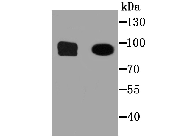
Western blot analysis of c-Kit on different lysates with Rabbit anti-c-Kit antibody (HA721154) at 1/1,000 dilution.
Lane 1: Saos-2 cell lysate
Lane 2: HL-60 cell lysate (low expression)
Lane 3: Jurkat cell lysate (low expression)
Lane 4: Human lung tissue lysate
Lysates/proteins at 20 µg/Lane.
Predicted band size: 110 kDa
Observed band size: 150 kDa
Exposure time: 2 minutes;
4-20% SDS-PAGE gel.
Proteins were transferred to a PVDF membrane and blocked with 5% NFDM/TBST for 1 hour at room temperature. The primary antibody (HA721154) at 1/1,000 dilution was used in 5% NFDM/TBST at 4℃ overnight. Goat Anti-Rabbit IgG - HRP Secondary Antibody (HA1001) at 1:100,000 dilution was used for 1 hour at room temperature.
Related Products
CD34 Recombinant Rabbit Monoclonal Antibody [SI16-01]
Application: WB,IF-Cell,IF-Tissue,IHC-P,IP,mIHC,FC
Reactivity: Human,Mouse,Rat
Conjugate: unconjugated
CD34 Rabbit Polyclonal Antibody
Application: WB,IHC-P
Reactivity: Human,Mouse
Conjugate: unconjugated
CD34 Rabbit Polyclonal Antibody
Application: WB,IF-Cell,IHC-P,FC
Reactivity: Human,Mouse,Rat,Zebrafish
Conjugate: unconjugated
CD34 Recombinant Mouse Monoclonal Antibody [PDM0-12]
Application: IHC-P,IF-Tissue
Reactivity: Human
Conjugate: unconjugated
CD34 Recombinant Rabbit Monoclonal Antibody
Application: mIHC
Reactivity: Human
Conjugate: unconjugated
CD34 Mouse Monoclonal Antibody [15H1]
Application: IHC-P,FC
Reactivity: Human,Mouse,Rat
Conjugate: unconjugated


