概述
产品名称
Phospho-STAT1 (S727) Recombinant Rabbit Monoclonal Antibody [SN67-04]
抗体类型
Recombinant Rabbit monoclonal Antibody
免疫原
Synthetic phospho-peptide corresponding to residues surrounding Ser727 of Human STAT1 aa 701-750 / 750.
种属反应性
Human, Mouse, Rat
验证应用
WB, IHC-P, IF-Cell, FC
分子量
Predicted band size: 87 kDa
阳性对照
HeLa cell lysate, mouse brain tissue lysate, rat brain tissue lysate, HeLa, human breast invasive ductal tumor tissue, rat colon tissue, mouse colon tissue, SiHa cell lysates, human liver carcinoma tissue, human lung carcinoma tissue.
偶联
unconjugated
克隆号
SN67-04
RRID
产品特性
形态
Liquid
存放说明
Shipped at 4℃. Store at +4℃ short term (1-2 weeks). It is recommended to aliquot into single-use upon delivery. Store at -20℃ long term.
存储缓冲液
1*TBS (pH7.4), 0.05% BSA, 40% Glycerol. Preservative: 0.05% Sodium Azide.
亚型
IgG
纯化方式
Protein A affinity purified.
应用稀释度
-
WB
-
1:2,000-1:5,000
-
IHC-P
-
1:200-1:1,000
-
IF-Cell
-
1:2,000
-
FC
-
1:1,000
发表文章中的应用
| WB | 查看 25 篇文献如下 |
| IHC-P | 查看 3 篇文献如下 |
| IF | 查看 2 篇文献如下 |
发表文章中的种属
| Mouse | 查看 18 篇文献如下 |
| Human | 查看 6 篇文献如下 |
| Rat | 查看 3 篇文献如下 |
| Rabbit | 查看 1 篇文献如下 |
靶点
功能
Membrane receptor signaling by various ligands, including interferons and growth hormones such as EGF, induces activation of Jak kinases which then leads to tyrosine phosphorylation of the various Stat transcription factors. Stat1 and Stat2 are induced by IFN-a and form a heterodimer which is part of the ISGF3 transcription factor complex. Although early reports indicate Stat3 activation by EGF and IL-6, it has been shown that Stat3β appears to be activated by both while Stat3α is activated by EGF, but not by IL-6. Highest expression of Stat4 is seen in testis and myeloid cells. IL-12 has been identified as an activator of Stat4. Stat5 has been shown to be activated by prolactin and by IL-3. Stat6 is involved in IL-4 activated signaling pathways.
背景文献
1. Chen L et al. Metastasis is regulated via microRNA-200/ZEB1 axis control of tumour cell PD-L1 expression and intratumoral immunosuppression. Nat Commun 5:5241 (2014).
2. Camicia R et al. BAL1/ARTD9 represses the anti-proliferative and pro-apoptotic IFN -STAT1-IRF1-p53 axis in diffuse large B-cell lymphoma. J Cell Sci 126:1969-80 (2013).
序列相似性
Belongs to the transcription factor STAT family.
翻译后修饰
Phosphorylated on tyrosine and serine residues in response to a variety of cytokines/growth hormones including IFN-alpha, IFN-gamma, PDGF and EGF. Activated KIT promotes phosphorylation on tyrosine residues and subsequent translocation to the nucleus. Upon EGF stimulation, phosphorylation on Tyr-701 (lacking in beta form) by JAK1, JAK2 or TYK2 promotes dimerization and subsequent translocation to the nucleus. Growth hormone (GH) activates STAT1 signaling only via JAK2. Tyrosine phosphorylated in response to constitutively activated FGFR1, FGFR2, FGFR3 and FGFR4. Phosphorylation on Ser-727 by several kinases including MAPK14, ERK1/2 and CAMKII on IFN-gamma stimulation, regulates STAT1 transcriptional activity. Phosphorylation on Ser-727 promotes sumoylation though increasing interaction with PIAS. Phosphorylation on Ser-727 by PRKCD induces apoptosis in response to DNA-damaging agents. Phosphorylated on tyrosine residues when PTK2/FAK1 is activated; most likely this is catalyzed by a SRC family kinase. Dephosphorylation on tyrosine residues by PTPN2 negatively regulates interferon-mediated signaling. Upon viral infection or IFN induction, phosphorylation on Ser-708 occurs much later than phosphorylation on Tyr-701 and is required for the binding of ISGF3 on the ISREs of a subset of IFN-stimulated genes IKBKE-dependent. Phosphorylation at Tyr-701 and Ser-708 are mutually exclusive, phosphorylation at Ser-708 requires previous dephosphorylation of Tyr-701.; Sumoylated with SUMO1, SUMO2 and SUMO3. Sumoylation is enhanced by IFN-gamma-induced phosphorylation on Ser-727, and by interaction with PIAS proteins. Enhances the transactivation activity.; ISGylated.; Mono-ADP-ribosylated at Glu-657 and Glu-705 by PARP14; ADP-ribosylation prevents phosphorylation at Tyr-701. However, the role of ADP-ribosylation in the prevention of phosphorylation has been called into question and the lack of phosphorylation may be due to sumoylation of Lys-703.; Monomethylated at Lys-525 by SETD2; monomethylation is necessary for phosphorylation at Tyr-701, translocation into the nucleus and activation of the antiviral defense.
亚细胞定位
Nucleus, Cytoplasm.
别名
Signal transducer and activator of transcription 1 91kD antibody
CANDF7 antibody
DKFZp686B04100 antibody
IMD31A antibody
IMD31B antibody
IMD31C antibody
ISGF 3 antibody
ISGF-3 antibody
OTTHUMP00000163552 antibody
OTTHUMP00000165046 antibody
展开Signal transducer and activator of transcription 1 91kD antibody
CANDF7 antibody
DKFZp686B04100 antibody
IMD31A antibody
IMD31B antibody
IMD31C antibody
ISGF 3 antibody
ISGF-3 antibody
OTTHUMP00000163552 antibody
OTTHUMP00000165046 antibody
OTTHUMP00000165047 antibody
OTTHUMP00000205845 antibody
Signal transducer and activator of transcription 1 91kDa antibody
Signal transducer and activator of transcription 1 antibody
Signal transducer and activator of transcription 1, 91kD antibody
Signal transducer and activator of transcription 1-alpha/beta antibody
STAT 1 antibody
Stat1 antibody
STAT1_HUMAN antibody
STAT91 antibody
Transcription factor ISGF 3 components p91 p84 antibody
Transcription factor ISGF-3 components p91/p84 antibody
折叠图片
-

☑ Cell treatment (CT)
Western blot analysis of Phospho-STAT1 (S727) on different lysates with Rabbit anti-Phospho-STAT1 (S727) antibody (ET1611-20) at 1/5,000 dilution.
Lane 1: HeLa cell lysate (15 µg/Lane)
Lane 2: Mouse brain tissue lysate (20 µg/Lane)
Lane 3: Rat brain tissue lysate (20 µg/Lane)
Lane 4: Mouse brain treated with λpp for 1 hour tissue lysate (20 µg/Lane)
Predicted band size: 87 kDa
Observed band size: 87 kDa
Exposure time: 3 minutes 10 seconds;
4-20% SDS-PAGE gel.
Proteins were transferred to a PVDF membrane and blocked with 5% NFDM/TBST for 1 hour at room temperature. The primary antibody (ET1611-20) at 1/5,000 dilution was used in 5% NFDM/TBST at 4℃ overnight. Goat Anti-Rabbit IgG - HRP Secondary Antibody (HA1001) at 1/50,000 dilution was used for 1 hour at room temperature. -

☑ Cell treatment (CT)
Immunocytochemistry analysis of HeLa cells treated with or without λpp labeling Phospho-STAT1 (S727) with Rabbit anti-Phospho-STAT1 (S727) antibody (ET1611-20) at 1/2,000 dilution.
Cells were fixed in 4% paraformaldehyde for 20 minutes at room temperature, permeabilized with 0.1% Triton X-100 in PBS for 5 minutes at room temperature, then blocked with 1% BSA in 10% negative goat serum for 1 hour at room temperature. Cells were then incubated with Rabbit anti-Phospho-STAT1 (S727) antibody (ET1611-20) at 1/2,000 dilution in 1% BSA in PBST overnight at 4 ℃. Goat Anti-Rabbit IgG H&L (iFluor™ 488, HA1121) was used as the secondary antibody at 1/1,000 dilution. PBS instead of the primary antibody was used as the secondary antibody only control. Nuclear DNA was labelled in blue with DAPI.
Beta tubulin (M1305-2, red) was stained at 1/100 dilution overnight at +4℃. Goat Anti-Mouse IgG H&L (iFluor™ 594, HA1126) was used as the secondary antibody at 1/1,000 dilution. -

☑ Cell treatment (CT),Relative expression (RE)
Immunohistochemical analysis of paraffin-embedded human breast invasive ductal tumor tissue with Rabbit anti-Phospho-STAT1 (S727) antibody (ET1611-20) at 1/200 dilution.
A: Untreated human breast invasive ductal tumor tissue
B: λ-PPase treated human breast invasive ductal tumor tissue
C: Negative control
The section was pre-treated using heat mediated antigen retrieval with Tris-EDTA buffer (pH 9.0) for 20 minutes. The tissues were blocked in 1% BSA for 20 minutes at room temperature, washed with ddH2O and PBS, and then probed with the primary antibody (ET1611-20) at 1/200 dilution for 1 hour at room temperature. The detection was performed using an HRP conjugated compact polymer system. DAB was used as the chromogen. Tissues were counterstained with hematoxylin and mounted with DPX. -

☑ Cell treatment (CT),Relative expression (RE)
Immunohistochemical analysis of paraffin-embedded rat colon tissue with Rabbit anti-Phospho-STAT1 (S727) antibody (ET1611-20) at 1/200 dilution.
A: Untreated rat colon tissue
B: λ-PPase treated rat colon tissue
C: Negative control
The section was pre-treated using heat mediated antigen retrieval with Tris-EDTA buffer (pH 9.0) for 20 minutes. The tissues were blocked in 1% BSA for 20 minutes at room temperature, washed with ddH2O and PBS, and then probed with the primary antibody (ET1611-20) at 1/200 dilution for 1 hour at room temperature. The detection was performed using an HRP conjugated compact polymer system. DAB was used as the chromogen. Tissues were counterstained with hematoxylin and mounted with DPX. -

Immunohistochemical analysis of paraffin-embedded mouse colon tissue with Rabbit anti-Phospho-STAT1 (S727) antibody (ET1611-20) at 1/1,000 dilution.
The section was pre-treated using heat mediated antigen retrieval with sodium citrate buffer (pH 6.0) (high pressure) for 2 minutes. The tissues were blocked in 1% BSA for 20 minutes at room temperature, washed with ddH2O and PBS, and then probed with the primary antibody (ET1611-20) at 1/1,000 dilution for 1 hour at room temperature. The detection was performed using an HRP conjugated compact polymer system. DAB was used as the chromogen. Tissues were counterstained with hematoxylin and mounted with DPX. -

☑ Cell treatment (CT)
Western blot analysis of Phospho-STAT1 (S727) on SiHa cell lysates.
Lane 1: SiHa cells, whole cell lysate, 10 μg /lane.
Lane 2: SiHa cells were treated with 2.8 μg/ul lambda-PP for 30 minutes, whole cell lysates, 10 μg/lane.
Proteins were transferred to a PVDF membrane and blocked with 5% BSA in PBS for 1 hour at room temperature. The primary antibody Anti-Phospho-STAT1 (S727) (ET1611-20, 1/500) , Anti-STAT1 antibody ( ET1606-39, 1/500) and Anti-HSP90 beta antibody (ET1605-56, 1/10,000)was used in 5% BSA at room temperature for 2 hours. Goat Anti-Rabbit IgG H&L (HRP) Secondary Antibody (HA1001) at 1:200,000 dilution was used for 1 hour at room temperature.
Predicted band size: 87 kDa
Observed band size: 87 kDa
Exposure time: 30 seconds -

Immunohistochemical analysis of paraffin-embedded human liver carcinoma tissue with Rabbit anti-Phospho-STAT1 (S727) antibody (ET1611-20) at 1/500 dilution.
The section was pre-treated using heat mediated antigen retrieval with Tris-EDTA buffer (pH 9.0) for 20 minutes. The tissues were blocked in 1% BSA for 20 minutes at room temperature, washed with ddH2O and PBS, and then probed with the primary antibody (ET1611-20) at 1/500 dilution for 1 hour at room temperature. The detection was performed using an HRP conjugated compact polymer system. DAB was used as the chromogen. Tissues were counterstained with hematoxylin and mounted with DPX. -

Immunohistochemical analysis of paraffin-embedded human lung carcinoma tissue with Rabbit anti-Phospho-STAT1 (S727) antibody (ET1611-20) at 1/500 dilution.
The section was pre-treated using heat mediated antigen retrieval with Tris-EDTA buffer (pH 9.0) for 20 minutes. The tissues were blocked in 1% BSA for 20 minutes at room temperature, washed with ddH2O and PBS, and then probed with the primary antibody (ET1611-20) at 1/500 dilution for 1 hour at room temperature. The detection was performed using an HRP conjugated compact polymer system. DAB was used as the chromogen. Tissues were counterstained with hematoxylin and mounted with DPX. -

Flow cytometric analysis of HeLa cells labeling Phospho-STAT1 (S727).
Cells were fixed and permeabilized. Then stained with the primary antibody (ET1611-20, 1μg/mL) (red) compared with Rabbit IgG Isotype Control (green). After incubation of the primary antibody at +4℃ for an hour, the cells were stained with a iFluor™ 488 conjugate-Goat anti-Rabbit IgG Secondary antibody (HA1121) at 1/1,000 dilution for 30 minutes at +4℃. Unlabelled sample was used as a control (cells without incubation with primary antibody; black).
请注意: All products are "FOR RESEARCH USE ONLY AND ARE NOT INTENDED FOR DIAGNOSTIC OR THERAPEUTIC USE"
引文
-
Rubia cordifolia L. extract ameliorates vitiligo by inhibiting the CXCL10/CXCL9/STAT1 signaling pathway
Author: Mengqi Xia, Zhiqiang Li, Cuiping Yang, Yilei Wang, Jianjian Zhang, Guoyue Zhong, Hui Ouyang, Yulin Feng
PMID: 40412781
期刊: Journal Of Ethnopharmacology
应用: WB
反应种属: Mouse
发表时间: 2025 May
-
Citation
-
CCL7 promotes macrophage polarization and synovitis to exacerbate rheumatoid arthritis
Author: Jun Chen, Shuo Shi, Xiaojia Li, Feng Gao, Xu Zhu, Ru Feng, Ke Hu, Yicheng Li, Shuiyuan Chen, Rongkai Zhang, Xiaoshuai Wang, Changhai Ding, Gang Liu, Tianyu Chen, Wenquan Liang
PMID: 40224025
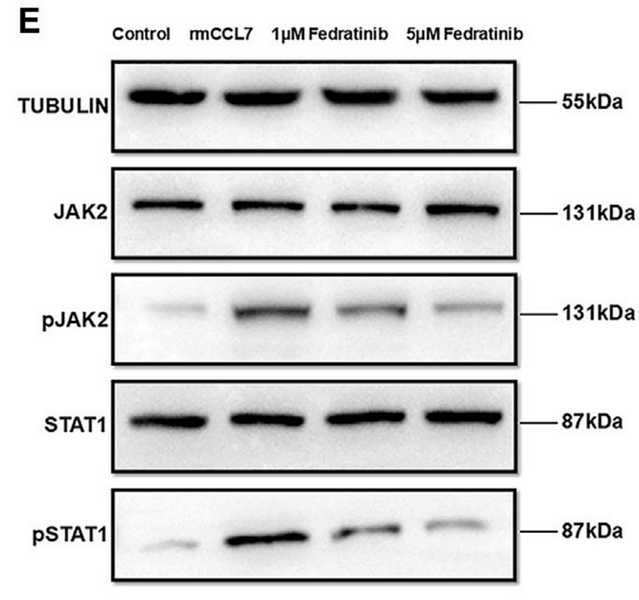
期刊: iScience
应用: WB
反应种属: Mouse
发表时间: 2025 Mar
-
Citation
-
KK2DP7 Stimulates CD11b+ Cell Populations in the Spleen to Elicit Trained Immunity for Anti-Tumor Therapy
Author: Rui Zhang, Lin Tang, Yusi Wang, Xuejing Zhou, Zhenyu Ding, Li Yang
PMID: 40145812
期刊: Advanced Science
应用: WB
反应种属: Mouse
发表时间: 2025 Mar
-
Citation
-
Treatment of rheumatoid arthritis via tetrahedral framework nucleic acid‑based efficient delivery of Jakinib: Synergistic anti-inflammatory and osteogenic effects
Author: Yichen Yang, Ruiqing Wang, Wumeng Yin, Ziqi Yue, Yuxuan Zhao, Zhou Jiang, Yunfeng Lin, Yao He, Shaojingya Gao, Xiaoxiao Cai
PMID: 10NOPMID25052701
期刊: Chemical Engineering Journal
应用: WB
反应种属: Rat
发表时间: 2025 Apr
-
Citation
-
Injectable ECM-mimetic dynamic hydrogels abolish ferroptosis-induced post-discectomy herniation through delivering nucleus pulposus progenitor cell-derived exosomes
Author: Wang Wenkai, Cheng Zhuo, Yu Miao, Liu Ke, Duan Hongli, Zhang Yang, Huang Xinle, Li Menghuan, Li Changqing, Hu Yan, Luo Zhong, Liu Minghan
PMID: 40169595
期刊: Nature Communications
应用: WB
反应种属: Rat
发表时间: 2025 Apr
-
Citation
-
Natural Product-Inspired Discovery of Naphthoquinone-Furo-Piperidine Derivatives as Novel STAT3 Inhibitors for the Treatment of Triple-Negative Breast Cancer
Author: Fan Chengcheng,et al
PMID: 39226127
期刊: Journal Of Medicinal Chemistry
应用: WB
反应种属: Human
发表时间: 2024 Sep
-
Citation
-
Long non-coding RNA SUN2-AS1 acts as a negative regulator of ISGs transcription to promote flavivirus infection
Author: Chao Yang,et al
PMID: 39288611
期刊: Virology
应用: WB
反应种属: Rabbit
发表时间: 2024 Sep
-
Citation
-
Blockade of the mitochondrial DNA release ameliorates hepatic ischemia-reperfusion injury through avoiding the activation of cGAS-Sting pathway
Author: Xiong Yi,et al
PMID: 39198913
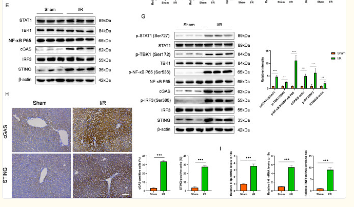
期刊: Journal Of Translational Medicine
应用: WB
反应种属: Mouse
发表时间: 2024 Sep
-
Citation
-
Gene Therapy for Inflammatory Cascade in Intrauterine Injury with Engineered Extracellular Vesicles Hybrid Snail Mucus-enhanced Adhesive Hydrogels
Author: Xiaotong Peng, Tao Wang, Bo Dai, Yiping Zhu, Mei Ji, Pusheng Yang, Jiaxin Zhang, Wenwen Liu, Yaxin Miao, Yonghang Liu, Shuo Wang, Jing Sun
PMID: 39454114
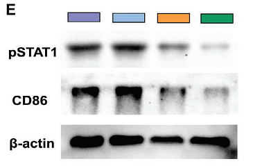
期刊: Advanced Science
应用: WB,IF
反应种属: Mouse
发表时间: 2024 Oct
-
Citation
-
MicroRNA-122 protects against interferon-α-induced hepatic inflammatory response via the Janus kinase–signal transducer and activator of transcription pathway
Author: Fanwei Liu ,et al
PMID: 39358210
期刊: Endocrine Journal
应用: WB
反应种属: Human
发表时间: 2024 Oct
-
Citation
-
Neurons upregulate PD-L1 via IFN/STAT1/IRF1 to alleviate damage by CD8+ T cells in cerebral malaria
Author: Wang Yi,et al
PMID: 38715061
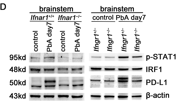
期刊: Journal Of Neuroinflammation
应用: WB,IHC-P
反应种属: Mouse
发表时间: 2024 May
-
Citation
-
Environmentally related microcystin-LR-induced ovarian dysfunction via the CCL2-CCR10 axis in mice ameliorated by dietary mulberry
Author: Du Xingde,et al
PMID: 38582190
期刊: Environmental Pollution
应用: WB
反应种属: Mouse
发表时间: 2024 May
-
Citation
-
Cediranib enhances the transcription of MHC-I by upregulating IRF-1
Author: Zhang Jie,et al
PMID: 38301967
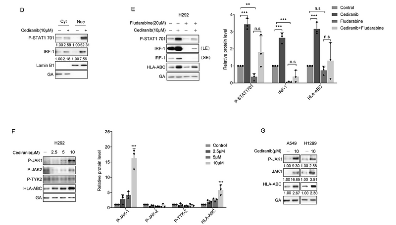
期刊: Biochemical Pharmacology
应用: WB
反应种属: Human,Mouse
发表时间: 2024 Mar
-
Citation
-
Inhibit of the cGAS-STING-STAT1 pathway protects heart from the Doxorubicin-induced cardiotoxicity
Author: Hou Ning,et al
PMID: NOPMID20240612
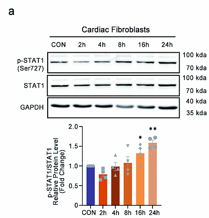
期刊: Preprint And Has Not Been Certified By Peer Review
应用: IF,WB
反应种属: Mouse
发表时间: 2024 Jun
-
Citation
-
Empagliflozin protects against heart failure with preserved ejection fraction partly by inhibiting the senescence-associated STAT1-STING axis
Author: Shi Ying,et al
PMID: 39044275
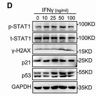
期刊: Cardiovasc Diabetol
应用: WB
反应种属: Mouse,Rat
发表时间: 2024 Jul
-
Citation
-
Linderae Radix extract attenuates ulcerative colitis by inhibiting the JAK/STAT signaling pathway
Author: Wang Yingying,et al
PMID: 39032278
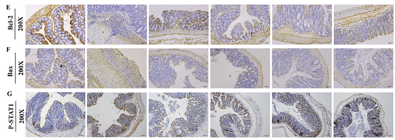
期刊: Phytomedicine
应用: WB,IHC-P
反应种属: Mouse
发表时间: 2024 Jul
-
Citation
-
IMPDH inhibitors upregulate PD-L1 in cancer cells without impairing immune checkpoint inhibitor efficacy
Author: Ming-Ming Zheng,et al
PMID: 39592732
期刊: Acta Pharmacologica Sinica
应用: WB
反应种属: Mouse
发表时间: 2024 Dec
-
Citation
-
CD8+ T cell infiltration and proliferation in the brainstem during experimental cerebral malaria
Author:
PMID: 37697956
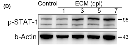
期刊: CNS Neuroscience & Therapeutics
应用: WB
反应种属: Mouse
发表时间: 2023 Sept
-
Citation
-
Filamin A is overexpressed in non-alcoholic steatohepatitis and contributes to the progression of inflammation and fibrosis
Author:
PMID: 36863213
期刊:
应用: WB
反应种属: Mouse
发表时间: 2023 Apr
-
Citation
-
Butein suppresses PD-L1 expression via downregulating STAT1 in non-small cell lung cancer
Author: Zhao, L., Zhang, W., Luan, F., Chen, X., Wu, H., He, Q., Weng, Q., Ding, L., & Yang, B.
PMID: 36455456

期刊: Biomedicine & Pharmacotherapy
应用: WB
反应种属: Human
发表时间: 2022 Nov
-
Citation
-
Activation of STAT6 by intranasal allergens correlated with the development of eosinophilic chronic rhinosinusitis in a mouse model
Author: Wei, H., Xu, L., Sun, P., Xing, H., Zhu, Z., & Liu, J.
PMID: 35726645
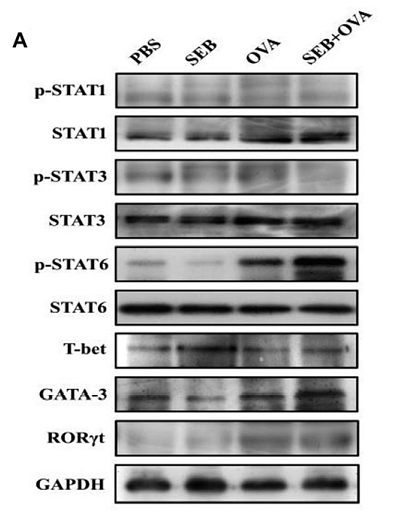
期刊: International Journal Of Immunopathology And Pharmacology
应用: IHC-P,WB
反应种属: Mouse
发表时间: 2022 Jun
-
Citation
-
The role of alcohol dehydrogenase 1C in regulating inflammatory responses in ulcerative colitis
Author:
PMID: 34293286
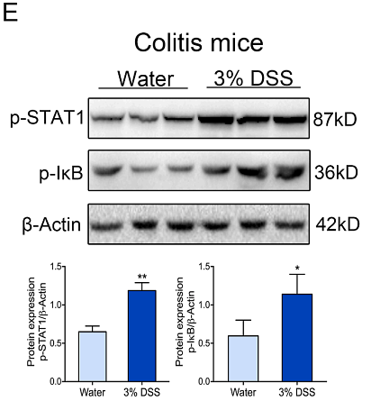
期刊: Biochemical Pharmacology
应用: WB
反应种属: Mouse,Human
发表时间: 2021 Oct
-
Citation
-
A 2-Benzylmalonate Derivative as STAT3 Inhibitor Suppresses Tumor Growth in Hepatocellular Carcinoma by Upregulating β-TrCP E3 Ubiquitin Ligase. International journal of molecular sciences, 22(7), 3354.
Author: Peng, T., Wonganan, O., Zhang, Z., Yu, J., Xi, R., Cao, Y., Suksamrarn, A., Zhang, G., & Wang, F.
PMID: 33805945
期刊: International Journal Of Molecular Sciences
应用: WB
反应种属: Mouse
发表时间: 2021 Mar
-
Citation
-
Combination Foretinib and Anti-PD-1 Antibody Immunotherapy for Colorectal Carcinoma. Frontiers in cell and developmental biology, 9, 689727.
Author: Fu, Y., Peng, Y., Zhao, S., Mou, J., Zeng, L., Jiang, X., Yang, C., Huang, C., Li, Y., Lu, Y., Wu, M., Yang, Y., Kong, T., Lai, Q., Wu, Y., Yao, Y., Wang, Y., Gou, L., & Yang, J.
PMID: 34307367
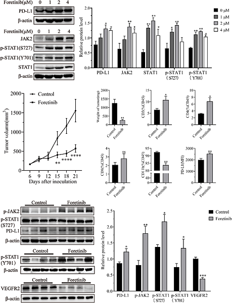
期刊: Frontiers In Cell And Developmental Biology
应用: WB
反应种属: Mouse
发表时间: 2021 Jul
-
Citation
-
Dexmedetomidine Directs T Helper Cells toward Th1 Cell Differentiation via the STAT1-T-Bet Pathway
Author:
PMID: 34414234
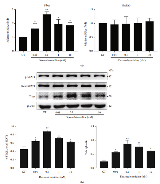
期刊: Biomed Research International
应用: WB
反应种属: Human
发表时间: 2021 Aug
-
Citation
同靶点 & 同通路的产品
Phospho-STAT1 (S727) Recombinant Rabbit Monoclonal Antibody [SN67-04] - BSA and Azide free
Application: WB,IHC-P,IF-Cell,FC
Reactivity: Human,Mouse,Rat
Conjugate: unconjugated
STAT1 Rabbit Polyclonal Antibody
Application: WB,IF-Cell
Reactivity: Human,Mouse,Rat,Zebrafish
Conjugate: unconjugated
Phospho-STAT1 (Y701) Recombinant Rabbit Monoclonal Antibody [PSH04-02]
Application: WB,IF-Cell
Reactivity: Human
Conjugate: unconjugated
Phospho-STAT1 (Y701) Recombinant Rabbit Monoclonal Antibody [PSH04-02] - BSA and Azide free
Application: WB,IF-Cell
Reactivity: Human
Conjugate: unconjugated
STAT1 Recombinant Mouse Monoclonal Antibody [G3-B11-R] - BSA and Azide free
Application: WB,IHC-P
Reactivity: Human
Conjugate: unconjugated
STAT1 Mouse Monoclonal Antibody [G3-B11]
Application: WB,IF-Cell,IHC-P,IF-Tissue
Reactivity: Human
Conjugate: unconjugated
STAT1 Recombinant Mouse Monoclonal Antibody [G3-B11-R]
Application: WB,IHC-P
Reactivity: Human
Conjugate: unconjugated
STAT1 Recombinant Rabbit Monoclonal Antibody [SD20-75]
Application: WB,IF-Cell,IF-Tissue,IHC-P,FC,IP
Reactivity: Human,Mouse,Rat
Conjugate: unconjugated


