概述
产品名称
Vimentin Recombinant Rabbit Monoclonal Antibody [PDH0-01]
抗体类型
Recombinant Rabbit monoclonal Antibody
免疫原
Synthetic peptide within C-terminal human Vimentin.
种属反应性
Human, Mouse, Rat
验证应用
WB, IHC-P, IF-Cell, FC, IF-Tissue, mIHC
分子量
Predicted band size: 54 kDa
阳性对照
L6 cell lysate, C2C12 cell lysate, Hela cell lysate, A549 cell lysate, human liver tissue, NIH/3T3 cell lysate, C6 cell lysate, human appendix tissue, human kidney tissue, human stomach carcinoma tissue, human endometrium tissue, HeLa, NIH/3T3, human skin tissue, mouse lung tissue.
偶联
unconjugated
克隆号
PDH0-01
RRID
产品特性
形态
Liquid
浓度
1.025ug/ul
存放说明
Store at +4℃ after thawing. Aliquot store at -20℃. Avoid repeated freeze / thaw cycles.
存储缓冲液
PBS (pH7.4), 0.1% BSA, 40% Glycerol. Preservative: 0.05% Sodium Azide.
亚型
IgG
纯化方式
Protein A affinity purified.
应用稀释度
-
WB
-
1:5,000
-
IHC-P
-
1:500-1:2,000
-
IF-Cell
-
1:200
-
FC
-
1:500-1:1,000
-
IF-Tissue
-
1:200
-
mIHC
-
1:10,000
发表文章中的应用
发表文章中的种属
| Mouse | See 1 publications below |
靶点
功能
Vimentin (57 kDa) is the most ubiquituos intermediate filament protein and the first to be expressed during cell differentiation. All primitive cell types express vimentin but in most non-mesenchymal cells it is replaced by other intermediate filament proteins during differentiation. Vimentin is expressed in a wide variety of mesenchymal cell types: fibroblasts, endothelial cells etc., and in a number of other cell types derived from mesoderm, e.g., mesothelium and ovarian granulosa cells. Vimentin is present in many different neoplasms but is particulary expressed in those originated from mesenchymal cells. Sarcomas e.g., fibrosarcoma, malignt fibrous histiocytoma, angiosarcoma, and leio- and rhabdomyosarcoma, as well as lymphomas, malignant melanoma and schwannoma, are virtually always vimentin positive. Mesoderm derived carcinomas like renal cell carcinoma, adrenal cortical carcinoma and adenocarcinomas from endometrium and ovary usually express vimentin. Also thyroid carcinomas are vimentin positive. Any low differentiated or sarcomatoid carcinoma may express some vimentin. Vimentin is frequently included in the so-called primary panel (together with CD45, cytokeratin, and S-100 protein): Intense staining reaction for vimentin without coexpression of other intermediate filament proteins is strongly suggestive of a mesenchymal tumour or a malignant melanoma. However, in biopsies representing only a sarcomatoid part of renal cell carcinoma a.o. a strong positivity for vimentin without cytokeratin expression may be seen. Tumours like lymphomas and seminomas have the same intermediate filament profile, but the vimentin expression is usually weaker.
背景文献
1. Ridge KM et al. Roles of vimentin in health and disease. Genes Dev. 2022 Apr
2. Kuburich NA et al. Vimentin and cytokeratin: Good alone, bad together. Semin Cancer Biol. 2022 Nov
亚细胞定位
Cytoplasm.
别名
CTRCT30 antibody
Epididymis luminal protein 113 antibody
FLJ36605 antibody
HEL113 antibody
VIM antibody
VIME_HUMAN antibody
Vimentin antibody
图片
-

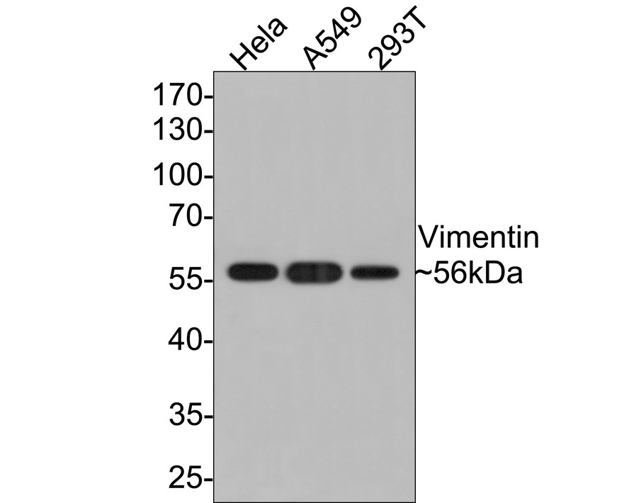
Western blot analysis of Vimentin on different lysates with Rabbit anti-Vimentin antibody (HA721174) at 1/5,000 dilution.
Lane 1: L6 cell lysate
Lane 2: C2C12 cell lysate
Lane 3: HeLa cell lysate
Lane 4: A549 cell lysate
Lysates/proteins at 10 µg/Lane.
Predicted band size: 54 kDa
Observed band size: 57 kDa
Exposure time: 2 minutes;
10% SDS-PAGE gel.
Proteins were transferred to a PVDF membrane and blocked with 5% NFDM/TBST for 1 hour at room temperature. The primary antibody (HA721174) at 1/5,000 dilution was used in 5% NFDM/TBST at room temperature for 2 hours. Goat Anti-Rabbit IgG - HRP Secondary Antibody (HA1001) at 1:300,000 dilution was used for 1 hour at room temperature. -

Immunohistochemical analysis of paraffin-embedded human liver tissue with Rabbit anti-Vimentin antibody (HA721174) at 1/2,000 dilution.
The section was pre-treated using heat mediated antigen retrieval with Tris-EDTA buffer (pH 9.0) for 20 minutes. The tissues were blocked in 1% BSA for 20 minutes at room temperature, washed with ddH2O and PBS, and then probed with the primary antibody (HA721174) at 1/2,000 dilution for 1 hour at room temperature. The detection was performed using an HRP conjugated compact polymer system. DAB was used as the chromogen. Tissues were counterstained with hematoxylin and mounted with DPX. -

Western blot analysis of Vimentin on different lysates with Rabbit anti-Vimentin antibody (HA721174) at 1/5,000 dilution.
Lane 1: HeLa cell lysate
Lane 2: NIH/3T3 cell lysate
Lane 3: C6 cell lysate
Lysates/proteins at 15 µg/Lane.
Predicted band size: 54 kDa
Observed band size: 54 kDa
Exposure time: 7 seconds;
4-20% SDS-PAGE gel.
Proteins were transferred to a PVDF membrane and blocked with 5% NFDM/TBST for 1 hour at room temperature. The primary antibody (HA721174) at 1/5,000 dilution was used in 5% NFDM/TBST at room temperature for 2 hours. Goat Anti-Rabbit IgG - HRP Secondary Antibody (HA1001) at 1:50,000 dilution was used for 1 hour at room temperature. -

Immunohistochemical analysis of paraffin-embedded human appendix tissue with Rabbit anti-Vimentin antibody (HA721174) at 1/2,000 dilution.
The section was pre-treated using heat mediated antigen retrieval with Tris-EDTA buffer (pH 9.0) for 20 minutes. The tissues were blocked in 1% BSA for 20 minutes at room temperature, washed with ddH2O and PBS, and then probed with the primary antibody (HA721174) at 1/2,000 dilution for 1 hour at room temperature. The detection was performed using an HRP conjugated compact polymer system. DAB was used as the chromogen. Tissues were counterstained with hematoxylin and mounted with DPX. -

Immunohistochemical analysis of paraffin-embedded human kidney tissue with Rabbit anti-Vimentin antibody (HA721174) at 1/2,000 dilution.
The section was pre-treated using heat mediated antigen retrieval with Tris-EDTA buffer (pH 9.0) for 20 minutes. The tissues were blocked in 1% BSA for 20 minutes at room temperature, washed with ddH2O and PBS, and then probed with the primary antibody (HA721174) at 1/2,000 dilution for 1 hour at room temperature. The detection was performed using an HRP conjugated compact polymer system. DAB was used as the chromogen. Tissues were counterstained with hematoxylin and mounted with DPX. -

Immunohistochemical analysis of paraffin-embedded human stomach carcinoma tissue with Rabbit anti-Vimentin antibody (HA721174) at 1/2,000 dilution.
The section was pre-treated using heat mediated antigen retrieval with Tris-EDTA buffer (pH 9.0) for 20 minutes. The tissues were blocked in 1% BSA for 20 minutes at room temperature, washed with ddH2O and PBS, and then probed with the primary antibody (HA721174) at 1/2,000 dilution for 1 hour at room temperature. The detection was performed using an HRP conjugated compact polymer system. DAB was used as the chromogen. Tissues were counterstained with hematoxylin and mounted with DPX. -

Immunohistochemical analysis of paraffin-embedded human endometrium tissue with Rabbit anti-Vimentin antibody (HA721174) at 1/500 dilution.
The section was pre-treated using heat mediated antigen retrieval with Tris-EDTA buffer (pH 9.0) for 20 minutes. The tissues were blocked in 1% BSA for 20 minutes at room temperature, washed with ddH2O and PBS, and then probed with the primary antibody (HA721174) at 1/500 dilution for 1 hour at room temperature. The detection was performed using an HRP conjugated compact polymer system. DAB was used as the chromogen. Tissues were counterstained with hematoxylin and mounted with DPX. -

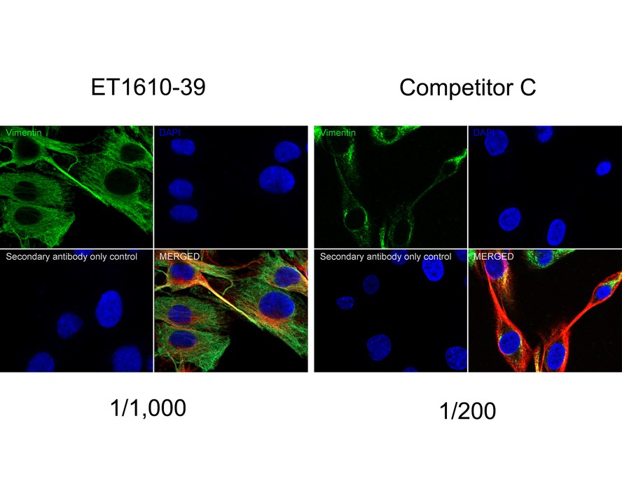
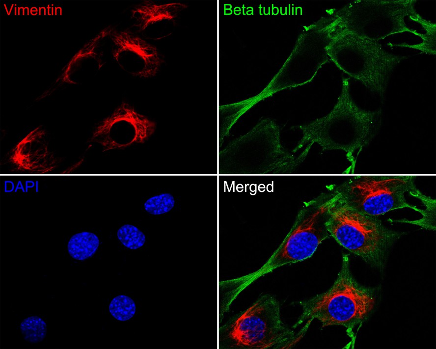
Immunocytochemistry analysis of HeLa cells labeling Vimentin with Rabbit anti-Vimentin antibody (HA721174) at 1/100 dilution.
Cells were fixed in 4% paraformaldehyde for 10 minutes at 37 ℃, permeabilized with 0.05% Triton X-100 in PBS for 20 minutes, and then blocked with 2% negative goat serum for 30 minutes at room temperature. Cells were then incubated with Rabbit anti-Vimentin antibody (HA721174) at 1/100 dilution in 2% negative goat serum overnight at 4 ℃. Goat Anti-Rabbit IgG H&L (iFluor™ 488, HA1121) was used as the secondary antibody at 1/1,000 dilution. PBS instead of the primary antibody was used as the secondary antibody only control. Nuclear DNA was labelled in blue with DAPI. -

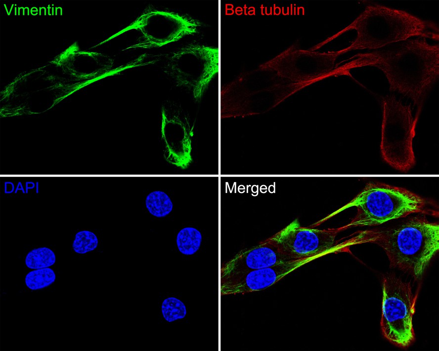
Immunocytochemistry analysis of NIH/3T3 cells labeling Vimentin with Rabbit anti-Vimentin antibody (HA721174) at 1/200 dilution.
Cells were fixed in 4% paraformaldehyde for 10 minutes at 37 ℃, permeabilized with 0.05% Triton X-100 in PBS for 20 minutes, and then blocked with 2% negative goat serum for 30 minutes at room temperature. Cells were then incubated with Rabbit anti-Vimentin antibody (HA721174) at 1/200 dilution in 2% negative goat serum overnight at 4 ℃. Goat Anti-Rabbit IgG H&L (iFluor™ 488, HA1121) was used as the secondary antibody at 1/1,000 dilution. PBS instead of the primary antibody was used as the secondary antibody only control. Nuclear DNA was labelled in blue with DAPI. -

Immunofluorescence analysis of paraffin-embedded human kidney tissue labeling Vimentin with Rabbit anti-Vimentin antibody (HA721174) at 1/200 dilution.
The section was pre-treated using heat mediated antigen retrieval with Tris-EDTA buffer (pH 9.0) for 20 minutes. The tissues were blocked in 10% negative goat serum for 1 hour at room temperature, washed with PBS, and then probed with the primary antibody (HA721174, green) at 1/200 dilution overnight at 4 ℃, washed with PBS.
Goat Anti-Rabbit IgG H&L (iFluor™ 488, HA1121) was used as the secondary antibody at 1/1,000 dilution. Nuclei were counterstained with DAPI (blue). -

Immunofluorescence analysis of paraffin-embedded human skin tissue labeling Vimentin with Rabbit anti-Vimentin antibody (HA721174) at 1/200 dilution.
The section was pre-treated using heat mediated antigen retrieval with Tris-EDTA buffer (pH 9.0) for 20 minutes. The tissues were blocked in 10% negative goat serum for 1 hour at room temperature, washed with PBS, and then probed with the primary antibody (HA721174, green) at 1/200 dilution overnight at 4 ℃, washed with PBS.
Goat Anti-Rabbit IgG H&L (iFluor™ 488, HA1121) was used as the secondary antibody at 1/1,000 dilution. Nuclei were counterstained with DAPI (blue). -

Immunofluorescence analysis of paraffin-embedded human appendix tissue labeling Vimentin with Rabbit anti-Vimentin antibody (HA721174) at 1/200 dilution.
The section was pre-treated using heat mediated antigen retrieval with Tris-EDTA buffer (pH 9.0) for 20 minutes. The tissues were blocked in 10% negative goat serum for 1 hour at room temperature, washed with PBS, and then probed with the primary antibody (HA721174, green) at 1/200 dilution overnight at 4 ℃, washed with PBS.
Goat Anti-Rabbit IgG H&L (iFluor™ 488, HA1121) was used as the secondary antibody at 1/1,000 dilution. Nuclei were counterstained with DAPI (blue). -

Flow cytometric analysis of Hela cells labeling Vimentin.
Cells were fixed and permeabilized. Then stained with the primary antibody (HA721174, 1ug/ml) (red) compared with Rabbit IgG Isotype Control (green). After incubation of the primary antibody at +4℃ for an hour, the cells were stained with a iFluor™ 488 conjugate-Goat anti-Rabbit IgG Secondary antibody (HA1121) at 1/1,000 dilution for 30 minutes at +4℃. Unlabelled sample was used as a control (cells without incubation with primary antibody; black). -

mIHC analysis of human skin tissue (Formalin/PFA-fixed paraffin-embedded sections) with Rabbit anti-Vimentin antibody (HA721174) at 1/10,000 dilution. The immunostaining was performed with the IRISKit® HyperView mTSA Kit (MH900206). Heat mediated antigen retrieval with Tris-EDTA buffer (pH 9.0) for 30 mins at 95℃. DAPI (blue) was used as a nuclear counter stain. Image acquisition was performed with Olympus VS200 Slide Scanner.
-

mIHC analysis of mouse lung tissue (Formalin/PFA-fixed paraffin-embedded sections) with Rabbit anti-Vimentin antibody (HA721174) at 1/10,000 dilution. The immunostaining was performed with the IRISKit® HyperView mTSA Kit (MH900206). Heat mediated antigen retrieval with Tris-EDTA buffer (pH 9.0) for 30 mins at 95℃. DAPI (blue) was used as a nuclear counter stain. Image acquisition was performed with Olympus VS200 Slide Scanner.
Please note: All products are "FOR RESEARCH USE ONLY AND ARE NOT INTENDED FOR DIAGNOSTIC OR THERAPEUTIC USE"
引文
-
Systemic mutation of Ncf1 ameliorates obstruction-induced renal fibrosis while macrophage-rescued NCF1 further alleviates renal fibrosis
Author:
PMID: 37392014

应用: WB
反应种属: Mouse
发表时间: 2023 Aug
-
Citation
同靶点&同通路的产品
Vimentin Recombinant Rabbit Monoclonal Antibody [PDH0-01]
Application: mIHC
Reactivity: Human
Conjugate: unconjugated
Vimentin Recombinant Rabbit Monoclonal Antibody [SC60-05]
Application: WB,IF-Cell,IF-Tissue,IHC-P,IP,FC,IHC-Fr
Reactivity: Human,Mouse,Rat
Conjugate: unconjugated
Vimentin Mouse Monoclonal Antibody [A6-C1]
Application: WB,IF-Cell,IHC-P
Reactivity: Human,Mouse,Rat
Conjugate: unconjugated
Vimentin Rabbit Polyclonal Antibody
Application: WB,IF-Cell,IHC-P
Reactivity: Human,Mouse,Rat
Conjugate: unconjugated
iFluor™ 647 Conjugated Vimentin Recombinant Rabbit Monoclonal Antibody [SC60-05]
Application: IF-Cell,IF-Tissue,FC
Reactivity: Human,Mouse,Rat
Conjugate: iFluor™ 647
Phospho-Vimentin (S39) Recombinant Rabbit Monoclonal Antibody [JE43-26]
Application: WB,IF-Cell
Reactivity: Human,Mouse,Rat
Conjugate: unconjugated
iFluor™ 488 Conjugated Vimentin Recombinant Rabbit Monoclonal Antibody [SC60-05]
Application: IF-Cell,IF-Tissue,FC
Reactivity: Human,Mouse,Rat
Conjugate: iFluor™ 488
Vimentin Recombinant Mouse Monoclonal Antibody [A6-C1-R]
Application: WB,IF-Cell,IHC-P
Reactivity: Human,Mouse,Rat
Conjugate: unconjugated
Vimentin Mouse Monoclonal Antibody [D4-B11]
Application: WB,IF-Cell,IHC-P,FC,IF-Tissue,IHC-Fr,IP
Reactivity: Human,Mouse,Rat
Conjugate: unconjugated


