概述
产品名称
Histone H3 (mono+di+tri methyl K79) Recombinant Rabbit Monoclonal Antibody [PSH09-10]
抗体类型
Recombinant Rabbit monoclonal Antibody
免疫原
Synthetic peptide within
种属反应性
Human, Mouse, Rat, Monkey
验证应用
WB, IHC-P, IF-Cell, ChIP
分子量
Predicted band size: 15 kDa
阳性对照
HeLa cell lysate, THP-1 cell lysate, NIH/3T3 cell lysate, RAW264.7 cell lysate, PC-12 cell lysate, C6 cell lysate, COS-1 cell lysate, human colon tissue, mouse colon tissue, rat colon tissue, HeLa, NIH/3T3, C6.
偶联
unconjugated
克隆号
PSH09-10
产品特性
形态
Liquid
浓度
1ug/ul
存放说明
Store at +4℃ after thawing. Aliquot store at -20℃. Avoid repeated freeze / thaw cycles.
存储缓冲液
PBS (pH7.4), 0.1% BSA, 40% Glycerol. Preservative: 0.05% Sodium Azide.
亚型
IgG
纯化方式
Protein A affinity purified.
应用稀释度
-
WB
-
1:2,000
-
IHC-P
-
1:1,000
-
IF-Cell
-
1:100-1:500
-
ChIP
-
Use 0.5~2 μg for 25 μg of chromatin.
靶点
功能
In eukaryotes, DNA is wrapped around histone octamers to form the basic unit of chromatin structure. The octamer is composed of histones H2A, H2B, H3 and H4, and it associates with approximately 200 base pairs of DNA to form the nucleosome. The association of DNA with histones results in dense packing of chromatin, which restricts proteins involved in gene transcription from binding to DNA. p300 preferentially acetylates Histone H3 at lysines 14 and 18 and Histone H4 at lysines 5 and 8. PCAF in its native form, primarily acetylates Histone H3 at lysine 14 to a monoacetylated form, and less efficiently acetylates Histone H4 at lysine 8. Histone H4 may also be acetylated at lysines 12 and 16, and the involvement of acetylated H4 with Histones H2A, H2B and H3 suggests that acetylated histones may be involved in dynamic chromatin remodeling.
背景文献
1. Wani S et al. Human SCP4 is a chromatin-associated CTD phosphatase and exhibits the dynamic translocation during erythroid differentiation. J Biochem 160:111-20 (2016).
2. Ni JZ et al. A transgenerational role of the germline nuclear RNAi pathway in repressing heat stress-induced transcriptional activation in C. elegans. Epigenetics Chromatin 9:3 (2016).
亚细胞定位
Nucleus, Chromosome.
UNIPROT #
别名
H3 histone family member E pseudogene antibody
H3 histone family, member A antibody
H3/A antibody
H31_HUMAN antibody
H3F3 antibody
H3FA antibody
Hist1h3a antibody
HIST1H3B antibody
HIST1H3C antibody
HIST1H3D antibody
展开H3 histone family member E pseudogene antibody
H3 histone family, member A antibody
H3/A antibody
H31_HUMAN antibody
H3F3 antibody
H3FA antibody
Hist1h3a antibody
HIST1H3B antibody
HIST1H3C antibody
HIST1H3D antibody
HIST1H3E antibody
HIST1H3F antibody
HIST1H3G antibody
HIST1H3H antibody
HIST1H3I antibody
HIST1H3J antibody
HIST3H3 antibody
histone 1, H3a antibody
Histone cluster 1, H3a antibody
Histone H3 3 pseudogene antibody
Histone H3.1 antibody
Histone H3/a antibody
Histone H3/b antibody
Histone H3/c antibody
Histone H3/d antibody
Histone H3/f antibody
Histone H3/h antibody
Histone H3/i antibody
Histone H3/j antibody
Histone H3/k antibody
Histone H3/l antibody
H3K27me3
折叠图片
-

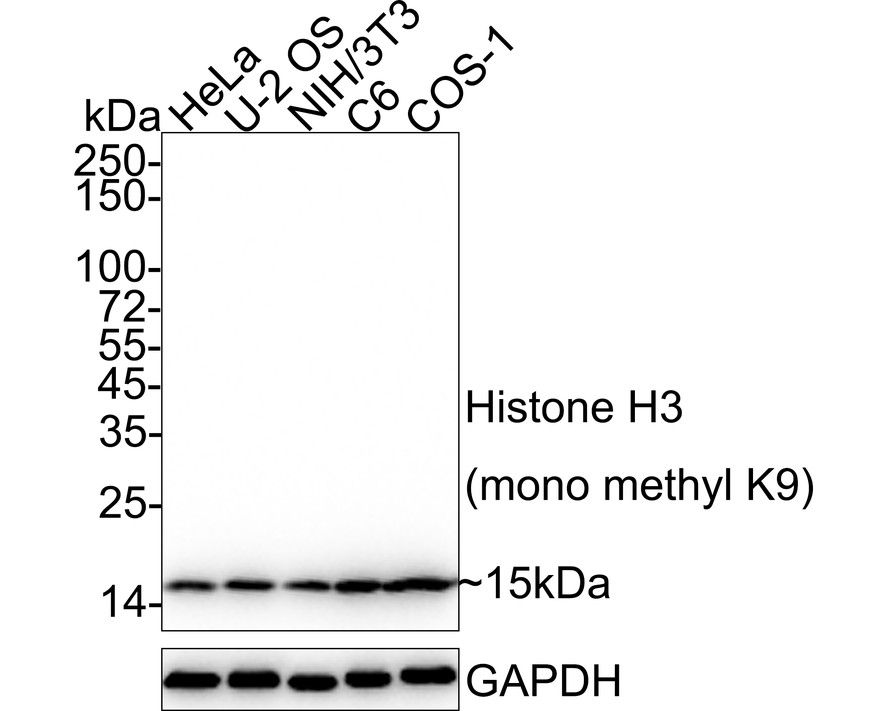
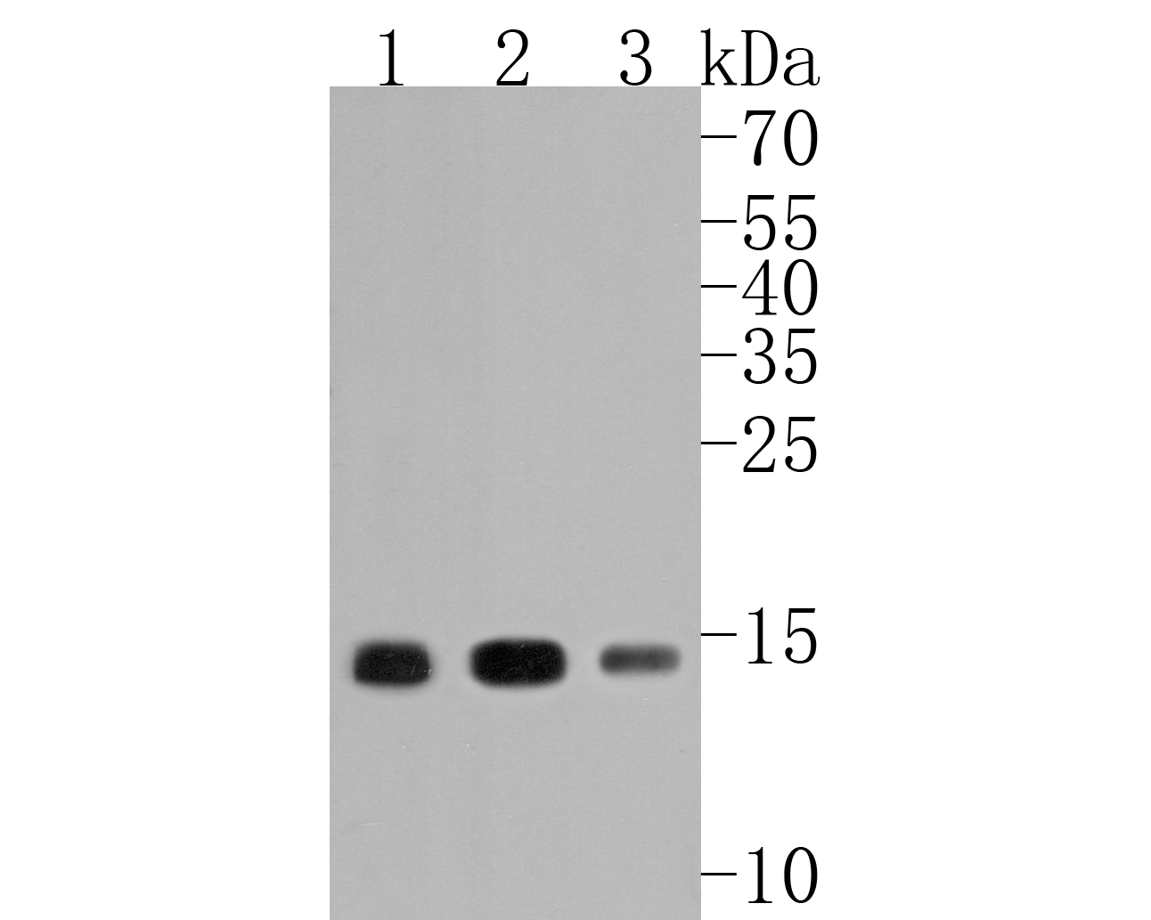
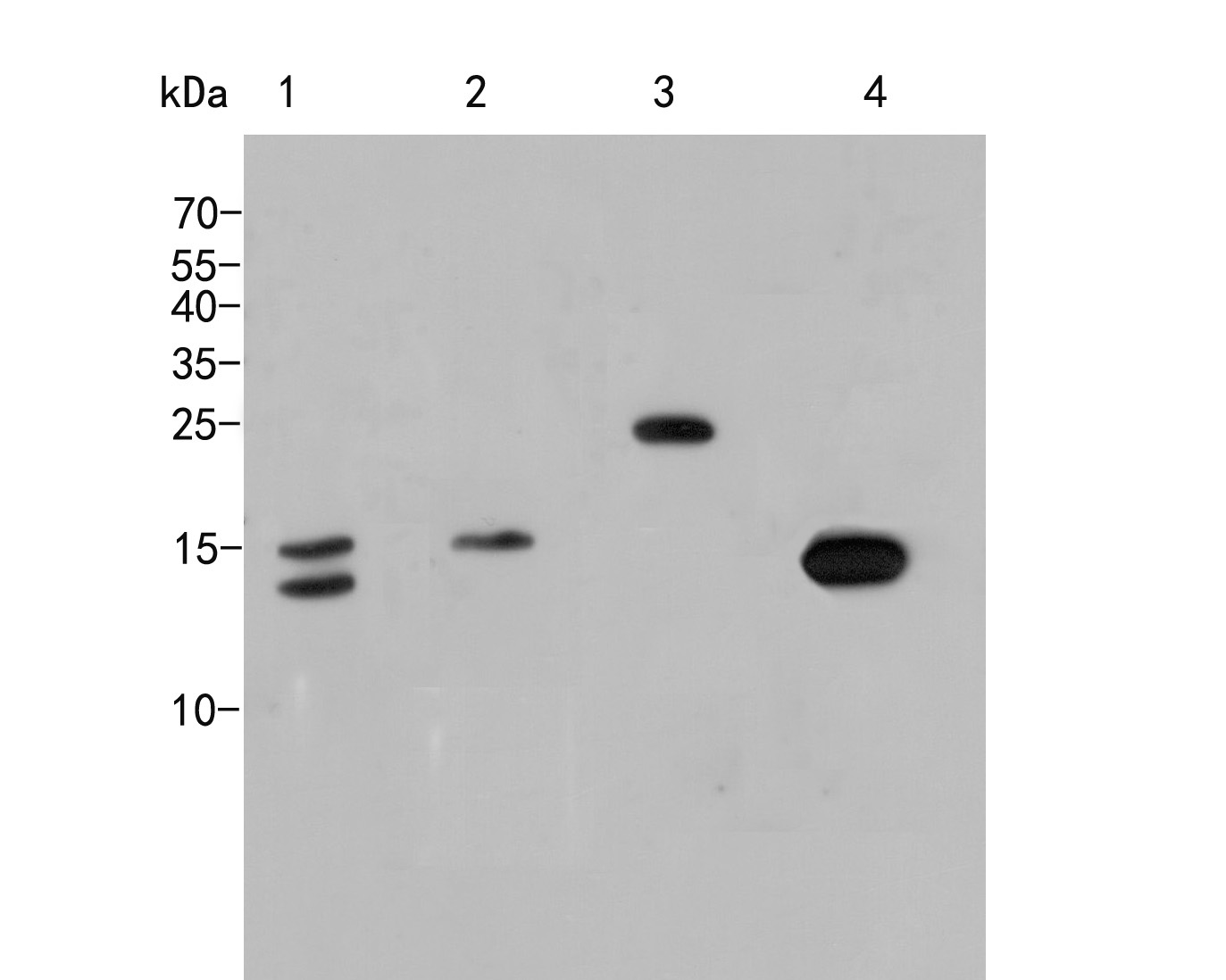
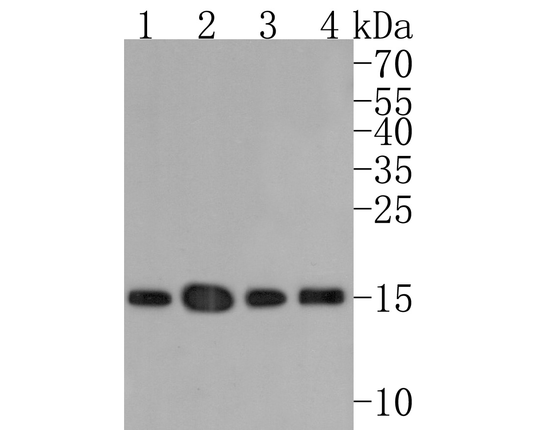
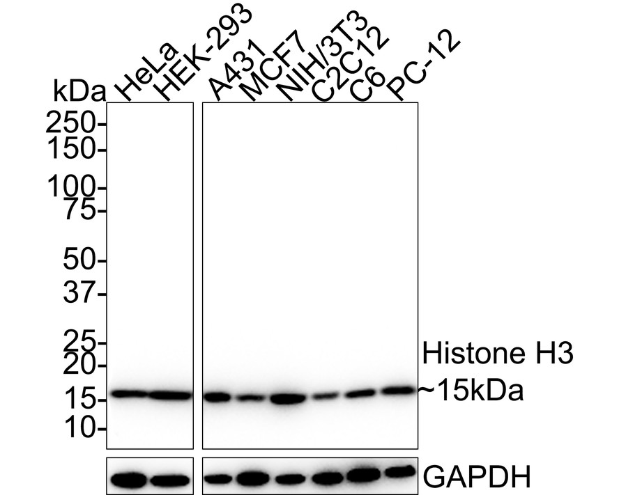
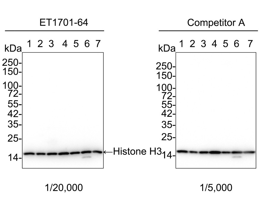
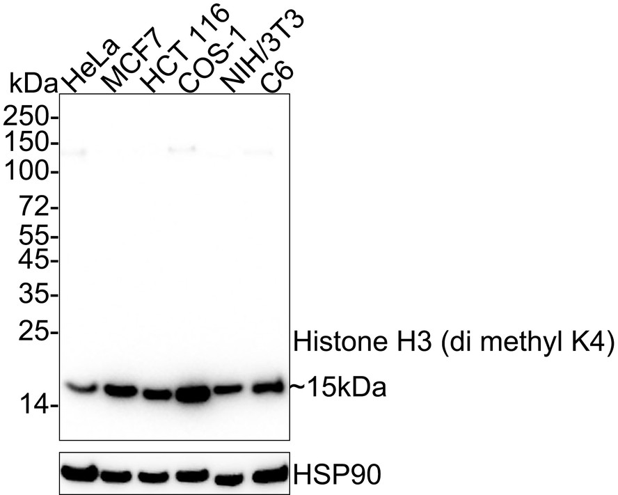
Western blot analysis of Histone H3 (mono+di+tri methyl K79) on different lysates with Rabbit anti-Histone H3 (mono+di+tri methyl K79) antibody (HA723068) at 1/2,000 dilution.
Lane 1: HeLa cell lysate
Lane 2: THP-1 cell lysate
Lane 3: NIH/3T3 cell lysate
Lane 4: RAW264.7 cell lysate
Lane 5: PC-12 cell lysate
Lane 6: C6 cell lysate
Lane 7: COS-1 cell lysate
Lysates/proteins at 20 µg/Lane.
Predicted band size: 15 kDa
Observed band size: 15 kDa
Exposure time: 10 seconds; ECL: K1801;
4-20% SDS-PAGE gel.
Proteins were transferred to a PVDF membrane and blocked with 5% NFDM/TBST for 1 hour at room temperature. The primary antibody (HA723068) at 1/2,000 dilution was used in 5% NFDM/TBST at 4℃ overnight. Goat Anti-Rabbit IgG - HRP Secondary Antibody (HA1001) at 1/50,000 dilution was used for 1 hour at room temperature. -

Immunohistochemical analysis of paraffin-embedded human colon tissue with Rabbit anti-Histone H3 (mono+di+tri methyl K79) antibody (HA723068) at 1/1,000 dilution.
The section was pre-treated using heat mediated antigen retrieval with sodium citrate buffer (pH 6.0) for 2 minutes. The tissues were blocked in 1% BSA for 20 minutes at room temperature, washed with ddH2O and PBS, and then probed with the primary antibody (HA723068) at 1/1,000 dilution for 1 hour at room temperature. The detection was performed using an HRP conjugated compact polymer system. DAB was used as the chromogen. Tissues were counterstained with hematoxylin and mounted with DPX. -

Immunohistochemical analysis of paraffin-embedded mouse colon tissue with Rabbit anti-Histone H3 (mono+di+tri methyl K79) antibody (HA723068) at 1/1,000 dilution.
The section was pre-treated using heat mediated antigen retrieval with sodium citrate buffer (pH 6.0) for 2 minutes. The tissues were blocked in 1% BSA for 20 minutes at room temperature, washed with ddH2O and PBS, and then probed with the primary antibody (HA723068) at 1/1,000 dilution for 1 hour at room temperature. The detection was performed using an HRP conjugated compact polymer system. DAB was used as the chromogen. Tissues were counterstained with hematoxylin and mounted with DPX. -

Immunohistochemical analysis of paraffin-embedded rat colon tissue with Rabbit anti-Histone H3 (mono+di+tri methyl K79) antibody (HA723068) at 1/1,000 dilution.
The section was pre-treated using heat mediated antigen retrieval with sodium citrate buffer (pH 6.0) for 2 minutes. The tissues were blocked in 1% BSA for 20 minutes at room temperature, washed with ddH2O and PBS, and then probed with the primary antibody (HA723068) at 1/1,000 dilution for 1 hour at room temperature. The detection was performed using an HRP conjugated compact polymer system. DAB was used as the chromogen. Tissues were counterstained with hematoxylin and mounted with DPX. -

Immunocytochemistry analysis of HeLa cells labeling Histone H3 (mono+di+tri methyl K79) with Rabbit anti-Histone H3 (mono+di+tri methyl K79) antibody (HA723068) at 1/100 dilution.
Cells were fixed in 4% paraformaldehyde for 15 minutes at room temperature, permeabilized with 0.1% Triton X-100 in PBS for 15 minutes at room temperature, then blocked with 1% BSA in 10% negative goat serum for 1 hour at room temperature. Cells were then incubated with Rabbit anti-Histone H3 (mono+di+tri methyl K79) antibody (HA723068) at 1/100 dilution in 1% BSA in PBST overnight at 4 ℃. Goat Anti-Rabbit IgG H&L (iFluor™ 488, HA1121) was used as the secondary antibody at 1/1,000 dilution. PBS instead of the primary antibody was used as the secondary antibody only control. Nuclear DNA was labelled in blue with DAPI.
Beta tubulin (HA601187, red) was stained at 1/100 dilution overnight at +4℃. Goat Anti-Mouse IgG H&L (iFluor™ 594, HA1126) was used as the secondary antibody at 1/1,000 dilution. -

Immunocytochemistry analysis of NIH/3T3 cells labeling Histone H3 (mono+di+tri methyl K79) with Rabbit anti-Histone H3 (mono+di+tri methyl K79) antibody (HA723068) at 1/500 dilution.
Cells were fixed in 4% paraformaldehyde for 15 minutes at room temperature, permeabilized with 0.1% Triton X-100 in PBS for 15 minutes at room temperature, then blocked with 1% BSA in 10% negative goat serum for 1 hour at room temperature. Cells were then incubated with Rabbit anti-Histone H3 (mono+di+tri methyl K79) antibody (HA723068) at 1/500 dilution in 1% BSA in PBST overnight at 4 ℃. Goat Anti-Rabbit IgG H&L (iFluor™ 488, HA1121) was used as the secondary antibody at 1/1,000 dilution. PBS instead of the primary antibody was used as the secondary antibody only control. Nuclear DNA was labelled in blue with DAPI.
Beta tubulin (HA601187, red) was stained at 1/100 dilution overnight at +4℃. Goat Anti-Mouse IgG H&L (iFluor™ 594, HA1126) was used as the secondary antibody at 1/1,000 dilution. -

Immunocytochemistry analysis of C6 cells labeling Histone H3 (mono+di+tri methyl K79) with Rabbit anti-Histone H3 (mono+di+tri methyl K79) antibody (HA723068) at 1/100 dilution.
Cells were fixed in 4% paraformaldehyde for 15 minutes at room temperature, permeabilized with 0.1% Triton X-100 in PBS for 15 minutes at room temperature, then blocked with 1% BSA in 10% negative goat serum for 1 hour at room temperature. Cells were then incubated with Rabbit anti-Histone H3 (mono+di+tri methyl K79) antibody (HA723068) at 1/100 dilution in 1% BSA in PBST overnight at 4 ℃. Goat Anti-Rabbit IgG H&L (iFluor™ 488, HA1121) was used as the secondary antibody at 1/1,000 dilution. PBS instead of the primary antibody was used as the secondary antibody only control. Nuclear DNA was labelled in blue with DAPI.
Beta tubulin (HA601187, red) was stained at 1/100 dilution overnight at +4℃. Goat Anti-Mouse IgG H&L (iFluor™ 594, HA1126) was used as the secondary antibody at 1/1,000 dilution. -

Chromatin immunoprecipitations were performed with cross-linked chromatin from HeLa cells and either Histone H3 (mono+di+tri methyl K79) (HA723068) or Normal Rabbit IgG according to the ChIP protocol. The enriched DNA was quantified by real-time PCR using indicated primers. The amount of immunoprecipitated DNA in each sample is represented as signal relative to the total amount of input chromatin, which is equivalent to one.
Please note: All products are "FOR RESEARCH USE ONLY AND ARE NOT INTENDED FOR DIAGNOSTIC OR THERAPEUTIC USE"
同靶点&同通路的产品
Histone H3 (mono methyl K79) Recombinant Rabbit Monoclonal Antibody [PSH07-66]
Application: WB,IF-Cell,IHC-P,FC,ChIP
Reactivity: Human,Mouse,Rat,Monkey
Conjugate: unconjugated
Phospho-Histone H3 (S28) Recombinant Rabbit Monoclonal Antibody [JE56-06]
Application: WB,IF-Cell,IHC-P,FC,ChIP
Reactivity: Human,Rat,Mouse
Conjugate: unconjugated
Histone H3 (mono methyl K36) Recombinant Rabbit Monoclonal Antibody [SR04-20]
Application: WB,IF-Cell,IF-Tissue,ChIP
Reactivity: Human,Mouse
Conjugate: unconjugated
Histone H3 (mono methyl R2) Recombinant Rabbit Monoclonal Antibody [ST0427]
Application: WB,IF-Cell,IF-Tissue,IHC-P,ChIP
Reactivity: Human,Mouse,Rat
Conjugate: unconjugated
Histone H3 (acetyl K56) Recombinant Rabbit Monoclonal Antibody [SU30-10]
Application: WB,IF-Cell,IF-Tissue,IHC-P,ChIP,CUT&Tag-seq
Reactivity: Human,Mouse,Rat
Conjugate: unconjugated
Histone H3 (tri methyl K9) Mouse Monoclonal Antibody [1-6]
Application: WB,IF-Cell,IHC-P,IF-Tissue
Reactivity: Human,Mouse,Rat
Conjugate: unconjugated
Histone H3 Mouse Monoclonal Antibody [A11-D7]
Application: WB,IF-Cell,IHC-P,IF-Tissue
Reactivity: Human,Mouse,Rat,Zebrafish
Conjugate: unconjugated
Histone H3 (tri methyl K27) Recombinant Rabbit Monoclonal Antibody [PSH05-15]
Application: WB,IF-Cell,IHC-P,IF-Tissue,ChIP
Reactivity: Human,Mouse,Rat
Conjugate: unconjugated
Histone H3 (di methyl K4) Rabbit Polyclonal Antibody
Application: WB,IF-Cell,IHC-P,FC,Dot Blot
Reactivity: Human,Mouse,Rat
Conjugate: unconjugated
Histone H3 (mono methyl K9) Recombinant Rabbit Monoclonal Antibody [JE43-27]
Application: WB,IF-Cell,IHC-P,FC,ChIP,IF-Tissue,Dot Blot
Reactivity: Human,Mouse,Rat,Monkey
Conjugate: unconjugated
Histone H3 (mono methyl K27) Recombinant Rabbit Monoclonal Antibody [PSH05-67]
Application: WB,IF-Cell,FC,ChIP
Reactivity: Human,Mouse,Rat
Conjugate: unconjugated
Histone H3 (acetyl K36) Recombinant Rabbit Monoclonal Antibody [JE77-32]
Application: WB,IHC-P,IF-Cell,ChIP
Reactivity: Human,Mouse,Rat
Conjugate: unconjugated
Histone H3 (mono+di+tri methyl K4) Recombinant Rabbit Monoclonal Antibody [PSH06-30]
Application: WB,IHC-P,IF-Cell,FC,ChIP
Reactivity: Human,Mouse,Rat
Conjugate: unconjugated
Histone H3 (di methyl K27) Recombinant Rabbit Monoclonal Antibody [PSH05-88]
Application: WB,IF-Cell,ChIP
Reactivity: Human,Mouse,Rat
Conjugate: unconjugated
Histone H3 (mono methyl K4) Recombinant Rabbit Monoclonal Antibody [PSH07-15]
Application: WB,IF-Cell,ChIP,Dot Blot
Reactivity: Human,Mouse,Rat
Conjugate: unconjugated
Histone H3 (acetyl K23) Recombinant Rabbit Monoclonal Antibody [JE46-39]
Application: WB,IF-Cell
Reactivity: Human,Mouse
Conjugate: unconjugated
Histone H3 (mono+di+tri methyl K36) Recombinant Rabbit Monoclonal Antibody [PSH07-14]
Application: WB,IHC-P,IF-Cell,FC,ChIP
Reactivity: Human,Mouse,Rat
Conjugate: unconjugated
Histone H3 Mouse Monoclonal Antibody [3-C4]
Application: WB,IHC-P,mIHC,IF-Tissue,ChIP
Reactivity: Human,Mouse,Rat
Conjugate: unconjugated
Histone H3 (mono methyl K18) Recombinant Rabbit Monoclonal Antibody [SA42-07]
Application: WB,IF-Cell,IF-Tissue,IHC-P
Reactivity: Human,Mouse,Rat
Conjugate: unconjugated
Phospho-Histone H3 (S10) Recombinant Rabbit Monoclonal Antibody [SA31-01]
Application: WB,IF-Cell,IF-Tissue,IHC-P,IP,FC,ChIP
Reactivity: Human,Mouse,Rat
Conjugate: unconjugated
Histone H3 (di methyl K9) Recombinant Rabbit Monoclonal Antibody [SN07-30]
Application: WB,IF-Cell,IF-Tissue,IHC-P
Reactivity: Human,Mouse,Rat
Conjugate: unconjugated
Histone H3 Recombinant Rabbit Monoclonal Antibody [JJ092-08]
Application: WB,IF-Tissue,IHC-P,ChIP
Reactivity: Human,Mouse,Rat
Conjugate: unconjugated
Histone H3 (acetyl K4) Recombinant Rabbit Monoclonal Antibody [PSH03-71]
Application: WB,IF-Cell,ChIP,Dot Blot
Reactivity: Human,Mouse,Rat
Conjugate: unconjugated
Histone H3 (acetyl K14) Recombinant Rabbit Monoclonal Antibody [JU43-26]
Application: WB,IF-Cell,IF-Tissue,IHC-P,IP,SNAP-ChIP,CUT&Tag-seq
Reactivity: Human,Mouse,Rat
Conjugate: unconjugated
Histone H3 (tri methyl K9) Mouse Monoclonal Antibody [2G1]
Application: WB,IF
Reactivity: Human
Conjugate: unconjugated
Histone H3 (acetyl K27) Rabbit Polyclonal Antibody
Application: WB,IHC-P,ChIP
Reactivity: Human,Mouse
Conjugate: unconjugated
Histone H3 (acetyl K18) Rabbit Polyclonal Antibody
Application: WB,IHC-P,IF-Cell
Reactivity: Human,Mouse
Conjugate: unconjugated
Histone H3 Rabbit Polyclonal Antibody
Application: WB
Reactivity: Zebrafish,Human,Mouse
Conjugate: unconjugated
Phospho-Histone H3 (T3) Recombinant Rabbit Monoclonal Antibody [JE42-48]
Application: WB,IHC-P
Reactivity: Human,Mouse,Rat
Conjugate: unconjugated
Histone H3 Mouse Monoclonal Antibody [6-A7]
Application: WB,IHC-P,IF-Tissue,ChIP
Reactivity: Human,Mouse,Rat
Conjugate: unconjugated
Histone H3 Rabbit Polyclonal Antibody
Application: WB
Reactivity: Human,Mouse,Rat
Conjugate: unconjugated
Histone H3 (mono methyl K4) Rabbit Polyclonal Antibody
Application: WB,IF-Cell,IHC-P,FC,Dot Blot
Reactivity: Human,Mouse,Rat
Conjugate: unconjugated
Histone H3 (acetyl K27) Mouse Monoclonal Antibody [A6D7]
Application: WB,IF-Cell,IHC-P,FC,ChIP
Reactivity: Human,Mouse,Rat
Conjugate: unconjugated
Histone H3 (acetyl K27) Mouse Monoclonal Antibody [A6D6]
Application: WB,IF-Cell,IHC-P,FC,ChIP
Reactivity: Human,Mouse,Rat
Conjugate: unconjugated
Histone H3 (acetyl K18) Mouse Monoclonal Antibody [A7B5]
Application: WB,IHC-P,ChIP
Reactivity: Human,Rat
Conjugate: unconjugated
Histone H3 Recombinant Mouse Monoclonal Antibody [A11-D7-R]
Application: WB,IF-Cell,IHC-P
Reactivity: Human,Mouse,Rat
Conjugate: unconjugated
Histone H3 Recombinant Mouse Monoclonal Antibody [6-A7-R] - Loading control
Application: WB,IHC-P,IF-Tissue,ChIP
Reactivity: Human,Mouse,Rat
Conjugate: unconjugated
Histone H3 Recombinant Rabbit Monoclonal Antibody [JJ090-07]
Application: WB,IF-Cell,IF-Tissue,IHC-P,ChIP,IP
Reactivity: Human,Mouse,Rat
Conjugate: unconjugated
Histone H3 (acetyl K9) Recombinant Rabbit Monoclonal Antibody [PSH04-47] - ChIP Grade
Application: WB,IF-Cell,IHC-P,IF-Tissue,FC,ChIP,Dot Blot,IP
Reactivity: Human,Mouse,Rat
Conjugate: unconjugated
Histone H3 (mono methyl K14) Recombinant Rabbit Monoclonal Antibody [JE43-29]
Application: WB,IF-Cell,ChIP,Dot Blot
Reactivity: Human,Mouse,Rat
Conjugate: unconjugated
Histone H3 (mono+di+tri methyl K79) Recombinant Rabbit Monoclonal Antibody [SR42-06]
Application: WB,IHC-P,ChIP
Reactivity: Human,Mouse,Rat
Conjugate: unconjugated
Histone H3 (acetyl K18) Mouse Monoclonal Antibody [A7B6]
Application: WB,IF-Cell
Reactivity: Human
Conjugate: unconjugated
Histone H3 (di methyl K4) Recombinant Rabbit Monoclonal Antibody [JE00-98]
Application: WB,IF-Cell,ChIP,Dot Blot,IHC-P
Reactivity: Human,Mouse,Rat,Monkey
Conjugate: unconjugated


