概述
产品名称
GAPDH Rabbit Polyclonal Antibody
抗体类型
Rabbit Polyclonal Antibody
免疫原
Recombinant protein within Human GAPDH aa 15-171.
种属反应性
Human, Mouse, Rat
验证应用
WB, IHC-P, IF-Cell, FC
分子量
36 kDa
阳性对照
PC-12 cell lysate, Hela cell lysate, mouse ovary tissue lysate, human placenta tissue lysate, rat brain tissue lysate, NCCIT cell lysate, A549, LoVo, MCF-7, rat kidney tissue, human colon cancer tissue, human spleen tissue, mouse testis tissue.
偶联
unconjugated
RRID
产品特性
形态
Liquid
浓度
1ug/ul
存放说明
Store at +4℃ after thawing. Aliquot store at -20℃. Avoid repeated freeze / thaw cycles.
存储缓冲液
1*PBS (pH7.4), 0.2% BSA, 50% Glycerol. Preservative: 0.05% Sodium Azide.
亚型
IgG
纯化方式
Protein A affinity purified.
应用稀释度
-
WB
-
1:500-1:2,000
-
IF-Cell
-
1:50-1:200
-
IHC-P
-
1:50-1:200
-
FC
-
1:50-1:100
发表文章中的应用
发表文章中的种属
靶点
功能
Has both glyceraldehyde-3-phosphate dehydrogenase and nitrosylase activities, thereby playing a role in glycolysis and nuclear functions, respectively. Participates in nuclear events including transcription, RNA transport, DNA replication and apoptosis. Nuclear functions are probably due to the nitrosylase activity that mediates cysteine S-nitrosylation of nuclear target proteins such as SIRT1, HDAC2 and PRKDC. Modulates the organization and assembly of the cytoskeleton. Facilitates the CHP1-dependent microtubule and membrane associations through its ability to stimulate the binding of CHP1 to microtubules. Glyceraldehyde-3- phosphate dehydrogenase is a key enzyme in glycolysis that catalyzes the first step of the pathway by converting D-glyceraldehyde 3-phosphate (G3P) into 3-phospho-D-glyceroyl phosphate. Component of the GAIT (gamma interferon-activated inhibitor of translation) complex which mediates interferon-gamma-induced transcript-selective translation inhibition in inflammatio n processes. Upon interferon-gamma treatment assembles into the GAIT complex which binds to stem loop-containing GAIT elements in the 3'-UTR of diverse inflammatory mRNAs (such as ceruplasmin) and suppresses their translation.
背景文献
1. Jenkins JL et al. High-resolution structure of human D-glyceraldehyde-3-phosphate dehydrogenase. Acta Crystallogr. D 62:290-301 (2006).
2. Ismail SA et al. Structural analysis of human liver glyceraldehyde-3-phosphate dehydrogenase. Acta Crystallogr. D 61:1508-1513 (2005).
序列相似性
Belongs to the glyceraldehyde-3-phosphate dehydrogenase family.
翻译后修饰
S-nitrosylation of Cys-152 leads to interaction with SIAH1, followed by translocation to the nucleus (By similarity). S-nitrosylation of Cys-247 is induced by interferon-gamma and LDL(ox) implicating the iNOS-S100A8/9 transnitrosylase complex and seems to prevent interaction with phosphorylated RPL13A and to interfere with GAIT complex activity.; ISGylated.; Sulfhydration at Cys-152 increases catalytic activity.; Oxidative stress can promote the formation of high molecular weight disulfide-linked GAPDH aggregates, through a process called nucleocytoplasmic coagulation. Such aggregates can be observed in vivo in the affected tissues of patients with Alzheimer disease or alcoholic liver cirrhosis, or in cell cultures during necrosis. Oxidation at Met-46 may play a pivotal role in the formation of these insoluble structures. This modification has been detected in vitro following treatment with free radical donor (+/-)-(E)-4-ethyl-2-[(E)-hydroxyimino]-5-nitro-3-hexenamide. It has been proposed to destabilize nearby residues, increasing the likelihood of secondary oxidative damages, including oxidation of Tyr-45 and Met-105. This cascade of oxidations may augment GAPDH misfolding, leading to intermolecular disulfide cross-linking and aggregation.; Succination of Cys-152 and Cys-247 by the Krebs cycle intermediate fumarate, which leads to S-(2-succinyl)cysteine residues, inhibits glyceraldehyde-3-phosphate dehydrogenase activity. Fumarate concentration as well as succination of cysteine residues in GAPDH is significantly increased in muscle of diabetic mammals. It was proposed that the S-(2-succinyl)cysteine chemical modification may be a useful biomarker of mitochondrial and oxidative stress in diabetes and that succination of GAPDH and other thiol proteins by fumarate may contribute to the metabolic changes underlying the development of diabetes complications.
亚细胞定位
Cytoplasm, Nucleus, Membrane.
别名
38 kDa BFA-dependent ADP-ribosylation substrate antibody
aging associated gene 9 protein antibody
Aging-associated gene 9 protein antibody
BARS-38 antibody
cb609 antibody
EC 1.2.1.12 antibody
Epididymis secretory sperm binding protein Li 162eP antibody
G3P_HUMAN antibody
G3PD antibody
G3PDH antibody
展开38 kDa BFA-dependent ADP-ribosylation substrate antibody
aging associated gene 9 protein antibody
Aging-associated gene 9 protein antibody
BARS-38 antibody
cb609 antibody
EC 1.2.1.12 antibody
Epididymis secretory sperm binding protein Li 162eP antibody
G3P_HUMAN antibody
G3PD antibody
G3PDH antibody
GAPD antibody
GAPDH antibody
Glyceraldehyde 3 phosphate dehydrogenase antibody
Glyceraldehyde-3-phosphate dehydrogenase antibody
HEL-S-162eP antibody
KNC-NDS6 antibody
MGC102544 antibody
MGC102546 antibody
MGC103190 antibody
MGC103191 antibody
MGC105239 antibody
MGC127711 antibody
MGC88685 antibody
OCAS, p38 component antibody
OCT1 coactivator in S phase, 38-KD component antibody
peptidyl cysteine S nitrosylase GAPDH antibody
Peptidyl-cysteine S-nitrosylase GAPDH antibody
wu:fb33a10 antibody
折叠图片
-

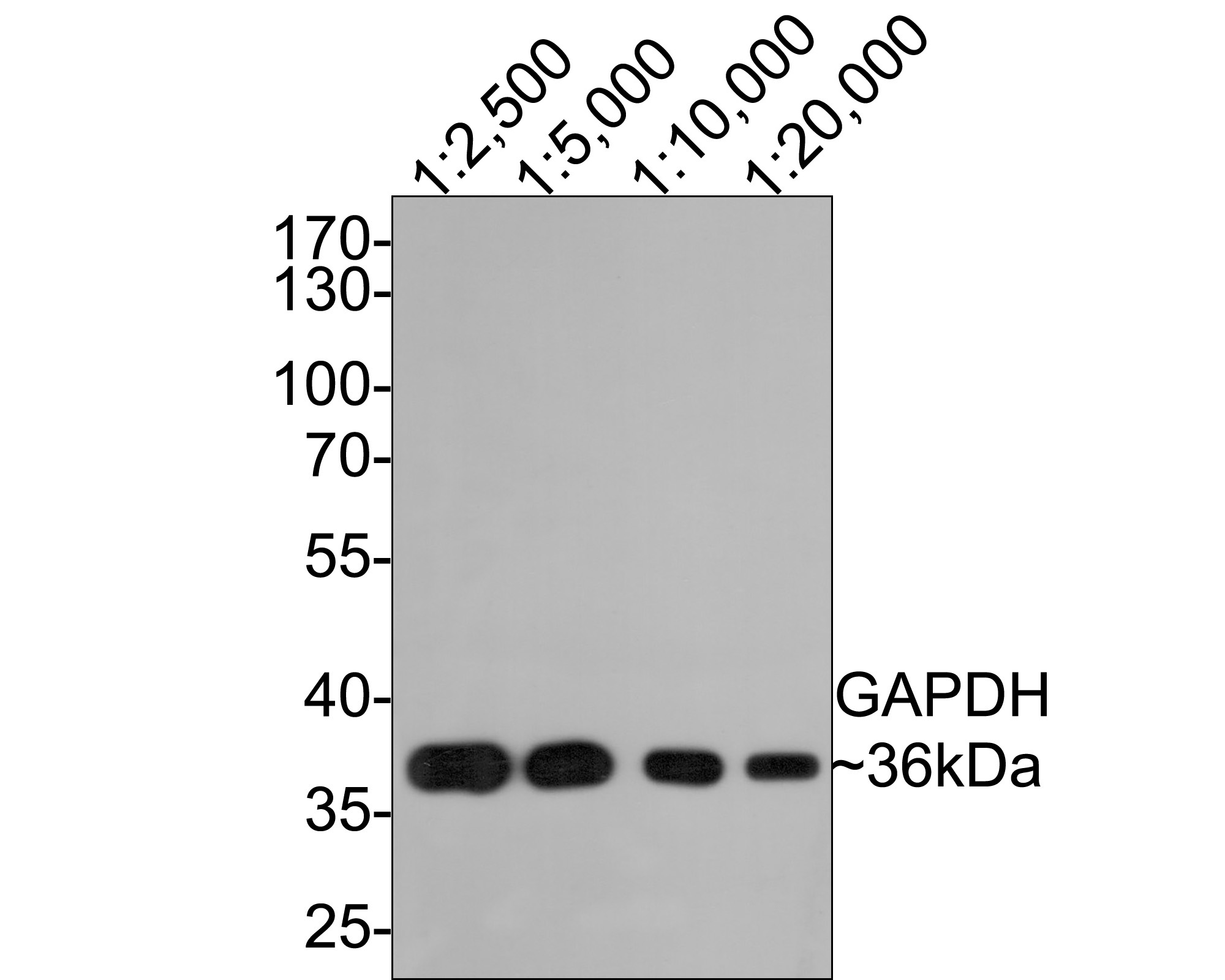
Western blot analysis of GAPDH on PC-12 cell lysate. Proteins were transferred to a PVDF membrane and blocked with 5% BSA in PBS for 1 hour at room temperature. The primary antibody was used at a 1:2,000 dilution in 5% BSA at room temperature for 2 hours. Goat Anti-Rabbit IgG - HRP Secondary Antibody (HA1001) at 1:5,000 dilution was used for 1 hour at room temperature.
-

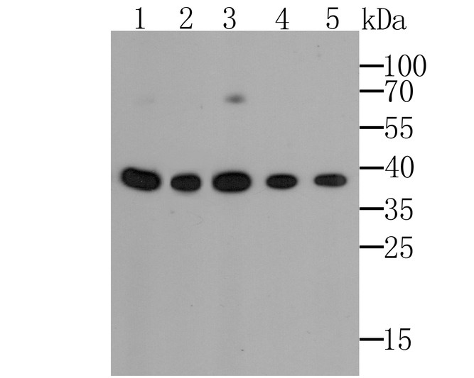
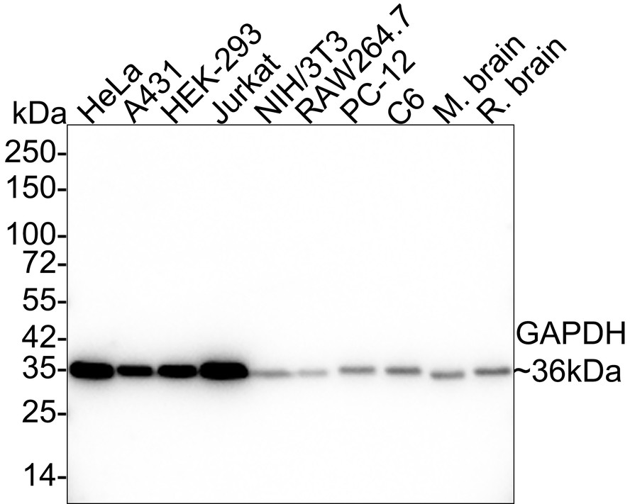
Western blot analysis of GAPDH on different lysates. Proteins were transferred to a PVDF membrane and blocked with 5% BSA in PBS for 1 hour at room temperature. The primary antibody (ER1901-65, 1/500) was used in 5% BSA at room temperature for 2 hours. Goat Anti-Rabbit IgG - HRP Secondary Antibody (HA1001) at 1:5,000 dilution was used for 1 hour at room temperature.
Positive control:
Lane 1: PC-12 cell lysate
Lane 2: Hela cell lysate
Lane 3: Mouse ovary tissue lysate
Lane 4: Human placenta tissue lysate
Lane 5: Rat brain tissue lysate
Lane 6: NCCIT cell lysate -

ICC staining of GAPDH in A549 cells (green). Formalin fixed cells were permeabilized with 0.1% Triton X-100 in TBS for 10 minutes at room temperature and blocked with 1% Blocker BSA for 15 minutes at room temperature. Cells were probed with the antibody (ER1901-65) at a dilution of 1:50 for 1 hour at room temperature, washed with PBS. Alexa Fluor®488 Goat anti-Rabbit IgG was used as the secondary antibody at 1/100 dilution. The nuclear counter stain is DAPI (blue).
-

ICC staining of GAPDH in LoVo cells (green). Formalin fixed cells were permeabilized with 0.1% Triton X-100 in TBS for 10 minutes at room temperature and blocked with 1% Blocker BSA for 15 minutes at room temperature. Cells were probed with the antibody (ER1901-65) at a dilution of 1:50 for 1 hour at room temperature, washed with PBS. Alexa Fluor®488 Goat anti-Rabbit IgG was used as the secondary antibody at 1/100 dilution. The nuclear counter stain is DAPI (blue).
-

ICC staining of GAPDH in MCF-7 cells (green). Formalin fixed cells were permeabilized with 0.1% Triton X-100 in TBS for 10 minutes at room temperature and blocked with 1% Blocker BSA for 15 minutes at room temperature. Cells were probed with the antibody (ER1901-65) at a dilution of 1:100 for 1 hour at room temperature, washed with PBS. Alexa Fluor®488 Goat anti-Rabbit IgG was used as the secondary antibody at 1/100 dilution. The nuclear counter stain is DAPI (blue).
-

Immunohistochemical analysis of paraffin-embedded rat kidney tissue using anti-GAPDH antibody. The section was pre-treated using heat mediated antigen retrieval with sodium citrate buffer (pH 6.0) for 20 minutes. The tissues were blocked in 5% BSA for 30 minutes at room temperature, washed with ddH2O and PBS, and then probed with the antibody (ER1901-65) at 1/50 dilution, for 30 minutes at room temperature and detected using an HRP conjugated compact polymer system. DAB was used as the chromogen. Counter stained with hematoxylin and mounted with DPX.
-

Immunohistochemical analysis of paraffin-embedded human colon cancer tissue using anti-GAPDH antibody. The section was pre-treated using heat mediated antigen retrieval with sodium citrate buffer (pH 6.0) for 20 minutes. The tissues were blocked in 5% BSA for 30 minutes at room temperature, washed with ddH2O and PBS, and then probed with the antibody (ER1901-65) at 1/50 dilution, for 30 minutes at room temperature and detected using an HRP conjugated compact polymer system. DAB was used as the chromogen. Counter stained with hematoxylin and mounted with DPX.
-

Immunohistochemical analysis of paraffin-embedded human spleen tissue using anti-GAPDH antibody. The section was pre-treated using heat mediated antigen retrieval with sodium citrate buffer (pH 6.0) for 20 minutes. The tissues were blocked in 5% BSA for 30 minutes at room temperature, washed with ddH2O and PBS, and then probed with the antibody (ER1901-65) at 1/50 dilution, for 30 minutes at room temperature and detected using an HRP conjugated compact polymer system. DAB was used as the chromogen. Counter stained with hematoxylin and mounted with DPX.
-

Immunohistochemical analysis of paraffin-embedded mouse testis tissue using anti-GAPDH antibody. The section was pre-treated using heat mediated antigen retrieval with sodium citrate buffer (pH 6.0) for 20 minutes. The tissues were blocked in 5% BSA for 30 minutes at room temperature, washed with ddH2O and PBS, and then probed with the antibody (ER1901-65) at 1/50 dilution, for 30 minutes at room temperature and detected using an HRP conjugated compact polymer system. DAB was used as the chromogen. Counter stained with hematoxylin and mounted with DPX.
-

Flow cytometric analysis of GAPDH was done on MCF-7 cells. The cells were fixed, permeabilized and stained with GAPDH antibody at 1/50 dilution (red) compared with an unlabelled control (cells without incubation with primary antibody; black). After incubation of the primary antibody on room temperature for an hour, the cells was stained with a Alexa Fluor 488-conjugated goat anti-rabbit IgG Secondary antibody at 1/500 dilution for 30 minutes.
Please note: All products are "FOR RESEARCH USE ONLY AND ARE NOT INTENDED FOR DIAGNOSTIC OR THERAPEUTIC USE"
引文
-
Prescription of Sageretia hamosa Brongn Relieved Goiter through Promoted Apoptosis of Thyroid Cells via miR-511-3p and PTEN/PI3K/Akt Pathway
Author: Deng, Y. L., Chen, S., Wang, H. T., Wang, B., & Xiao, K.
PMID: 34630982

应用: WB
反应种属: Rat
发表时间: 2021 Sep
-
Citation
-
<html>NF-κB-Induced Upregulation of miR-146a-5p Promoted Hippocampal Neuronal Oxidative Stress and Pyroptosis <i>via</i> TIGAR in a Model of Alzheimer's Disease. Frontiers in cellular neuroscience, 15, 653881.</html>
Author: Lei, B., Liu, J., Yao, Z., Xiao, Y., Zhang, X., Zhang, Y., & Xu, J.
PMID: 33935653
应用: WB
反应种属: Human
发表时间: 2021 Apr
-
Citation
Alternative Products
GAPDH Recombinant Rabbit Monoclonal Antibody [SA30-01] - Loading control
Application: WB,IF-Cell,IF-Tissue,IHC-P,FC,IP
Reactivity: Human,Mouse,Rat,Chicken,Drosophila melanogaster,Zebrafish
Conjugate: unconjugated
GAPDH Recombinant Rabbit Monoclonal Antibody [PD00-07]
Application: WB,IHC-P,IF-Cell,FC
Reactivity: Human,Mouse,Rat,Escherichia coli,Zebrafish,Drosophila melanogaster
Conjugate: unconjugated
同靶点&同通路的产品
GAPDH Mouse Monoclonal Antibody [5-E10]
Application: WB,IF-Cell,IHC-P
Reactivity: Human,Mouse,Rat,Zebrafish,Rabbit
Conjugate: unconjugated
GAPDH Recombinant Rabbit Monoclonal Antibody [SA30-01] - Loading control
Application: WB,IF-Cell,IF-Tissue,IHC-P,FC,IP
Reactivity: Human,Mouse,Rat,Chicken,Drosophila melanogaster,Zebrafish
Conjugate: unconjugated
GAPDH Mouse Monoclonal Antibody [12D6]
Application: WB,IF-Cell,IHC-P
Reactivity: Human,Mouse,Rat,Zebrafish,Escherichia coli
Conjugate: unconjugated
GAPDH Rabbit Polyclonal Antibody
Application: WB
Reactivity: Magnaporthe oryzae,Zebrafish
Conjugate: unconjugated
GAPDH Rabbit Polyclonal Antibody
Application: WB,IF-Cell,IHC-P,FC
Reactivity: Human,Mouse,Rat,Oryza sativa
Conjugate: unconjugated
HRP Conjugated GAPDH Recombinant Rabbit Monoclonal Antibody [SA30-01]
Application: WB,IHC
Reactivity: Human,Mouse,Rat
Conjugate: HRP
GAPDH Mouse Monoclonal Antibody [12D7]
Application: WB,IHC-P,FC
Reactivity: Human,Mouse,Rat,Zebrafish
Conjugate: unconjugated
GAPDH Rabbit Polyclonal Antibody
Application: WB,IF-Cell,IHC-P,FC
Reactivity: Human,Mouse,Rat
Conjugate: unconjugated
GAPDH Rabbit Polyclonal Antibody
Application: WB
Reactivity: Human,Mouse,Rat
Conjugate: unconjugated
HRP Conjugated GAPDH Recombinant Rabbit Monoclonal Antibody [JF81-04]
Application: WB
Reactivity: Human,Mouse,Rat,Monkey,Chicken,Fish
Conjugate: HRP
GAPDH Recombinant Rabbit Monoclonal Antibody [PD00-07]
Application: WB,IHC-P,IF-Cell,FC
Reactivity: Human,Mouse,Rat,Escherichia coli,Zebrafish,Drosophila melanogaster
Conjugate: unconjugated


