概述
产品名称
Phospho-Creb (S133) Recombinant Rabbit Monoclonal Antibody [JB25-40]
抗体类型
Recombinant Rabbit monoclonal Antibody
免疫原
Synthetic phospho-peptide corresponding to residues surrounding Ser133 of human Creb.
种属反应性
Human, Mouse, Rat
验证应用
WB, IF-Cell, IHC-P, IP, FC, IF-Tissue
分子量
Predicted band size: 35 kDa
阳性对照
HeLa treated with 25μg/mL anisomycin for 30 minutes whole cell lysate, SH-SY5Y, HUVEC, human spleen tissue, mouse colon tissue, human colon carcinoma tissue, human lymph nodes tissue, mouse large intestine tissue, HeLa.
偶联
unconjugated
克隆号
JB25-40
RRID
产品特性
形态
Liquid
浓度
1ug/ul
存放说明
Store at +4℃ after thawing. Aliquot store at -20℃ or -80℃. Avoid repeated freeze / thaw cycles.
存储缓冲液
1*TBS (pH7.4), 0.05% BSA, 40% Glycerol. Preservative: 0.05% Sodium Azide.
亚型
IgG
纯化方式
Protein A affinity purified.
应用稀释度
-
WB
-
1:1,000
-
IF-Cell
-
1:50-1:100
-
IHC-P
-
1:50-1:1,000
-
FC
-
1:50-1:100
-
IP
-
Use at an assay dependent concentration.
-
IF-Tissue
-
1:200
发表文章中的应用
发表文章中的种属
| Mouse | See 4 publications below |
| Human | See 1 publications below |
| Rat | See 1 publications below |
靶点
功能
Phosphorylation-dependent transcription factor that stimulates transcription upon binding to the DNA cAMP response element (CRE), a sequence present in many viral and cellular promoters. Transcription activation is enhanced by the TORC coactivators which act independently of Ser-133 phosphorylation. Involved in different cellular processes including the synchronization of circadian rhythmicity and the differentiation of adipose cells.
背景文献
1. Comerford K M et al. Small ubiquitin-related modifier-1 modification mediates resolution of CREB-dependent responses to hypoxia. Proc Natl Acad Sci USA 100:986-991 (2003).
2. Kitazawa S et al. A p.D116G mutation in CREB1 leads to novel multiple malformation syndrome resembling CrebA knockout mouse. Hum Mutat 33:651-654 (2012).
序列相似性
Belongs to the bZIP family.
翻译后修饰
Stimulated by phosphorylation. Phosphorylation of both Ser-133 and Ser-142 in the SCN regulates the activity of CREB and participates in circadian rhythm generation. Phosphorylation of Ser-133 allows CREBBP binding. In liver, phosphorylation is induced by fasting or glucagon in a circadian fashion (By similarity). CREBL2 positively regulates phosphorylation at Ser-133 thereby stimulating CREB1 transcriptional activity (By similarity). Phosphorylated upon calcium influx by CaMK4 and CaMK2 on Ser-133. CaMK4 is much more potent than CaMK2 in activating CREB. Phosphorylated by CaMK2 on Ser-142. Phosphorylation of Ser-142 blocks CREB-mediated transcription even when Ser-133 is phosphorylated. Phosphorylated by CaMK1 (By similarity). Phosphorylation of Ser-271 by HIPK2 in response to genotoxic stress promotes CREB1 activity, facilitating the recruitment of the coactivator CBP. Phosphorylated at Ser-133 by RPS6KA3, RPS6KA4 and RPS6KA5 in response to mitogenic or stress stimuli. Phosphorylated by TSSK4 on Ser-133.; Sumoylated with SUMO1. Sumoylation on Lys-304, but not on Lys-285, is required for nuclear localization of this protein. Sumoylation is enhanced under hypoxia, promoting nuclear localization and stabilization.
亚细胞定位
Nucleus.
别名
Active transcription factor CREB antibody
cAMP response element binding protein 1 antibody
cAMP response element binding protein antibody
cAMP responsive element binding protein 1 antibody
cAMP-responsive element-binding protein 1 antibody
CREB antibody
CREB-1 antibody
CREB1 antibody
CREB1_HUMAN antibody
Cyclic AMP-responsive element-binding protein 1 antibody
展开Active transcription factor CREB antibody
cAMP response element binding protein 1 antibody
cAMP response element binding protein antibody
cAMP responsive element binding protein 1 antibody
cAMP-responsive element-binding protein 1 antibody
CREB antibody
CREB-1 antibody
CREB1 antibody
CREB1_HUMAN antibody
Cyclic AMP-responsive element-binding protein 1 antibody
MGC9284 antibody
OTTHUMP00000163864 antibody
OTTHUMP00000163865 antibody
OTTHUMP00000206660 antibody
OTTHUMP00000206662 antibody
OTTHUMP00000206667 antibody
Transactivator protein antibody
折叠图片
-

☑ Cell treatment (CT)
Western blot analysis of Phospho-Creb (S133) on different lysates with Rabbit anti-Phospho-Creb (S133) antibody (ET7107-93) at 1/1,000 dilution.
Lane 1: HeLa whole cell lysate
Lane 2: HeLa treated with 25μg/mL anisomycin for 30 minutes whole cell lysate
Lysates/proteins at 20 µg/Lane.
Predicted band size: 35 kDa
Observed band size: 40 kDa
Exposure time: 3 minutes;
4-20% SDS-PAGE gel.
Proteins were transferred to a PVDF membrane and blocked with 5% NFDM/TBST for 1 hour at room temperature. The primary antibody (ET7107-93) at 1/1,000 dilution was used in 5% NFDM/TBST at room temperature for 2 hours. Goat Anti-Rabbit IgG - HRP Secondary Antibody (HA1001) at 1/100,000 dilution was used for 1 hour at room temperature. -

ICC staining of Phospho-Creb (S133) in SH-SY5Y cells (green). Formalin fixed cells were permeabilized with 0.1% Triton X-100 in TBS for 10 minutes at room temperature and blocked with 1% Blocker BSA for 15 minutes at room temperature. Cells were probed with the primary antibody (ET7107-93, 1/50) for 1 hour at room temperature, washed with PBS. Alexa Fluor®488 Goat anti-Rabbit IgG was used as the secondary antibody at 1/1,000 dilution. The nuclear counter stain is DAPI (blue).
-

ICC staining of Phospho-Creb (S133) in HUVEC cells (green). Formalin fixed cells were permeabilized with 0.1% Triton X-100 in TBS for 10 minutes at room temperature and blocked with 1% Blocker BSA for 15 minutes at room temperature. Cells were probed with the primary antibody (ET7107-93, 1/50) for 1 hour at room temperature, washed with PBS. Alexa Fluor®488 Goat anti-Rabbit IgG was used as the secondary antibody at 1/1,000 dilution. The nuclear counter stain is DAPI (blue).
-

Immunohistochemical analysis of paraffin-embedded human spleen tissue using anti-Phospho-Creb (S133) antibody. The section was pre-treated using heat mediated antigen retrieval with Tris-EDTA buffer (pH 8.0-8.4) for 20 minutes.The tissues were blocked in 5% BSA for 30 minutes at room temperature, washed with ddH2O and PBS, and then probed with the primary antibody (ET7107-93, 1/50) for 30 minutes at room temperature. The detection was performed using an HRP conjugated compact polymer system. DAB was used as the chromogen. Tissues were counterstained with hematoxylin and mounted with DPX.
-

Immunohistochemical analysis of paraffin-embedded mouse colon tissue using anti-Phospho-Creb (S133) antibody. The section was pre-treated using heat mediated antigen retrieval with Tris-EDTA buffer (pH 8.0-8.4) for 20 minutes.The tissues were blocked in 5% BSA for 30 minutes at room temperature, washed with ddH2O and PBS, and then probed with the primary antibody (ET7107-93, 1/50) for 30 minutes at room temperature. The detection was performed using an HRP conjugated compact polymer system. DAB was used as the chromogen. Tissues were counterstained with hematoxylin and mounted with DPX.
-

Immunohistochemical analysis of paraffin-embedded human lymph nodes tissue with Rabbit anti-Phospho-Creb (S133) antibody (ET7107-93) at 1/400 dilution.
The section was pre-treated using heat mediated antigen retrieval with sodium citrate buffer (pH 6.0) for 2 minutes. The tissues were blocked in 1% BSA for 20 minutes at room temperature, washed with ddH2O and PBS, and then probed with the primary antibody (ET7107-93) at 1/400 dilution for 1 hour at room temperature. The detection was performed using an HRP conjugated compact polymer system. DAB was used as the chromogen. Tissues were counterstained with hematoxylin and mounted with DPX. -

Immunohistochemical analysis of paraffin-embedded mouse large intestine tissue with Rabbit anti-Phospho-Creb (S133) antibody (ET7107-93) at 1/400 dilution.
The section was pre-treated using heat mediated antigen retrieval with sodium citrate buffer (pH 6.0) for 2 minutes. The tissues were blocked in 1% BSA for 20 minutes at room temperature, washed with ddH2O and PBS, and then probed with the primary antibody (ET7107-93) at 1/400 dilution for 1 hour at room temperature. The detection was performed using an HRP conjugated compact polymer system. DAB was used as the chromogen. Tissues were counterstained with hematoxylin and mounted with DPX. -

Immunohistochemical analysis of paraffin-embedded human colon carcinoma tissue with Rabbit anti-Phospho-Creb (S133) antibody (ET7107-93) at 1/200 dilution.
The section was pre-treated using heat mediated antigen retrieval with sodium citrate buffer (pH 6.0) for 2 minutes. The tissues were blocked in 1% BSA for 20 minutes at room temperature, washed with ddH2O and PBS, and then probed with the primary antibody (ET7107-93) at 1/200 dilution for 1 hour at room temperature. The detection was performed using an HRP conjugated compact polymer system. DAB was used as the chromogen. Tissues were counterstained with hematoxylin and mounted with DPX. -

Flow cytometric analysis of Phospho-Creb (S133) was done on HUVEC cells. The cells were fixed, permeabilized and stained with the primary antibody (ET7107-93, 1/50) (red). After incubation of the primary antibody at room temperature for an hour, the cells were stained with a Alexa Fluor 488-conjugated Goat anti-Rabbit IgG Secondary antibody at 1/1000 dilution for 30 minutes.Unlabelled sample was used as a control (cells without incubation with primary antibody; black).
-

☑ Cell treatment (CT)
Immunocytochemistry analysis of HeLa cells treated with or without Lambda Protein Phosphatase for 1 hour labeling Phospho-Creb (S133) with Rabbit anti-Phospho-Creb (S133) antibody (ET7107-93) at 1/100 dilution.
Cells were fixed in 4% paraformaldehyde for 10 minutes at 37 ℃, permeabilized with 0.05% Triton X-100 in PBS for 20 minutes, and then blocked with 2% negative goat serum for 30 minutes at room temperature. Cells were then incubated with Rabbit anti-Phospho-Creb (S133) antibody (ET7107-93) at 1/100 dilution in 2% negative goat serum overnight at 4 ℃. Goat Anti-Rabbit IgG H&L (iFluor™ 488, HA1121) was used as the secondary antibody at 1/1,000 dilution. Nuclear DNA was labelled in blue with DAPI.
Beta tubulin (M1305-2, red) was stained at 1/100 dilution overnight at +4℃. Goat Anti-Mouse IgG H&L (iFluor™ 594, HA1126) was used as the secondary antibody at 1/1,000 dilution. -

Immunohistochemical analysis of paraffin-embedded rat brain tissue with Rabbit anti-Phospho-Creb (S133) antibody (ET7107-93) at 1/1,000 dilution.
The section was pre-treated using heat mediated antigen retrieval with sodium citrate buffer (pH 6.0) for 2 minutes. The tissues were blocked in 1% BSA for 20 minutes at room temperature, washed with ddH2O and PBS, and then probed with the primary antibody (ET7107-93) at 1/1,000 dilution for 1 hour at room temperature. The detection was performed using an HRP conjugated compact polymer system. DAB was used as the chromogen. Tissues were counterstained with hematoxylin and mounted with DPX. -

Immunohistochemical analysis of paraffin-embedded rat colon tissue with Rabbit anti-Phospho-Creb (S133) antibody (ET7107-93) at 1/1,000 dilution.
The section was pre-treated using heat mediated antigen retrieval with sodium citrate buffer (pH 6.0) for 2 minutes. The tissues were blocked in 1% BSA for 20 minutes at room temperature, washed with ddH2O and PBS, and then probed with the primary antibody (ET7107-93) at 1/1,000 dilution for 1 hour at room temperature. The detection was performed using an HRP conjugated compact polymer system. DAB was used as the chromogen. Tissues were counterstained with hematoxylin and mounted with DPX. -

Immunocytochemistry analysis of NIH/3T3 cells labeling Phospho-Creb (S133) with Rabbit anti-Phospho-Creb (S133) antibody (ET7107-93) at 1/100 dilution.
Cells were fixed in 4% paraformaldehyde for 20 minutes at room temperature, permeabilized with 0.1% Triton X-100 in PBS for 5 minutes at room temperature, then blocked with 1% BSA in 10% negative goat serum for 1 hour at room temperature. Cells were then incubated with Rabbit anti-Phospho-Creb (S133) antibody (ET7107-93) at 1/100 dilution in 1% BSA in PBST overnight at 4 ℃. Goat Anti-Rabbit IgG H&L (iFluor™ 488, HA1121) was used as the secondary antibody at 1/1,000 dilution. PBS instead of the primary antibody was used as the secondary antibody only control. Nuclear DNA was labelled in blue with DAPI.
Beta tubulin (M1305-2, red) was stained at 1/100 dilution overnight at +4℃. Goat Anti-Mouse IgG H&L (iFluor™ 594, HA1126) was used as the secondary antibody at 1/1,000 dilution. -

Immunocytochemistry analysis of C6 cells labeling Phospho-Creb (S133) with Rabbit anti-Phospho-Creb (S133) antibody (ET7107-93) at 1/100 dilution.
Cells were fixed in 4% paraformaldehyde for 20 minutes at room temperature, permeabilized with 0.1% Triton X-100 in PBS for 5 minutes at room temperature, then blocked with 1% BSA in 10% negative goat serum for 1 hour at room temperature. Cells were then incubated with Rabbit anti-Phospho-Creb (S133) antibody (ET7107-93) at 1/100 dilution in 1% BSA in PBST overnight at 4 ℃. Goat Anti-Rabbit IgG H&L (iFluor™ 488, HA1121) was used as the secondary antibody at 1/1,000 dilution. PBS instead of the primary antibody was used as the secondary antibody only control. Nuclear DNA was labelled in blue with DAPI.
Beta tubulin (M1305-2, red) was stained at 1/100 dilution overnight at +4℃. Goat Anti-Mouse IgG H&L (iFluor™ 594, HA1126) was used as the secondary antibody at 1/1,000 dilution.
Please note: All products are "FOR RESEARCH USE ONLY AND ARE NOT INTENDED FOR DIAGNOSTIC OR THERAPEUTIC USE"
引文
-
Exploring the mechanism of action of Huoermai Essential Oil for plateau insomnia based on the cAMP/CREB/BDNF/GABAergic pathway
Author: JianhaoYang,et al
PMID: 39532223
应用: WB
反应种属: Mouse
发表时间: 2024 Nov
-
Citation
-
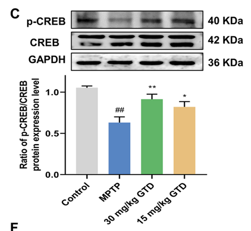
Gastrodin relieves Parkinson's disease-related motor deficits by facilitating the MEK-dependent VMAT2 to maintain dopamine homeostasis
Author: Zhao Meihuan,et al
PMID: 38885579

应用: WB
反应种属: Mouse
发表时间: 2024 Jun
-
Citation
-
Cholesterol Sulfate Exerts Protective Effect on Pancreatic β-Cells by Regulating β-Cell Mass and Insulin Secretion
Author: Zhang, X., Deng, D., Cui, D., Liu, Y., He, S., Zhang, H., Xie, Y., Yu, X., Yang, S., Chen, Y., & Su, Z.
PMID: 35308228

应用: WB
反应种属: Mouse,Rat
发表时间: 2022 Mar
-
Citation
-
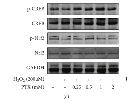
Pentoxifylline Enhances Antioxidative Capability and Promotes Mitochondrial Biogenesis in D-Galactose-Induced Aging Mice by Increasing Nrf2 and PGC-1α through the cAMP-CREB Pathway
Author:
PMID: 34257818

应用: WB
反应种属: Human
发表时间: 2021 Jun
-
Citation
-
Regulation of hepatic gluconeogenesis by nuclear factor Y transcription factor in mice
Author: Zhiguang Su
PMID: 29530977
应用: WB
反应种属: Mouse
发表时间: 2018 May
-
Citation
同靶点&同通路的产品
CREB Recombinant Rabbit Monoclonal Antibody [SA04-04]
Application: WB,IF-Cell,IF-Tissue,IHC-P,IP,FC
Reactivity: Human,Mouse,Zebrafish,Rat
Conjugate: unconjugated


