概述
产品名称
Cytokeratin 19 Recombinant Rabbit Monoclonal Antibody [SA30-06]
抗体类型
Recombinant Rabbit monoclonal Antibody
免疫原
Synthetic peptide within Human Cytokeratin 19 aa 348-400 / 400.
种属反应性
Human, Mouse, Rat
验证应用
WB, IF-Cell, IF-Tissue, IHC-P, FC, mIHC
分子量
Predicted band size: 44 kDa
阳性对照
MCF-7 cell lysates, AGS, human liver tissue, human breast carcinoma tissue, human breast tissue, mouse liver tissue, human kidney tissue, human placenta tissue, MCF-7, human stomach carcinoma tissue, human colon tissue, mouse pancreas.
偶联
unconjugated
克隆号
SA30-06
RRID
产品特性
形态
Liquid
浓度
1ug/ul
存放说明
Store at +4℃ after thawing. Aliquot store at -20℃ or -80℃. Avoid repeated freeze / thaw cycles.
存储缓冲液
1*TBS (pH7.4), 0.05% BSA, 40% Glycerol. Preservative: 0.05% Sodium Azide.
亚型
IgG
纯化方式
Protein A affinity purified.
应用稀释度
-
WB
-
1:500-1:5,000
-
IF-Cell
-
1:500
-
IF-Tissue
-
1:500
-
IHC-P
-
1:1,000
-
FC
-
1:1,000
-
mIHC
-
1:3,000-1:10,000
发表文章中的应用
发表文章中的种属
| Human | See 2 publications below |
| human | See 1 publications below |
靶点
功能
Keratin, type I cytoskeletal 19 also known as cytokeratin-19 (CK-19) or keratin-19 (K19) is a 40 kDa protein that in humans is encoded by the KRT19 gene. Keratin 19 is a type I keratin. Keratin 19 is a member of the keratin family. The keratins are intermediate filament proteins responsible for the structural integrity of epithelial cells and are subdivided into cytokeratins and hair keratins. The type I cytokeratins consist of acidic proteins which are arranged in pairs of heterotypic keratin chains. Unlike its related family members, this smallest known acidic cytokeratin is not paired with a basic cytokeratin in epithelial cells. It is specifically found in the periderm, the transiently superficial layer that envelops the developing epidermis.
背景文献
1. Guye P et al. Genetically engineering self-organization of human pluripotent stem cells into a liver bud-like tissue using Gata6. Nat Commun 7:10243 (2016).
2. Cui M et al. PTEN is a potent suppressor of small cell lung cancer. Mol Cancer Res 12:654-9 (2014).
序列相似性
Belongs to the intermediate filament family.
组织特异性
Expressed in a defined zone of basal keratinocytes in the deep outer root sheath of hair follicles. Also observed in sweat gland and mammary gland ductal and secretory cells, bile ducts, gastrointestinal tract, bladder urothelium, oral epithelia, esophagus, ectocervical epithelium (at protein level). Expressed in epidermal basal cells, in nipple epidermis and a defined region of the hair follicle. Also seen in a subset of vascular wall cells in both the veins and artery of human umbilical cord, and in umbilical cord vascular smooth muscle. Observed in muscle fibers accumulating in the costameres of myoplasm at the sarcolemma in structures that contain dystrophin and spectrin.
亚细胞定位
Cytoplasm
别名
40 kDa keratin intermediate filament antibody
CK 19 antibody
CK-19 antibody
CK19 antibody
Cytokeratin 19 antibody
Cytokeratin-19 antibody
K19 antibody
K1C19_HUMAN antibody
K1CS antibody
Keratin 19 antibody
展开40 kDa keratin intermediate filament antibody
CK 19 antibody
CK-19 antibody
CK19 antibody
Cytokeratin 19 antibody
Cytokeratin-19 antibody
K19 antibody
K1C19_HUMAN antibody
K1CS antibody
Keratin 19 antibody
Keratin type I 40 kD antibody
Keratin type I 40kD antibody
Keratin type I cytoskeletal 19 antibody
Keratin, type I cytoskeletal 19 antibody
Keratin, type I, 40 kd antibody
Keratin-19 antibody
KRT19 antibody
MGC15366 antibody
折叠图片
-

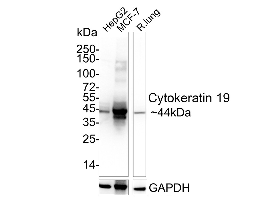
Western blot analysis of Cytokeratin 19 on different lysates with Rabbit anti-Cytokeratin 19 antibody (ET1601-6) at 1/1,000 dilution.
Lane 1: HepG2 cell lysate
Lane 1: MCF-7 cell lysate
Lane 3: Rat lung tissue lysate
Lysates/proteins at 20 µg/Lane.
Predicted band size: 44 kDa
Observed band size: 44 kDa
Exposure time: 9 seconds;
4-20% SDS-PAGE gel.
Proteins were transferred to a PVDF membrane and blocked with 5% NFDM/TBST for 1 hour at room temperature. The primary antibody (ET1601-6) at 1/1,000 dilution was used in 5% NFDM/TBST at 4℃ overnight. Goat Anti-Rabbit IgG - HRP Secondary Antibody (HA1001) at 1/50,000 dilution was used for 1 hour at room temperature. -
![<span style="font-weight: bold;">☑ Knockout (KO)</span><br /><br />All lanes: Western blot analysis of Cytokeratin 19 with anti-Cytokeratin 19 antibody [SA30-06] (<a href="/products/ET1601-6" style="font-weight: bold;text-decoration: underline;">ET1601-6</a>) at 1:5,000 dilution.<br />Lane 1: Wild-type HepG2 whole cell lysate (20 µg).<br />Lane 2/3: Cytokeratin 19 knockout HepG2 whole cell lysate (20 µg).<br /><br /><a href="/products/ET1601-6" style="font-weight: bold;text-decoration: underline;">ET1601-6</a> was shown to specifically react with Cytokeratin 19 in wild-type HepG2 cells. No band was observed when Cytokeratin 19 knockout samples were tested. Wild-type and Cytokeratin 19 knockout samples were subjected to SDS-PAGE. Proteins were transferred to a PVDF membrane and blocked with 5% NFDM in TBST for 1 hour at room temperature. The primary Anti-Cytokeratin 19 Antibody (<a href="/products/ET1601-6" style="font-weight: bold;text-decoration: underline;">ET1601-6</a>, 1/5,000) and anti-HSP90 antibody (<a href="/products/ET1605-56" style="font-weight: bold;text-decoration: underline;">ET1605-56</a>, 1/10,000) were used in 5% BSA at room temperature for 2 hours. Goat Anti-Rabbit IgG H&L (HRP) Secondary Antibody (<a href="/products/HA1001" style="font-weight: bold;text-decoration: underline;">HA1001</a>) at 1:200,000 dilution was used for 1 hour at room temperature.](http://storage.huabio.cn/huabio/productImg/ET1601-6_2.jpg?v=20241015165530)
☑ Knockout (KO)
All lanes: Western blot analysis of Cytokeratin 19 with anti-Cytokeratin 19 antibody [SA30-06] (ET1601-6) at 1:5,000 dilution.
Lane 1: Wild-type HepG2 whole cell lysate (20 µg).
Lane 2/3: Cytokeratin 19 knockout HepG2 whole cell lysate (20 µg).
ET1601-6 was shown to specifically react with Cytokeratin 19 in wild-type HepG2 cells. No band was observed when Cytokeratin 19 knockout samples were tested. Wild-type and Cytokeratin 19 knockout samples were subjected to SDS-PAGE. Proteins were transferred to a PVDF membrane and blocked with 5% NFDM in TBST for 1 hour at room temperature. The primary Anti-Cytokeratin 19 Antibody (ET1601-6, 1/5,000) and anti-HSP90 antibody (ET1605-56, 1/10,000) were used in 5% BSA at room temperature for 2 hours. Goat Anti-Rabbit IgG H&L (HRP) Secondary Antibody (HA1001) at 1:200,000 dilution was used for 1 hour at room temperature. -

Fluorescence multiplex immunohistochemical analysis of mouse pancreas (Formalin/PFA-fixed paraffin-embedded sections). Panel A: the merged image of anti-β-catenin (ET1601-5, Red), anti-Glucagon (ET1702-20, Green), anti-Insulin (ET1601-12, White), anti-CK19 (ET1601-6, Magenta) and anti-aSMA (ET1607-53, Yellow) on mouse pancreas. HRP Conjugated UltraPolymer Goat Polyclonal Antibody HA1119/HA1120 was used as a secondary antibody. The immunostaining was performed with the Sequential Immuno-staining Kit (IRISKit™MH010101, www.luminiris.cn). The section was incubated in five rounds of staining: in the order of ET1601-5 (1/2,000 dilution), ET1702-20 (1/6,000 dilution), ET1601-12 (1/8,000 dilution), ET1601-6 (1/5,000 dilution) and ET1607-53 (1/10,000 dilution) for 20 mins at room temperature. Each round was followed by a separate fluorescent tyramide signal amplification system. Heat mediated antigen retrieval with Tris-EDTA buffer (pH 9.0) for 30 mins at 95℃. DAPI (blue) was used as a nuclear counter stain. Image acquisition was performed with Olympus VS200 Slide Scanner.
-

Fluorescence multiplex immunohistochemical analysis of mouse liver (Formalin/PFA-fixed paraffin-embedded sections). Panel A: the merged image of anti-HNF4α (HA721006, Cyan), anti-CK19 (ET1601-6, Magenta) and anti-aSMA (ET1607-53, Yellow) on mouse liver. HRP Conjugated UltraPolymer Goat Polyclonal Antibody HA1119/HA1120 was used as a secondary antibody. The immunostaining was performed with the Sequential Immuno-staining Kit (IRISKit™MH010101, www.luminiris.cn). The section was incubated in three rounds of staining: in the order of HA721006 (1/5,000 dilution), ET1601-6 (1/10,000 dilution) and ET1607-53 (1/10,000 dilution) for 20 mins at room temperature. Each round was followed by a separate fluorescent tyramide signal amplification system. Heat mediated antigen retrieval with Tris-EDTA buffer (pH 9.0) for 30 mins at 95℃. DAPI (blue) was used as a nuclear counter stain. Image acquisition was performed with Olympus VS200 Slide Scanner.
-

Fluorescence multiplex immunohistochemical analysis of mouse pancreas (Formalin/PFA-fixed paraffin-embedded sections). Panel A: the merged image of anti-Cytokeratin 19 (ET1601-6, White) and anti-PAX6 (ET1612-58, Violet) on pancreas. HRP Conjugated UltraPolymer Goat Polyclonal Antibody HA1119/HA1120 was used as a secondary antibody. The immunostaining was performed with the Sequential Immuno-staining Kit (IRISKit™MH010101, www.luminiris.cn). The section was incubated in two rounds of staining: in the order of ET1601-6 (1/5,000 dilution) and ET1612-58 (1/1,000 dilution) for 20 mins at room temperature. Each round was followed by a separate fluorescent tyramide signal amplification system. Heat mediated antigen retrieval with Tris-EDTA buffer (pH 9.0) for 30 mins at 95℃. DAPI (blue) was used as a nuclear counter stain. Image acquisition was performed with Zeiss Observer 7 Inverted Fluorescence Microscope.
-

Fluorescence multiplex immunohistochemical analysis of mouse liver (Formalin/PFA-fixed paraffin-embedded sections). Panel A: the merged image of anti-Th (ET1611-12, Green), anti-HNF4a (HA721006, Magenta), anti-CK19 (ET1601-6, Cyan), anti-α-sma (ET1607-53, Red) and anti-β-catenin (ET1601-5, Yellow) on liver. HRP Conjugated UltraPolymer Goat Polyclonal Antibody HA1119/HA1120 was used as a secondary antibody. The immunostaining was performed with the Sequential Immuno-staining Kit (IRISKit™MH010101, www.luminiris.cn). The section was incubated in three rounds of staining: in the order of ET1611-12 (1/1,000 dilution), HA721006 (1/2,000 dilution), ET1601-6 (1/3,000 dilution), ET1607-53 (1/10,000 dilution) and ET1601-5 (1/2,000 dilution) for 20 mins at room temperature. Each round was followed by a separate fluorescent tyramide signal amplification system. Heat mediated antigen retrieval with Tris-EDTA buffer (pH 9.0) for 30 mins at 95℃. DAPI (blue) was used as a nuclear counter stain. Image acquisition was performed with Olympus VS200 Slide Scanner.
-

Immunohistochemical analysis of paraffin-embedded human liver tissue with Rabbit anti-Cytokeratin 19 antibody (ET1601-6) at 1/1,000 dilution.
The section was pre-treated using heat mediated antigen retrieval with Tris-EDTA buffer (pH 9.0) for 20 minutes. The tissues were blocked in 1% BSA for 20 minutes at room temperature, washed with ddH2O and PBS, and then probed with the primary antibody (ET1601-6) at 1/1,000 dilution for 1 hour at room temperature. The detection was performed using an HRP conjugated compact polymer system. DAB was used as the chromogen. Tissues were counterstained with hematoxylin and mounted with DPX. -

Immunohistochemical analysis of paraffin-embedded human breast carcinoma tissue with Rabbit anti-Cytokeratin 19 antibody (ET1601-6) at 1/1,500 dilution.
The section was pre-treated using heat mediated antigen retrieval with Tris-EDTA buffer (pH 9.0) for 20 minutes. The tissues were blocked in 1% BSA for 20 minutes at room temperature, washed with ddH2O and PBS, and then probed with the primary antibody (ET1601-6) at 1/1,500 dilution for 1 hour at room temperature. The detection was performed using an HRP conjugated compact polymer system. DAB was used as the chromogen. Tissues were counterstained with hematoxylin and mounted with DPX. -

Immunohistochemical analysis of paraffin-embedded human breast carcinoma tissue with Rabbit anti-Cytokeratin 19 antibody (ET1601-6) at 1/1,500 dilution.
The section was not undergone antigen retrieval. The tissues were blocked in 1% BSA for 20 minutes at room temperature, washed with ddH2O and PBS, and then probed with the primary antibody (ET1601-6) at 1/1,500 dilution for 1 hour at room temperature. The detection was performed using an HRP conjugated compact polymer system. DAB was used as the chromogen. Tissues were counterstained with hematoxylin and mounted with DPX. -

Immunohistochemical analysis of paraffin-embedded human breast tissue with Rabbit anti-Cytokeratin 19 antibody (ET1601-6) at 1/1,500 dilution.
The section was pre-treated using heat mediated antigen retrieval with Tris-EDTA buffer (pH 9.0) for 20 minutes. The tissues were blocked in 1% BSA for 20 minutes at room temperature, washed with ddH2O and PBS, and then probed with the primary antibody (ET1601-6) at 1/1,500 dilution for 1 hour at room temperature. The detection was performed using an HRP conjugated compact polymer system. DAB was used as the chromogen. Tissues were counterstained with hematoxylin and mounted with DPX. -

Immunohistochemical analysis of paraffin-embedded human breast tissue with Rabbit anti-Cytokeratin 19 antibody (ET1601-6) at 1/1,500 dilution.
The section was not undergone antigen retrieval. The tissues were blocked in 1% BSA for 20 minutes at room temperature, washed with ddH2O and PBS, and then probed with the primary antibody (ET1601-6) at 1/1,500 dilution for 1 hour at room temperature. The detection was performed using an HRP conjugated compact polymer system. DAB was used as the chromogen. Tissues were counterstained with hematoxylin and mounted with DPX. -

Immunohistochemical analysis of paraffin-embedded human stomach carcinoma tissue with Rabbit anti-Cytokeratin 19 antibody (ET1601-6) at 1/1,500 dilution.
The section was pre-treated using heat mediated antigen retrieval with Tris-EDTA buffer (pH 9.0) for 20 minutes. The tissues were blocked in 1% BSA for 20 minutes at room temperature, washed with ddH2O and PBS, and then probed with the primary antibody (ET1601-6) at 1/1,500 dilution for 1 hour at room temperature. The detection was performed using an HRP conjugated compact polymer system. DAB was used as the chromogen. Tissues were counterstained with hematoxylin and mounted with DPX. -

Immunohistochemical analysis of paraffin-embedded human kidney tissue with Rabbit anti-Cytokeratin 19 antibody (ET1601-6) at 1/1,000 dilution.
The section was pre-treated using heat mediated antigen retrieval with Tris-EDTA buffer (pH 9.0) for 20 minutes. The tissues were blocked in 1% BSA for 20 minutes at room temperature, washed with ddH2O and PBS, and then probed with the primary antibody (ET1601-6) at 1/1,000 dilution for 1 hour at room temperature. The detection was performed using an HRP conjugated compact polymer system. DAB was used as the chromogen. Tissues were counterstained with hematoxylin and mounted with DPX. -

Immunocytochemistry analysis of MCF-7 cells labeling Cytokeratin 19 with Rabbit anti-Cytokeratin 19 antibody (ET1601-6) at 1/500 dilution.
Cells were fixed in 4% paraformaldehyde for 20 minutes at room temperature, permeabilized with 0.1% Triton X-100 in PBS for 5 minutes at room temperature, then blocked with 1% BSA in 10% negative goat serum for 1 hour at room temperature. Cells were then incubated with Rabbit anti-Cytokeratin 19 antibody (ET1601-6) at 1/500 dilution in 1% BSA in PBST overnight at 4 ℃. Goat Anti-Rabbit IgG H&L (iFluor™ 488, HA1121) was used as the secondary antibody at 1/1,000 dilution. PBS instead of the primary antibody was used as the secondary antibody only control. Nuclear DNA was labelled in blue with DAPI.
Beta tubulin (M1305-2, red) was stained at 1/100 dilution overnight at +4℃. Goat Anti-Mouse IgG H&L (iFluor™ 594, HA1126) was used as the secondary antibody at 1/1,000 dilution. -

Immunohistochemical analysis of paraffin-embedded rat kidney tissue with Rabbit anti-Cytokeratin 19 antibody (ET1601-6) at 1/1,000 dilution.
The section was pre-treated using heat mediated antigen retrieval with Tris-EDTA buffer (pH 9.0) for 20 minutes. The tissues were blocked in 1% BSA for 20 minutes at room temperature, washed with ddH2O and PBS, and then probed with the primary antibody (ET1601-6) at 1/1,000 dilution for 1 hour at room temperature. The detection was performed using an HRP conjugated compact polymer system. DAB was used as the chromogen. Tissues were counterstained with hematoxylin and mounted with DPX. -

Immunofluorescence analysis of paraffin-embedded human kidney tissue labeling Cytokeratin 19 (ET1601-6).
The section was pre-treated using heat mediated antigen retrieval with Tris-EDTA buffer (pH 9.0) for 20 minutes. The tissues were blocked in 10% negative goat serum for 1 hour at room temperature, washed with PBS. And then probed with the primary antibodies Cytokeratin 19 (ET1601-6, red) at 1/500 dilution at +4℃ overnight, washed with PBS.
Goat Anti-Rabbit IgG H&L (iFluor™ 594, HA1122) was used as the secondary antibodies at 1/1,000 dilution. Nuclei were counterstained with DAPI (blue).
Please note: All products are "FOR RESEARCH USE ONLY AND ARE NOT INTENDED FOR DIAGNOSTIC OR THERAPEUTIC USE"
引文
-
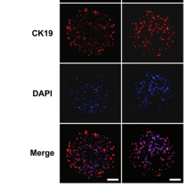
Bioprinting of hydrogel beads to engineer pancreatic tumor-stroma microtissues for drug screening
Author:
PMID: 37273977

应用: IF
反应种属: Human
发表时间: 2023 Feb
-
Citation
-
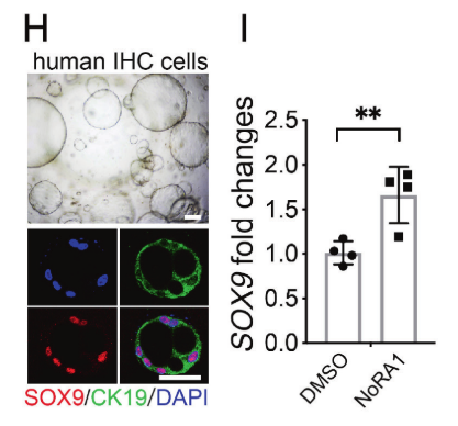
Regenerative failure of intrahepatic biliary cells in Alagille syndrome rescued by elevated Jagged/Notch/Sox9 signaling
Author: Zhao, C., Matalonga, J., Lancman, J. J., Liu, L., Xiao, C., Kumar, S., Gates, K. P., He, J., Graves, A., Huisken, J., Azuma, M., Lu, Z., Chen, C., Ding, B. S., & Dong, P. D. S.
PMID: 36469766

应用: IF
反应种属: Human
发表时间: 2022 Dec
-
Citation
-
Loss of FoxA2 accelerates neoplastic changes in the intrahepatic bile duct partly via the MAPK signaling pathway
Author: Tianfu Wen,Chuan Li;
PMID: 31689237
应用: WB,IHC
反应种属: human
发表时间: 2019 Nov
-
Citation


