概述
产品名称
ZEB Recombinant Rabbit Monoclonal Antibody [PSH0-61]
抗体类型
Recombinant Rabbit monoclonal Antibody
免疫原
Recombinant protein within human ZEB1 aa 1-400 / 1,124.
种属反应性
Human, Mouse, Rat
验证应用
WB, IHC-P, FC
分子量
Predicted band size: 124 kDa
阳性对照
Jurkat cell lysate, SK-OV-3 cell lysate, HeLa cell lysate, HEK-293 cell lysate, U87-MG cell lysate, MG-63 cell lysate, human colon tissue, human kidney tissue, human ovary carcinoma tissue, mouse heart tissue, mouse kidney tissue, mouse liver tissue, mouse lung tissue, mouse stomach tissue, mouse testis tissue, rat kidney tissue, rat liver tissue, rat lung tissue, rat small intestine tissue, rat testis tissue, Jurkat.
偶联
unconjugated
克隆号
PSH0-61
RRID
产品特性
形态
Liquid
存放说明
Shipped at 4℃. Store at +4℃ short term (1-2 weeks). It is recommended to aliquot into single-use upon delivery. Store at -20℃ long term.
存储缓冲液
PBS (pH7.4), 0.1% BSA, 40% Glycerol. Preservative: 0.05% Sodium Azide.
亚型
IgG
纯化方式
Protein A affinity purified.
应用稀释度
-
WB
-
1:1,000
-
IHC-P
-
1:1,000
-
FC
-
1:500-1:1,000
发表文章中的应用
| WB | 查看 1 篇文献如下 |
发表文章中的种属
| Human | 查看 1 篇文献如下 |
靶点
功能
Zinc finger E-box-binding homeobox 1 is a protein that in humans is encoded by the ZEB1 gene. ZEB1 (previously known as TCF8) encodes a zinc finger and homeodomain transcription factor that represses T-lymphocyte-specific IL2 gene expression by binding to a negative regulatory domain 100 nucleotides 5-prime of the IL2 transcription start site. ZEB1 and its mammalian paralog ZEB2 belongs to the Zeb family within the ZF (zinc finger) class of homeodomain transcription factors. ZEB1 protein has 7 zinc fingers and 1 homeodomain. The structure of the homeodomain is shown on the right. Mutations of the gene are linked to posterior polymorphous corneal dystrophy 3. ZEB1 downregulates E-cadherin and induces epithelial to mesenchymal transition in breast and other carcinomas A recent study suggested its contributing role in lung cancer invasiveness and metastasis development. Overexpression of ZEB1 has been identified as a potential risk factor for recurrence and poor prognosis in several types of cancers.
背景文献
1. Zhang Y et al. Expression and Function of ZEB1 in the Cornea. Cells. 2021 Apr
2. Cheng P et al. ZEB2 Shapes the Epigenetic Landscape of Atherosclerosis. Circulation. 2022 Feb
亚细胞定位
Nucleus.
UNIPROT
别名
AREB 6 antibody
AREB6 antibody
BZP antibody
Delta crystallin enhancer binding factor 1 antibody
DELTA EF1 antibody
FECD6 antibody
MGC133261 antibody
Negative regulator of IL 2 antibody
Negative regulator of IL2 antibody
NIL 2 A antibody
展开AREB 6 antibody
AREB6 antibody
BZP antibody
Delta crystallin enhancer binding factor 1 antibody
DELTA EF1 antibody
FECD6 antibody
MGC133261 antibody
Negative regulator of IL 2 antibody
Negative regulator of IL2 antibody
NIL 2 A antibody
NIL 2 A zinc finger protein antibody
NIL 2A antibody
NIL-2-A zinc finger protein antibody
NIL2A antibody
Posterior polymorphous corneal dystrophy 3 antibody
PPCD3 antibody
Represses interleukin 2 expression antibody
TCF 8 antibody
TCF-8 antibody
TCF8 antibody
Transcription factor 8 (represses interleukin 2 expression) antibody
Transcription factor 8 antibody
ZEB 1 antibody
ZEB antibody
ZEB1 antibody
ZEB1_HUMAN antibody
ZFHEP antibody
ZFHX 1A antibody
ZFHX1A antibody
Zinc finger E box binding homeobox 1 antibody
Zinc finger E-box-binding homeobox 1 antibody
Zinc finger homeodomain enhancer binding protein antibody
FLJ42816 antibody
HSPC082 antibody
KIAA0569 antibody
SIP 1 antibody
SIP1 antibody
Smad Interacting Protein 1 antibody
Smad-interacting protein 1 antibody
SMADIP 1 antibody
SMADIP1 antibody
ZEB 2 antibody
Zeb2 antibody
ZEB2_HUMAN antibody
Zfhx1b antibody
ZFHX1B protein antibody
Zfx1b antibody
Zinc finger E box binding protein 2 antibody
Zinc finger E-box-binding homeobox 2 antibody
Zinc finger homeobox 1b antibody
zinc finger homeobox protein 1 antibody
Zinc finger homeobox protein 1b antibody
折叠图片
-

Western blot analysis of ZEB on different lysates with Rabbit anti-ZEB antibody (HA721438) at 1/1,000 dilution.
Lane 1: Jurkat cell lysate
Lane 2: SK-OV-3 cell lysate
Lane 3: HeLa cell lysate
Lane 4: HEK-293 cell lysate
Lane 5: U87-MG cell lysate
Lane 6: MG-63 cell lysate
Lysates/proteins at 30 µg/Lane.
Predicted band size: 124 kDa
Observed band size: 200 kDa
Exposure time: 2 minutes;
8% SDS-PAGE gel.
Proteins were transferred to a PVDF membrane and blocked with 5% NFDM/TBST for 1 hour at room temperature. The primary antibody (HA721438) at 1/1,000 dilution was used in 5% NFDM/TBST at room temperature for 2 hours. Goat Anti-Rabbit IgG - HRP Secondary Antibody (HA1001) at 1:200,000 dilution was used for 1 hour at room temperature. -

Immunohistochemical analysis of paraffin-embedded human colon tissue with Rabbit anti-ZEB antibody (HA721438) at 1/1,000 dilution.
The section was pre-treated using heat mediated antigen retrieval with sodium citrate buffer (pH 6.0) (high pressure) for 2 minutes. The tissues were blocked in 1% BSA for 20 minutes at room temperature, washed with ddH2O and PBS, and then probed with the primary antibody (HA721438) at 1/1,000 dilution for 1 hour at room temperature. The detection was performed using an HRP conjugated compact polymer system. DAB was used as the chromogen. Tissues were counterstained with hematoxylin and mounted with DPX. -

Immunohistochemical analysis of paraffin-embedded human kidney tissue with Rabbit anti-ZEB antibody (HA721438) at 1/1,000 dilution.
The section was pre-treated using heat mediated antigen retrieval with sodium citrate buffer (pH 6.0) (high pressure) for 2 minutes. The tissues were blocked in 1% BSA for 20 minutes at room temperature, washed with ddH2O and PBS, and then probed with the primary antibody (HA721438) at 1/1,000 dilution for 1 hour at room temperature. The detection was performed using an HRP conjugated compact polymer system. DAB was used as the chromogen. Tissues were counterstained with hematoxylin and mounted with DPX. -

Immunohistochemical analysis of paraffin-embedded human ovary carcinoma tissue with Rabbit anti-ZEB antibody (HA721438) at 1/1,000 dilution.
The section was pre-treated using heat mediated antigen retrieval with sodium citrate buffer (pH 6.0) (high pressure) for 2 minutes. The tissues were blocked in 1% BSA for 20 minutes at room temperature, washed with ddH2O and PBS, and then probed with the primary antibody (HA721438) at 1/1,000 dilution for 1 hour at room temperature. The detection was performed using an HRP conjugated compact polymer system. DAB was used as the chromogen. Tissues were counterstained with hematoxylin and mounted with DPX. -

Immunohistochemical analysis of paraffin-embedded mouse heart tissue with Rabbit anti-ZEB antibody (HA721438) at 1/1,000 dilution.
The section was pre-treated using heat mediated antigen retrieval with sodium citrate buffer (pH 6.0) (high pressure) for 2 minutes. The tissues were blocked in 1% BSA for 20 minutes at room temperature, washed with ddH2O and PBS, and then probed with the primary antibody (HA721438) at 1/1,000 dilution for 1 hour at room temperature. The detection was performed using an HRP conjugated compact polymer system. DAB was used as the chromogen. Tissues were counterstained with hematoxylin and mounted with DPX. -

Immunohistochemical analysis of paraffin-embedded mouse kidney tissue with Rabbit anti-ZEB antibody (HA721438) at 1/1,000 dilution.
The section was pre-treated using heat mediated antigen retrieval with sodium citrate buffer (pH 6.0) (high pressure) for 2 minutes. The tissues were blocked in 1% BSA for 20 minutes at room temperature, washed with ddH2O and PBS, and then probed with the primary antibody (HA721438) at 1/1,000 dilution for 1 hour at room temperature. The detection was performed using an HRP conjugated compact polymer system. DAB was used as the chromogen. Tissues were counterstained with hematoxylin and mounted with DPX. -

Immunohistochemical analysis of paraffin-embedded mouse liver tissue with Rabbit anti-ZEB antibody (HA721438) at 1/1,000 dilution.
The section was pre-treated using heat mediated antigen retrieval with sodium citrate buffer (pH 6.0) (high pressure) for 2 minutes. The tissues were blocked in 1% BSA for 20 minutes at room temperature, washed with ddH2O and PBS, and then probed with the primary antibody (HA721438) at 1/1,000 dilution for 1 hour at room temperature. The detection was performed using an HRP conjugated compact polymer system. DAB was used as the chromogen. Tissues were counterstained with hematoxylin and mounted with DPX. -

Immunohistochemical analysis of paraffin-embedded mouse lung tissue with Rabbit anti-ZEB antibody (HA721438) at 1/1,000 dilution.
The section was pre-treated using heat mediated antigen retrieval with sodium citrate buffer (pH 6.0) (high pressure) for 2 minutes. The tissues were blocked in 1% BSA for 20 minutes at room temperature, washed with ddH2O and PBS, and then probed with the primary antibody (HA721438) at 1/1,000 dilution for 1 hour at room temperature. The detection was performed using an HRP conjugated compact polymer system. DAB was used as the chromogen. Tissues were counterstained with hematoxylin and mounted with DPX. -

Immunohistochemical analysis of paraffin-embedded mouse stomach tissue with Rabbit anti-ZEB antibody (HA721438) at 1/1,000 dilution.
The section was pre-treated using heat mediated antigen retrieval with sodium citrate buffer (pH 6.0) (high pressure) for 2 minutes. The tissues were blocked in 1% BSA for 20 minutes at room temperature, washed with ddH2O and PBS, and then probed with the primary antibody (HA721438) at 1/1,000 dilution for 1 hour at room temperature. The detection was performed using an HRP conjugated compact polymer system. DAB was used as the chromogen. Tissues were counterstained with hematoxylin and mounted with DPX. -

Immunohistochemical analysis of paraffin-embedded mouse testis tissue with Rabbit anti-ZEB antibody (HA721438) at 1/1,000 dilution.
The section was pre-treated using heat mediated antigen retrieval with sodium citrate buffer (pH 6.0) (high pressure) for 2 minutes. The tissues were blocked in 1% BSA for 20 minutes at room temperature, washed with ddH2O and PBS, and then probed with the primary antibody (HA721438) at 1/1,000 dilution for 1 hour at room temperature. The detection was performed using an HRP conjugated compact polymer system. DAB was used as the chromogen. Tissues were counterstained with hematoxylin and mounted with DPX. -

Immunohistochemical analysis of paraffin-embedded rat kidney tissue with Rabbit anti-ZEB antibody (HA721438) at 1/1,000 dilution.
The section was pre-treated using heat mediated antigen retrieval with sodium citrate buffer (pH 6.0) (high pressure) for 2 minutes. The tissues were blocked in 1% BSA for 20 minutes at room temperature, washed with ddH2O and PBS, and then probed with the primary antibody (HA721438) at 1/1,000 dilution for 1 hour at room temperature. The detection was performed using an HRP conjugated compact polymer system. DAB was used as the chromogen. Tissues were counterstained with hematoxylin and mounted with DPX. -

Immunohistochemical analysis of paraffin-embedded rat liver tissue with Rabbit anti-ZEB antibody (HA721438) at 1/1,000 dilution.
The section was pre-treated using heat mediated antigen retrieval with sodium citrate buffer (pH 6.0) (high pressure) for 2 minutes. The tissues were blocked in 1% BSA for 20 minutes at room temperature, washed with ddH2O and PBS, and then probed with the primary antibody (HA721438) at 1/1,000 dilution for 1 hour at room temperature. The detection was performed using an HRP conjugated compact polymer system. DAB was used as the chromogen. Tissues were counterstained with hematoxylin and mounted with DPX. -

Immunohistochemical analysis of paraffin-embedded rat lung tissue with Rabbit anti-ZEB antibody (HA721438) at 1/1,000 dilution.
The section was pre-treated using heat mediated antigen retrieval with sodium citrate buffer (pH 6.0) (high pressure) for 2 minutes. The tissues were blocked in 1% BSA for 20 minutes at room temperature, washed with ddH2O and PBS, and then probed with the primary antibody (HA721438) at 1/1,000 dilution for 1 hour at room temperature. The detection was performed using an HRP conjugated compact polymer system. DAB was used as the chromogen. Tissues were counterstained with hematoxylin and mounted with DPX. -

Immunohistochemical analysis of paraffin-embedded rat small intestine tissue with Rabbit anti-ZEB antibody (HA721438) at 1/1,000 dilution.
The section was pre-treated using heat mediated antigen retrieval with sodium citrate buffer (pH 6.0) (high pressure) for 2 minutes. The tissues were blocked in 1% BSA for 20 minutes at room temperature, washed with ddH2O and PBS, and then probed with the primary antibody (HA721438) at 1/1,000 dilution for 1 hour at room temperature. The detection was performed using an HRP conjugated compact polymer system. DAB was used as the chromogen. Tissues were counterstained with hematoxylin and mounted with DPX. -

Immunohistochemical analysis of paraffin-embedded rat testis tissue with Rabbit anti-ZEB antibody (HA721438) at 1/1,000 dilution.
The section was pre-treated using heat mediated antigen retrieval with sodium citrate buffer (pH 6.0) (high pressure) for 2 minutes. The tissues were blocked in 1% BSA for 20 minutes at room temperature, washed with ddH2O and PBS, and then probed with the primary antibody (HA721438) at 1/1,000 dilution for 1 hour at room temperature. The detection was performed using an HRP conjugated compact polymer system. DAB was used as the chromogen. Tissues were counterstained with hematoxylin and mounted with DPX. -

Flow cytometric analysis of Jurkat cells labeling ZEB.
Cells were fixed and permeabilized. Then stained with the primary antibody (HA721438, 1ug/ml) (red) compared with Rabbit IgG Isotype Control (green). After incubation of the primary antibody at +4℃ for an hour, the cells were stained with a iFluor™ 488 conjugate-Goat anti-Rabbit IgG Secondary antibody (HA1121) at 1/1,000 dilution for 30 minutes at +4℃. Unlabelled sample was used as a control (cells without incubation with primary antibody; black).
请注意: All products are "FOR RESEARCH USE ONLY AND ARE NOT INTENDED FOR DIAGNOSTIC OR THERAPEUTIC USE"
引文
-
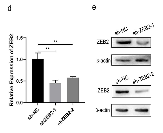
MiR-454-3p promotes apoptosis and autophagy of AML cells by targeting ZEB2 and regulating AKT/mTOR pathway
Author:
PMID: 37313984

期刊: Hematology
应用: WB
反应种属: Human
发表时间: 2023 Dec
-
Citation


