MSH6 Recombinant Rabbit Monoclonal Antibody [PD00-26]


Catalog# HA721164
MSH6 Recombinant Rabbit Monoclonal Antibody [PD00-26]
-
WB
-
IHC-P
-
IF-Cell
-
IF-Tissue
-
Human
-
Mouse
-
Rat
概述
产品名称
MSH6 Recombinant Rabbit Monoclonal Antibody [PD00-26]
抗体类型
Recombinant Rabbit monoclonal Antibody
免疫原
Synthetic peptide within Human MSH6 aa 350-450 (internal sequence).
种属反应性
Human, Mouse, Rat
验证应用
WB, IHC-P, IF-Cell, IF-Tissue
分子量
Predicted band size: 153 kDa
阳性对照
A549 cell lysate, HepG2 cell lysate, human tonsil tissue, human colon carcinoma tissue, human stomach carcinoma tissue, mouse small intestine tissue.
偶联
unconjugated
克隆号
PD00-26
RRID
产品特性
形态
Liquid
存放说明
Shipped at 4℃. Store at +4℃ short term (1-2 weeks). It is recommended to aliquot into single-use upon delivery. Store at -20℃ long term.
存储缓冲液
PBS (pH7.4), 0.1% BSA, 40% Glycerol. Preservative: 0.05% Sodium Azide.
亚型
IgG
纯化方式
Protein A affinity purified.
应用稀释度
-
WB
-
1:1,000
-
IHC-P
-
1:1,000
-
IF-Cell
-
1:100
-
IF-Tissue
-
1:200
靶点
功能
This gene encodes a member of the DNA mismatch repair MutS family. In E. coli, the MutS protein helps in the recognition of mismatched nucleotides prior to their repair. A highly conserved region of approximately 150 aa, called the Walker-A adenine nucleotide binding motif, exists in MutS homologs. The encoded protein heterodimerizes with MSH2 to form a mismatch recognition complex that functions as a bidirectional molecular switch that exchanges ADP and ATP as DNA mismatches are bound and dissociated. Mutations in this gene may be associated with hereditary nonpolyposis colon cancer, colorectal cancer, and endometrial cancer. Transcripts variants encoding different isoforms have been described. Tonsil is found to be a recommendable positive tissue control for MSH6. Virtually all mantle zone B-cells must show an at least weak to moderate nuclear staining reaction, while a moderate to strong nuclear staining reaction must be seen in the proliferating germinal centre B-cells. Colon adenocarcinoma with loss of MSH6 expression is recommended as negative tissue control. No nuclear staining reaction should be seen in the neoplastic cells, whereas a nuclear staining reaction must be seen in stromal cells serving as internal positive tissue control.
背景文献
1. Frederiksen JH. et. al. Classification of MSH6 Variants of Uncertain Significance Using Functional Assays. Int J Mol Sci. 2021 Aug
2. Salem ME. et. al. Relationship between MLH1, PMS2, MSH2 and MSH6 gene-specific alterations and tumor mutational burden in 1057 microsatellite instability-high solid tumors. Int J Cancer. 2020 Nov
亚细胞定位
Nucleus, Chromosome.
别名
DNA mismatch repair protein Msh6 antibody
G/T mismatch binding protein antibody
G/T mismatch-binding protein antibody
GTBP antibody
GTMBP antibody
hMSH6 antibody
HNPCC 5 antibody
HNPCC5 antibody
HSAP antibody
MSH 6 antibody
展开图片
-
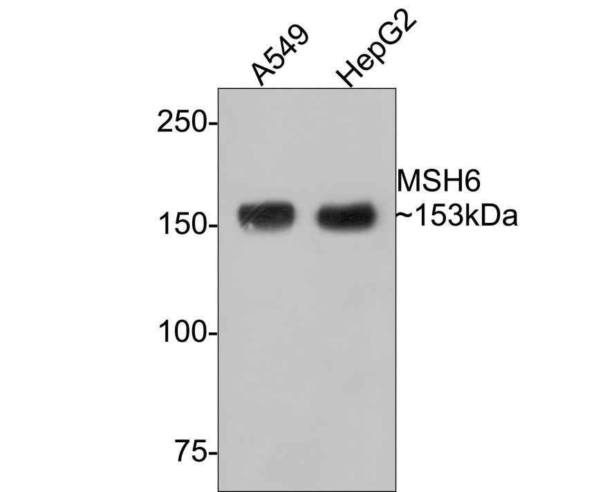
Western blot analysis of MSH6 on different lysates with Rabbit anti-MSH6 antibody (HA721164) at 1/1,000 dilution.
Lane 1: A549 cell lysate
Lane 2: HepG2 cell lysate
Lysates/proteins at 10 µg/Lane.
Predicted band size: 153 kDa
Observed band size: 153 kDa
Exposure time: 2 minutes;
6% SDS-PAGE gel.
Proteins were transferred to a PVDF membrane and blocked with 5% NFDM/TBST for 1 hour at room temperature. The primary antibody (HA721164) at 1/1,000 dilution was used in 5% NFDM/TBST at room temperature for 2 hours. Goat Anti-Rabbit IgG - HRP Secondary Antibody (HA1001) at 1:300,000 dilution was used for 1 hour at room temperature. -
Immunohistochemical analysis of paraffin-embedded human tonsil tissue with Rabbit anti-MSH6 antibody (HA721164) at 1/1,000 dilution.
The section was pre-treated using heat mediated antigen retrieval with sodium citrate buffer (pH 6.0) (high pressure) for 2 minutes. The tissues were blocked in 1% BSA for 20 minutes at room temperature, washed with ddH2O and PBS, and then probed with the primary antibody (HA721164) at 1/1,000 dilution for 1 hour at room temperature. The detection was performed using an HRP conjugated compact polymer system. DAB was used as the chromogen. Tissues were counterstained with hematoxylin and mounted with DPX. -
Immunohistochemical analysis of paraffin-embedded human colon carcinoma tissue with Rabbit anti-MSH6 antibody (HA721164) at 1/1,000 dilution.
The section was pre-treated using heat mediated antigen retrieval with sodium citrate buffer (pH 6.0) (high pressure) for 2 minutes. The tissues were blocked in 1% BSA for 20 minutes at room temperature, washed with ddH2O and PBS, and then probed with the primary antibody (HA721164) at 1/1,000 dilution for 1 hour at room temperature. The detection was performed using an HRP conjugated compact polymer system. DAB was used as the chromogen. Tissues were counterstained with hematoxylin and mounted with DPX. -
Immunohistochemical analysis of paraffin-embedded human stomach carcinoma tissue with Rabbit anti-MSH6 antibody (HA721164) at 1/1,000 dilution.
The section was pre-treated using heat mediated antigen retrieval with sodium citrate buffer (pH 6.0) (high pressure) for 2 minutes. The tissues were blocked in 1% BSA for 20 minutes at room temperature, washed with ddH2O and PBS, and then probed with the primary antibody (HA721164) at 1/1,000 dilution for 1 hour at room temperature. The detection was performed using an HRP conjugated compact polymer system. DAB was used as the chromogen. Tissues were counterstained with hematoxylin and mounted with DPX. -
Immunohistochemical analysis of paraffin-embedded mouse small intestine tissue with Rabbit anti-MSH6 antibody (HA721164) at 1/1,000 dilution.
The section was pre-treated using heat mediated antigen retrieval with sodium citrate buffer (pH 6.0) (high pressure) for 2 minutes. The tissues were blocked in 1% BSA for 20 minutes at room temperature, washed with ddH2O and PBS, and then probed with the primary antibody (HA721164) at 1/1,000 dilution for 1 hour at room temperature. The detection was performed using an HRP conjugated compact polymer system. DAB was used as the chromogen. Tissues were counterstained with hematoxylin and mounted with DPX. -
Immunohistochemical analysis of paraffin-embedded rat colon tissue with Rabbit anti-MSH6 antibody (HA721164) at 1/1,000 dilution.
The section was pre-treated using heat mediated antigen retrieval with sodium citrate buffer (pH 6.0) (high pressure) for 2 minutes. The tissues were blocked in 1% BSA for 20 minutes at room temperature, washed with ddH2O and PBS, and then probed with the primary antibody (HA721164) at 1/1,000 dilution for 1 hour at room temperature. The detection was performed using an HRP conjugated compact polymer system. DAB was used as the chromogen. Tissues were counterstained with hematoxylin and mounted with DPX. -
Immunohistochemical analysis of paraffin-embedded rat testis tissue with Rabbit anti-MSH6 antibody (HA721164) at 1/1,000 dilution.
The section was pre-treated using heat mediated antigen retrieval with sodium citrate buffer (pH 6.0) (high pressure) for 2 minutes. The tissues were blocked in 1% BSA for 20 minutes at room temperature, washed with ddH2O and PBS, and then probed with the primary antibody (HA721164) at 1/1,000 dilution for 1 hour at room temperature. The detection was performed using an HRP conjugated compact polymer system. DAB was used as the chromogen. Tissues were counterstained with hematoxylin and mounted with DPX. -
Immunocytochemistry analysis of A549 cells labeling MSH6 with Rabbit anti-MSH6 antibody (HA721164) at 1/100 dilution.
Cells were fixed in 4% paraformaldehyde for 20 minutes at room temperature, permeabilized with 0.1% Triton X-100 in PBS for 5 minutes at room temperature, then blocked with 1% BSA in 10% negative goat serum for 1 hour at room temperature. Cells were then incubated with Rabbit anti-MSH6 antibody (HA721164) at 1/100 dilution in 1% BSA in PBST overnight at 4 ℃. Goat Anti-Rabbit IgG H&L (iFluor™ 488, HA1121) was used as the secondary antibody at 1/1,000 dilution. PBS instead of the primary antibody was used as the secondary antibody only control. Nuclear DNA was labelled in blue with DAPI.
Beta tubulin (M1305-2, red) was stained at 1/100 dilution overnight at +4℃. Goat Anti-Mouse IgG H&L (iFluor™ 594, HA1126) was used as the secondary antibody at 1/1,000 dilution. -
Immunohistochemical analysis of paraffin embedded human colon cancer tissue using anti-MSH6 antibody (1/200) performed on the Ventana® BenchMark ULTRA.
请注意: All products are "FOR RESEARCH USE ONLY AND ARE NOT INTENDED FOR DIAGNOSTIC OR THERAPEUTIC USE"