概述
产品名称
alpha Tubulin 4A Recombinant Rabbit Monoclonal Antibody [JM73-24]
抗体类型
Recombinant Rabbit monoclonal Antibody
免疫原
Synthetic peptide within Human alpha Tubulin 4A aa 399-448 / 448.
种属反应性
Human, Mouse, Rat
验证应用
WB, IF-Cell, IF-Tissue, IHC-P, FC
分子量
Predicted band size: 50 kDa
阳性对照
HeLa cell lysate, PC-12 cell lysate, Mouse brain tissue lysate, Rat brain tissue lysate, HeLa, human tonsil tissue, human thyroid tissue, mouse brain tissue, human placenta tissue, mouse testis tissue.
偶联
unconjugated
克隆号
JM73-24
RRID
产品特性
形态
Liquid
浓度
1ug/ul
存放说明
Store at +4℃ after thawing. Aliquot store at -20℃ or -80℃. Avoid repeated freeze / thaw cycles.
存储缓冲液
1*TBS (pH7.4), 0.05% BSA, 40% Glycerol. Preservative: 0.05% Sodium Azide.
亚型
IgG
纯化方式
Protein A affinity purified.
应用稀释度
-
WB
-
1:500-1:2,000
-
IF-Cell
-
1:500
-
IF-Tissue
-
1:100-1:200
-
IHC-P
-
1:100-1:200
-
FC
-
1:1,000
发表文章中的应用
WB | 查看 8 篇文献如下 |
IF | 查看 2 篇文献如下 |
发表文章中的种属
Human | 查看 4 篇文献如下 |
Mouse | 查看 2 篇文献如下 |
Rat | 查看 2 篇文献如下 |
靶点
功能
Tubulin alpha-4A chain is a protein that in humans is encoded by the TUBA4A gene. Microtubules of the eukaryotic cytoskeleton perform essential and diverse functions and are composed of a heterodimer of alpha and beta tubulin. The genes encoding these microtubule constituents are part of the tubulin superfamily, which is composed of six distinct families. Genes from the alpha, beta and gamma tubulin families are found in all eukaryotes. The alpha and beta tubulins represent the major components of microtubules, while gamma tubulin plays a critical role in the nucleation of microtubule assembly. There are multiple alpha and beta tubulin genes and they are highly conserved among and between species. This gene encodes an alpha tubulin that is a highly conserved homolog of a rat testis-specific alpha tubulin.
背景文献
1. Monterisi S et al. PDE2A2 regulates mitochondria morphology and apoptotic cell death via local modulation of cAMP/PKA signalling. Elife 6. pii: e21374 (2017).
2. Jiang YF et al. Electron tomographic analysis reveals ultrastructural features of mitochondrial cristae architecture which reflect energetic state and aging. Sci Rep 7:45474 (2017).
序列相似性
Belongs to the tubulin family.
翻译后修饰
Some glutamate residues at the C-terminus are polyglutamylated, resulting in polyglutamate chains on the gamma-carboxyl group. Polyglutamylation plays a key role in microtubule severing by spastin (SPAST). SPAST preferentially recognizes and acts on microtubules decorated with short polyglutamate tails: severing activity by SPAST increases as the number of glutamates per tubulin rises from one to eight, but decreases beyond this glutamylation threshold.; Some glutamate residues at the C-terminus are monoglycylated but not polyglycylated due to the absence of functional TTLL10 in human. Monoglycylation is mainly limited to tubulin incorporated into axonemes (cilia and flagella). Both polyglutamylation and monoglycylation can coexist on the same protein on adjacent residues, and lowering glycylation levels increases polyglutamylation, and reciprocally. The precise function of monoglycylation is still unclear (Probable).; Acetylation of alpha chains at Lys-40 is located inside the microtubule lumen. This modification has been correlated with increased microtubule stability, intracellular transport and ciliary assembly.; Methylation of alpha chains at Lys-40 is found in mitotic microtubules and is required for normal mitosis and cytokinesis contributing to genomic stability.
亚细胞定位
Cytoskeleton.
别名
Alpha tubulin 1 antibody
Alpha-tubulin 1 antibody
FLJ30169 antibody
H2 alpha antibody
TBA4A_HUMAN antibody
Testis specific alpha tubulin antibody
Testis-specific alpha-tubulin antibody
TUBA 4A antibody
TUBA1 antibody
Tuba4a antibody
展开Alpha tubulin 1 antibody
Alpha-tubulin 1 antibody
FLJ30169 antibody
H2 alpha antibody
TBA4A_HUMAN antibody
Testis specific alpha tubulin antibody
Testis-specific alpha-tubulin antibody
TUBA 4A antibody
TUBA1 antibody
Tuba4a antibody
Tubulin alpha 1 (testis specific) antibody
Tubulin alpha 1 antibody
Tubulin alpha 1 chain antibody
Tubulin alpha 4a antibody
Tubulin alpha 4A chain antibody
Tubulin alpha-1 chain antibody
Tubulin alpha-4A chain antibody
Tubulin H2 alpha antibody
Tubulin H2-alpha antibody
折叠图片
-
Western blot analysis of alpha Tubulin 4A on different lysates with Rabbit anti-alpha Tubulin 4A antibody (ET1705-31) at 1/1,000 dilution.
Lane 1: HeLa cell lysate (20 µg/Lane)
Lane 2: PC-12 cell lysate (20 µg/Lane)
Lane 3: Mouse brain tissue lysate (40 µg/Lane)
Lane 4: Rat brain tissue lysate (40 µg/Lane)
Predicted band size: 50 kDa
Observed band size: 50 kDa
Exposure time: Lane 1-2: 8 seconds; Lane 3-4: 2 seconds; ECL: K1801;
4-20% SDS-PAGE gel.
Proteins were transferred to a PVDF membrane and blocked with 5% NFDM/TBST for 1 hour at room temperature. The primary antibody (ET1705-31) at 1/1,000 dilution was used in 5% NFDM/TBST at 4℃ overnight. Goat Anti-Rabbit IgG - HRP Secondary Antibody (HA1001) at 1/50,000 dilution was used for 1 hour at room temperature. -
☑ Knockdown (KD)
Western blot analysis of alpha Tubulin 4A on different lysates with Rabbit anti-alpha Tubulin 4A antibody (ET1705-31) at 1/2,000 dilution.
Lane 1: HAP1-parental cell lysate
Lane 2: HAP1-alpha Tubulin 4A KD cell lysate
Lysates/proteins at 10 µg/Lane.
Predicted band size: 50 kDa
Observed band size: 50 kDa
Exposure time: 10 seconds; ECL: K1801;
4-20% SDS-PAGE gel.
Proteins were transferred to a PVDF membrane and blocked with 5% NFDM/TBST for 1 hour at room temperature. The primary antibody (ET1705-31) at 1/2,000 dilution was used in primary antibody dilution (K1803) at 4℃ overnight. Goat Anti-Rabbit IgG - HRP Secondary Antibody (HA1001) at 1/50,000 dilution was used for 1 hour at room temperature. -
Immunocytochemistry analysis of HeLa cells labeling alpha Tubulin 4A with Rabbit anti-alpha Tubulin 4A antibody (ET1705-31) at 1/500 dilution.
Cells were fixed in 4% paraformaldehyde for 15 minutes at room temperature, permeabilized with 0.1% Triton X-100 in PBS for 15 minutes at room temperature, then blocked with 1% BSA in 10% negative goat serum for 1 hour at room temperature. Cells were then incubated with Rabbit anti-alpha Tubulin 4A antibody (ET1705-31) at 1/500 dilution in 1% BSA in PBST overnight at 4 ℃. Goat Anti-Rabbit IgG H&L (iFluor™ 488, HA1121) was used as the secondary antibody at 1/1,000 dilution. PBS instead of the primary antibody was used as the secondary antibody only control. Nuclear DNA was labelled in blue with DAPI.
Beta tubulin (HA601187, red) was stained at 1/100 dilution overnight at +4℃. Goat Anti-Mouse IgG H&L (iFluor™ 594, HA1126) was used as the secondary antibody at 1/1,000 dilution. -
Immunohistochemical analysis of paraffin-embedded human tonsil tissue using anti-alpha Tubulin 4A antibody. The section was pre-treated using heat mediated antigen retrieval with Tris-EDTA buffer (pH 8.0-8.4) for 20 minutes.The tissues were blocked in 5% BSA for 30 minutes at room temperature, washed with ddH2O and PBS, and then probed with the primary antibody (ET1705-31, 1/50) for 30 minutes at room temperature. The detection was performed using an HRP conjugated compact polymer system. DAB was used as the chromogen. Tissues were counterstained with hematoxylin and mounted with DPX.
-
Immunohistochemical analysis of paraffin-embedded human thyroid tissue using anti-alpha Tubulin 4A antibody. The section was pre-treated using heat mediated antigen retrieval with Tris-EDTA buffer (pH 8.0-8.4) for 20 minutes.The tissues were blocked in 5% BSA for 30 minutes at room temperature, washed with ddH2O and PBS, and then probed with the primary antibody (ET1705-31, 1/50) for 30 minutes at room temperature. The detection was performed using an HRP conjugated compact polymer system. DAB was used as the chromogen. Tissues were counterstained with hematoxylin and mounted with DPX.
-
Immunohistochemical analysis of paraffin-embedded mouse brain tissue using anti-alpha Tubulin 4A antibody. The section was pre-treated using heat mediated antigen retrieval with Tris-EDTA buffer (pH 8.0-8.4) for 20 minutes.The tissues were blocked in 5% BSA for 30 minutes at room temperature, washed with ddH2O and PBS, and then probed with the primary antibody (ET1705-31, 1/50) for 30 minutes at room temperature. The detection was performed using an HRP conjugated compact polymer system. DAB was used as the chromogen. Tissues were counterstained with hematoxylin and mounted with DPX.
-
Immunohistochemical analysis of paraffin-embedded human placenta tissue using anti-alpha Tubulin 4A antibody. The section was pre-treated using heat mediated antigen retrieval with Tris-EDTA buffer (pH 8.0-8.4) for 20 minutes.The tissues were blocked in 5% BSA for 30 minutes at room temperature, washed with ddH2O and PBS, and then probed with the primary antibody (ET1705-31, 1/50) for 30 minutes at room temperature. The detection was performed using an HRP conjugated compact polymer system. DAB was used as the chromogen. Tissues were counterstained with hematoxylin and mounted with DPX.
-
Immunohistochemical analysis of paraffin-embedded mouse testis tissue using anti-alpha Tubulin 4A antibody. The section was pre-treated using heat mediated antigen retrieval with Tris-EDTA buffer (pH 8.0-8.4) for 20 minutes.The tissues were blocked in 5% BSA for 30 minutes at room temperature, washed with ddH2O and PBS, and then probed with the primary antibody (ET1705-31, 1/50) for 30 minutes at room temperature. The detection was performed using an HRP conjugated compact polymer system. DAB was used as the chromogen. Tissues were counterstained with hematoxylin and mounted with DPX.
-
Flow cytometric analysis of HeLa cells labeling alpha Tubulin 4A.
Cells were fixed and permeabilized. Then stained with the primary antibody (ET1705-31, 1/1,000) (red) compared with Rabbit IgG Isotype Control (green). After incubation of the primary antibody at +4℃ for an hour, the cells were stained with a iFluor™ 488 conjugate-Goat anti-Rabbit IgG Secondary antibody (HA1121) at 1/1,000 dilution for 30 minutes at +4℃. Unlabelled sample was used as a control (cells without incubation with primary antibody; black).
请注意: All products are "FOR RESEARCH USE ONLY AND ARE NOT INTENDED FOR DIAGNOSTIC OR THERAPEUTIC USE"
引文
-
H3K18 lactylation-mediated nucleotide-binding oligomerization domain-2 (NOD2) expression promotes bilirubin-induced pyroptosis of astrocytes
Author: Jing Li ,et al
PMID: NO PMID 20240926
应用: WB
反应种属: Rat
发表时间: 2024 Sep
-
Citation
-
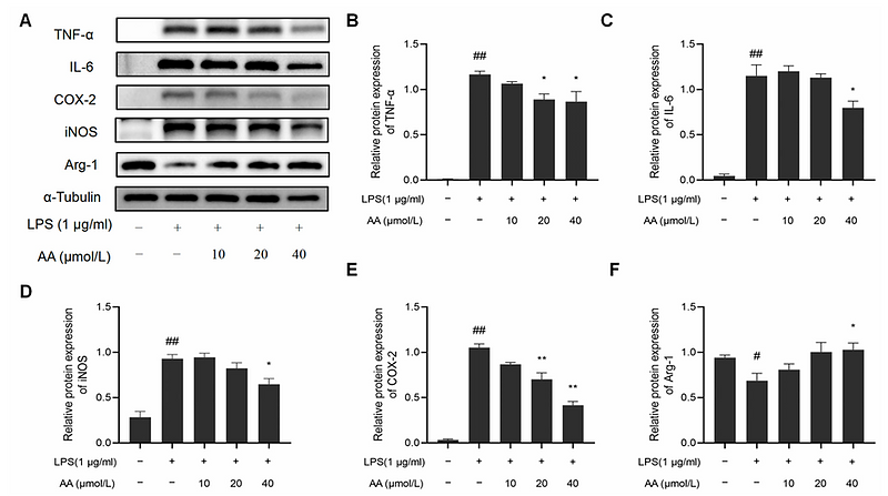
Exploring the Mechanism of Asiatic Acid against Atherosclerosis Based on Molecular Docking, Molecular Dynamics, and Experimental Verification
Author: Wu Zhihao,et al
PMID: NOPIMD202407C
应用: WB
反应种属: Mouse
发表时间: 2024 Jul
-
Citation
-
Identification and functional characterization of novel variants of MAPT and GRN in Chinese patients with frontotemporal dementia
Author:
PMID: 36641371
应用: IF,WB
反应种属: Human
发表时间: 2023 Mar
-
Citation
-
A cytological revisit on parthenogenetic Artemia and the deficiency of a meiosis-specific recombinase DMC1 in the possible transition from bisexuality to parthenogenesis
Author:
PMID: 36939898
应用: IF
反应种属: Species independent
发表时间: 2023 Jun
-
Citation
-
Microcystin-LR-induced nuclear translocation of cGAS promotes mutagenesis in human hepatocytes by impeding homologous recombination repair
Author:
PMID: 36435412
应用: WB
反应种属: Human
发表时间: 2023 Jan
-
Citation
-
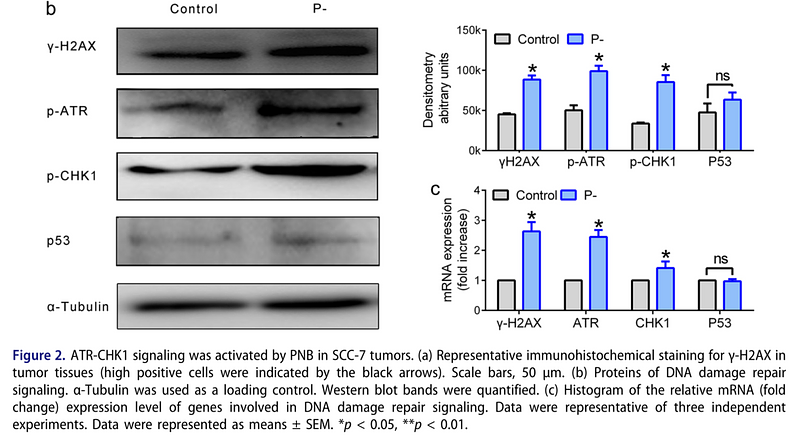
Neisseria sicca and Corynebacterium matruchotii inhibited oral squamous cell carcinomas by regulating genome stability
Author:
PMID: 35734856
应用: WB
反应种属: Mouse
发表时间: 2022 Jul
-
Citation
-
Sp1 S-Sulfhydration Induced by Hydrogen Sulfide Inhibits Inflammation via HDAC6/MyD88/NF-κB Signaling Pathway in Adjuvant-Induced Arthritis
Author:
PMID: 35453416
应用: WB
反应种属: Rat
发表时间: 2022 Apr
-
Citation
-
Ubiquitin-conjugating enzyme UBE2O regulates cellularclock function by promoting the degradation of thetranscription factor BMAL1
Author:
PMID: 29871923
应用: WB
反应种属: Human
发表时间: 2018 Jul
-
Citation
-
Inflammatory MAPK and NF-κB signaling pathways differentiated hepatitis potential of two agglomerated titanium dioxide particles.
Author: Gan J
PMID: 26590873
应用: WB
反应种属: Human
发表时间: 2016 Mar
-
Citation

Western blot analysis of alpha Tubulin 4A on different lysates with Rabbit anti-alpha Tubulin 4A antibody (ET1705-31) at 1/2,000 dilution.
Lane 1: HAP1-parental cell lysate
Lane 2: HAP1-alpha Tubulin 4A KD cell lysate
Lysates/proteins at 10 µg/Lane.
Predicted band size: 50 kDa
Observed band size: 50 kDa
Exposure time: 10 seconds; ECL: K1801;
4-20% SDS-PAGE gel.
Proteins were transferred to a PVDF membrane and blocked with 5% NFDM/TBST for 1 hour at room temperature. The primary antibody (ET1705-31) at 1/2,000 dilution was used in primary antibody dilution (K1803) at 4℃ overnight. Goat Anti-Rabbit IgG - HRP Secondary Antibody (HA1001) at 1/50,000 dilution was used for 1 hour at room temperature.