Lactate Dehydrogenase Recombinant Rabbit Monoclonal Antibody [SU39-06]


Catalog# ET1608-57
Lactate Dehydrogenase Recombinant Rabbit Monoclonal Antibody [SU39-06]
-
WB
-
IF-Cell
-
IF-Tissue
-
IHC-P
-
FC
-
IP
-
Human
-
Mouse
-
Rat
-
Zebrafish
概述
产品名称
Lactate Dehydrogenase Recombinant Rabbit Monoclonal Antibody [SU39-06]
抗体类型
Recombinant Rabbit monoclonal Antibody
免疫原
Synthetic peptide within human LDHB aa 270-310.
种属反应性
Human, Mouse (Predicted: Rat, Zebrafish)
验证应用
WB, IF-Cell, IF-Tissue, IHC-P, FC, IP
分子量
Predicted band size: 37 kDa
阳性对照
HeLa cell lysate, HEK-293 cell lysate, A549 cell lysate, hybrid fish (crucian-carp) brain tissue lysates, A549, A431, human liver tissue, human liver carcinoma tissue, human breast carcinoma tissue, mouse liver tissue, mouse testis tissue, mouse skeletal muscle tissue, Hela, LOVO.
偶联
unconjugated
克隆号
SU39-06
RRID
产品特性
形态
Liquid
存放说明
Shipped at 4℃. Store at +4℃ short term (1-2 weeks). It is recommended to aliquot into single-use upon delivery. Store at -20℃ long term.
存储缓冲液
1*TBS (pH7.4), 0.05% BSA, 40% Glycerol. Preservative: 0.05% Sodium Azide.
亚型
IgG
纯化方式
Protein A affinity purified.
应用稀释度
-
WB
-
1:1,000-1:5,000
-
IF-Cell
-
1:50-1:200
-
IF-Tissue
-
1:50-1:200
-
IHC-P
-
1:200-1:2,000
-
FC
-
1:50-1:100
-
IP
-
Use at an assay dependent concentration.
发表文章中的应用
| WB | 查看 10 篇文献如下 |
| IHC-P | 查看 3 篇文献如下 |
发表文章中的种属
| Human | 查看 9 篇文献如下 |
| Mouse | 查看 3 篇文献如下 |
| Chicken | 查看 1 篇文献如下 |
靶点
功能
Lactate dehydrogenase (LDH or LD) is an enzyme found in nearly all living cells. LDH catalyzes the conversion of lactate to pyruvate and back, as it converts NAD+ to NADH and back. A dehydrogenase is an enzyme that transfers a hydride from one molecule to another. LDH exists in four distinct enzyme classes. LDH is expressed extensively in body tissues, such as blood cells and heart muscle. Because it is released during tissue damage, it is a marker of common injuries and disease such as heart failure.
背景文献
1. Rocha CS et al. Melatonin alters the glycolytic profile of Sertoli cells: implications for male fertility. Mol Hum Reprod 20:1067-76 (2014).
2. Mallat Y et al. Proteome modulation in H9c2 cardiac cells by microRNAs miR-378 and miR-378. Mol Cell Proteomics 13:18-29 (2014).
序列相似性
Belongs to the LDH/MDH superfamily. LDH family.
翻译后修饰
ISGylated.
亚细胞定位
Cytoplasm.
UNIPROT
别名
Cell proliferation-inducing gene 19 protein antibody
GSD11 antibody
L lactate dehydrogenase B chain antibody
L-lactate dehydrogenase A chain antibody
Lactate dehydrogenase A antibody
Lactate dehydrogenase B antibody
Lactate dehydrogenase H chain antibody
Lactate dehydrogenase M antibody
LDH A antibody
LDH B antibody
展开图片
-
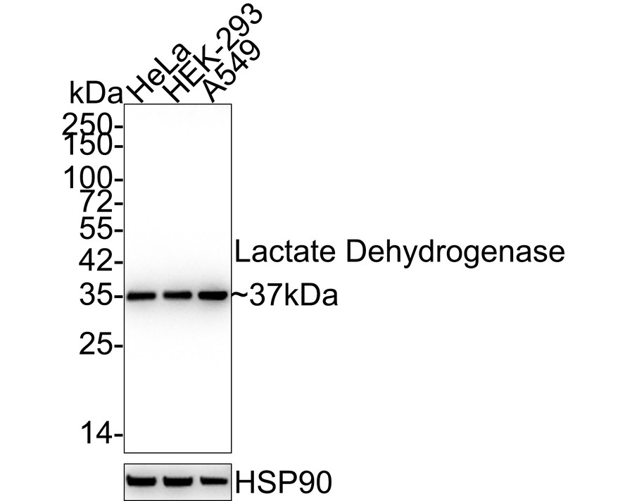
Western blot analysis of Lactate Dehydrogenase on different lysates with Rabbit anti-Lactate Dehydrogenase antibody (ET1608-57) at 1/5,000 dilution.
Lane 1: HeLa cell lysate (15 µg/Lane)
Lane 2: HEK-293 cell lysate (15 µg/Lane)
Lane 3: A549 cell lysate (15 µg/Lane)
Predicted band size: 37 kDa
Observed band size: 37 kDa
Exposure time: 1 minute 36 seconds;
4-20% SDS-PAGE gel.
Proteins were transferred to a PVDF membrane and blocked with 5% NFDM/TBST for 1 hour at room temperature. The primary antibody (ET1608-57) at 1/5,000 dilution was used in 5% NFDM/TBST at 4℃ overnight. Goat Anti-Rabbit IgG - HRP Secondary Antibody (HA1001) at 1:50,000 dilution was used for 1 hour at room temperature. -
☑ Knockdown (KD)
Western blot analysis of Lactate Dehydrogenase on different lysates with Rabbit anti-Lactate Dehydrogenase antibody (ET1608-57) at 1/1,000 dilution.
Lane 1: Hela-si NT cell lysate (10 µg/Lane)
Lane 2: Hela-si Lactate Dehydrogenase cell lysate (10 µg/Lane)
Predicted band size: 37 kDa
Observed band size: 37 kDa
Exposure time: 25 seconds;
4-20% SDS-PAGE gel.
ET1608-57 was shown to specifically react with Lactate Dehydrogenase in Hela-si NT cells. Weakened band was observed when Hela-si Lactate Dehydrogenase sample was tested. Hela-si NT and Hela-si Lactate Dehydrogenase samples were subjected to SDS-PAGE. Proteins were transferred to a PVDF membrane and blocked with 5% NFDM in TBST for 1 hour at room temperature. The primary antibody (ET1608-57, 1/1,000) and Loading control antibody (Rabbit anti-GAPDH, ET1601-4, 1/10,000) were used in 5% BSA at room temperature for 2 hours. Goat Anti-rabbit IgG-HRP Secondary Antibody (HA1001) at 1:100,000 dilution was used for 1 hour at room temperature. -
Western blot analysis of Lactate Dehydrogenase on hybrid fish (crucian-carp) brain tissue lysates. Proteins were transferred to a PVDF membrane and blocked with 5% BSA in PBS for 1 hour at room temperature. The primary antibody (ET1608-57, 1/500) was used in 5% BSA at room temperature for 2 hours. Goat Anti-Rabbit IgG - HRP Secondary Antibody (HA1001) at 1:200,000 dilution was used for 1 hour at room temperature.
-
ICC staining of Lactate Dehydrogenase in A549 cells (green). Formalin fixed cells were permeabilized with 0.1% Triton X-100 in TBS for 10 minutes at room temperature and blocked with 10% negative goat serum for 15 minutes at room temperature. Cells were probed with the primary antibody (ET1608-57, 1/50) for 1 hour at room temperature, washed with PBS. Alexa Fluor®488 conjugate-Goat anti-Rabbit IgG was used as the secondary antibody at 1/1,000 dilution. The nuclear counter stain is DAPI (blue).
-
ICC staining of Lactate Dehydrogenase in A431 cells (green). Formalin fixed cells were permeabilized with 0.1% Triton X-100 in TBS for 10 minutes at room temperature and blocked with 10% negative goat serum for 15 minutes at room temperature. Cells were probed with the primary antibody (ET1608-57, 1/50) for 1 hour at room temperature, washed with PBS. Alexa Fluor®488 conjugate-Goat anti-Rabbit IgG was used as the secondary antibody at 1/1,000 dilution. The nuclear counter stain is DAPI (blue).
-
Immunohistochemical analysis of paraffin-embedded human liver tissue using anti-Lactate Dehydrogenase antibody. The section was pre-treated using heat mediated antigen retrieval with Tris-EDTA buffer (pH 9.0) for 20 minutes.The tissues were blocked in 5% BSA for 30 minutes at room temperature, washed with ddH2O and PBS, and then probed with the primary antibody (ET1608-57, 1/50) for 30 minutes at room temperature. The detection was performed using an HRP conjugated compact polymer system. DAB was used as the chromogen. Tissues were counterstained with hematoxylin and mounted with DPX.
-
Immunohistochemical analysis of paraffin-embedded human liver carcinoma tissue using anti-Lactate Dehydrogenase antibody. The section was pre-treated using heat mediated antigen retrieval with Tris-EDTA buffer (pH 9.0) for 20 minutes.The tissues were blocked in 5% BSA for 30 minutes at room temperature, washed with ddH2O and PBS, and then probed with the primary antibody (ET1608-57, 1/50) for 30 minutes at room temperature. The detection was performed using an HRP conjugated compact polymer system. DAB was used as the chromogen. Tissues were counterstained with hematoxylin and mounted with DPX.
-
Immunohistochemical analysis of paraffin-embedded human breast carcinoma tissue with Rabbit anti-Lactate Dehydrogenase antibody (ET1608-57) at 1/2,000 dilution.
The section was pre-treated using heat mediated antigen retrieval with Tris-EDTA buffer (pH 9.0) for 20 minutes. The tissues were blocked in 1% BSA for 20 minutes at room temperature, washed with ddH2O and PBS, and then probed with the primary antibody (ET1608-57) at 1/2,000 dilution for 1 hour at room temperature. The detection was performed using an HRP conjugated compact polymer system. DAB was used as the chromogen. Tissues were counterstained with hematoxylin and mounted with DPX. -
Immunohistochemical analysis of paraffin-embedded mouse liver tissue using anti-Lactate Dehydrogenase antibody. The section was pre-treated using heat mediated antigen retrieval with Tris-EDTA buffer (pH 9.0) for 20 minutes.The tissues were blocked in 5% BSA for 30 minutes at room temperature, washed with ddH2O and PBS, and then probed with the primary antibody (ET1608-57, 1/50) for 30 minutes at room temperature. The detection was performed using an HRP conjugated compact polymer system. DAB was used as the chromogen. Tissues were counterstained with hematoxylin and mounted with DPX.
-
Immunohistochemical analysis of paraffin-embedded mouse testis tissue using anti-Lactate Dehydrogenase antibody. The section was pre-treated using heat mediated antigen retrieval with Tris-EDTA buffer (pH 9.0) for 20 minutes.The tissues were blocked in 5% BSA for 30 minutes at room temperature, washed with ddH2O and PBS, and then probed with the primary antibody (ET1608-57, 1/50) for 30 minutes at room temperature. The detection was performed using an HRP conjugated compact polymer system. DAB was used as the chromogen. Tissues were counterstained with hematoxylin and mounted with DPX.
-
Immunohistochemical analysis of paraffin-embedded mouse skeletal muscle tissue using anti-Lactate Dehydrogenase antibody. The section was pre-treated using heat mediated antigen retrieval with Tris-EDTA buffer (pH 9.0) for 20 minutes.The tissues were blocked in 5% BSA for 30 minutes at room temperature, washed with ddH2O and PBS, and then probed with the primary antibody (ET1608-57, 1/50) for 30 minutes at room temperature. The detection was performed using an HRP conjugated compact polymer system. DAB was used as the chromogen. Tissues were counterstained with hematoxylin and mounted with DPX.
-
Flow cytometric analysis of Lactate Dehydrogenase was done on Hela cells. The cells were fixed, permeabilized and stained with the primary antibody (ET1608-57, 1/50) (red). After incubation of the primary antibody at room temperature for an hour, the cells were stained with a Alexa Fluor®488 conjugate-Goat anti-Rabbit IgG Secondary antibody at 1/1,000 dilution for 30 minutes.Unlabelled sample was used as a control (cells without incubation with primary antibody; black).
-
Immunocytochemistry analysis of LOVO cells labeling Lactate Dehydrogenase with Rabbit anti-Lactate Dehydrogenase antibody (ET1608-57) at 1/50 dilution.
Cells were fixed in 4% paraformaldehyde for 10 minutes at 37 ℃, permeabilized with 0.05% Triton X-100 in PBS for 20 minutes, and then blocked with 2% negative goat serum for 30 minutes at room temperature. Cells were then incubated with Rabbit anti-Lactate Dehydrogenase antibody (ET1608-57) at 1/50 dilution in 2% negative goat serum overnight at 4 ℃. Goat Anti-Rabbit IgG H&L (iFluor™ 488, HA1121) was used as the secondary antibody at 1/1,000 dilution. PBS instead of the primary antibody was used as the secondary antibody only control. Nuclear DNA was labelled in blue with DAPI.
请注意: All products are "FOR RESEARCH USE ONLY AND ARE NOT INTENDED FOR DIAGNOSTIC OR THERAPEUTIC USE"
引文
-
Lactylation orchestrates ubiquitin-independent degradation of cGAS and promotes tumor growth
Author: Keqiang Rao, Xinchao Zhang, Yi Luo, Qiang Xia, Yuting Jin, Jing He
PMID: 40106438

期刊: Cell Reports
应用: WB
反应种属: Human
发表时间: 2025 Mar
-
 Citation
-
Ciprofol suppresses glycolysis and EMT in colorectal cancer cells by activating APC to modulate the Wnt/β-catenin signaling pathway
Author: Wu Han, Gao Jiening, Wang Yong, Zhang Yao, Jia Li, Li Weijing
PMID: 40685385
期刊: Scientific Reports
应用: WB
反应种属: Mouse,Human
发表时间: 2025 Jul
-
Citation
-
Crotonylation deficiency of S100A7 K49 promotes psoriatic keratinocyte proliferation through enhanced interaction with RAGE
Author: Liang Huifang, Wang Ying, Li Junqin, Zhang Kaiming
PMID: 40287453
期刊: Scientific Reports
应用: WB
反应种属: Human
发表时间: 2025 Apr
-
Citation
-
Upregulated expression of ubiquitin ligase TRIM21 promotes PKM2 nuclear translocation and astrocyte activation in experimental autoimmune encephalomyelitis
Author: Luting Yang, Chunqing Hu, Xiaowen Chen, Jie Zhang, Zhe Feng, Yanxin Xiao, Weitai He, Tingting Cui, Xin Zhang, Yang Yang, Yaling Zhang, Yaping Yan
PMID: 39264698

期刊: eLife
应用: WB
反应种属: Mouse
发表时间: 2024 Sept
-
Citation
-
Single-cell sequencing and multiple machine learning algorithms to identify key T-cell differentiation gene for progression of NAFLD cirrhosis to hepatocellular carcinoma
Author: Wang De-hua, Ye Li-hong, Ning Jing-yuan, Zhang Xiao-kuan, Lv Ting-ting, Li Zi-jie, Wang Zhi-yu
PMID: 38993839

期刊: Frontiers In Molecular Biosciences
应用:
反应种属:
发表时间: 2024 Jun
-
Citation
-
MYG1 drives glycolysis and colorectal cancer development through nuclear-mitochondrial collaboration
Author: Chen Jianxiong,et al
PMID: 38862489

期刊: Nature Communications
应用: WB
反应种属: Human
发表时间: 2024 Jun
-
Citation
-
CCT6A regulates cervical cancer cell glycolysis and proliferation under hypoxic conditions via the TCAB1/TERT
Author: Wang Yu, Wang Hongli
PMID: 38657573

期刊: Gynecologic And Obstetric Investigation
应用: WB,IHC-P
反应种属: Mouse,Human
发表时间: 2024 Apr
-
Citation
-
PBRM1 deficiency oncogenic addiction is associated with activated AKT–mTOR signalling and aerobic glycolysis in clear cell renal cell carcinoma cells
Author: Tang, Y., Jin, Y. H., Li, H. L., Xin, H., Chen, J. D., Li, X. Y., & Pan, Y. F.
PMID: 35672925

期刊: Journal Of Cellular And Molecular Medicine
应用: WB
反应种属: Human
发表时间: 2022 Jul
-
Citation
-
Autophagy participates in germline cyst breakdown and follicular formation by modulating glycolysis switch via Akt signaling in newly-hatched chicken ovaries
Author:
PMID: 35525303

期刊: Developmental Biology
应用: IHC-P,WB
反应种属: Chicken
发表时间: 2022 Jul
-
Citation
-
circMYC promotes cell proliferation, metastasis, and glycolysis in cervical cancer by up-regulating MET and sponging miR-577
Author:
PMID: 34306343

期刊: American Journal Of Translational Research
应用: WB
反应种属: Human
发表时间: 2021 Jun
-
Citation
-
DNA demethylase ALKBH1 promotes adipogenic differentiation via regulation of HIF-1 signaling
Author: Liu, Y., Chen, Y., Wang, Y., Jiang, S., Lin, W., Wu, Y., Li, Q., Guo, Y., Liu, W., & Yuan, Q.
PMID: 34922943

期刊: Journal Of Biological Chemistry
应用: WB
反应种属: Human
发表时间: 2021 Jan
-
Citation
-
Aberrant lactate dehydrogenase A signaling contributes metabolic signatures in pancreatic cancer
Author: Wenna Jiang, Lu Qiao, Duo Zuo, Di Qin, Jiawei Xiao, Haohua An, Yanhui Wang, Xinwei Zhang, Yu Jin, Li Ren
PMID: 33708985

期刊: Annals Of Translational Medicine
应用: IHC-P
反应种属: Human
发表时间: 2021 Feb
-
Citation
同靶点 & 同通路的产品
