ATG5 Recombinant Rabbit Monoclonal Antibody [SN73-07]


-
-
-
-
-
-
-
-
-
-
6+
Catalog# ET1611-38
ATG5 Recombinant Rabbit Monoclonal Antibody [SN73-07]
-
WB
-
IF-Cell
-
IF-Tissue
-
IHC-P
-
IP
-
FC
-
Human
-
Mouse
-
Rat
-
Monkey
概述
产品名称
ATG5 Recombinant Rabbit Monoclonal Antibody [SN73-07]
抗体类型
Recombinant Rabbit monoclonal Antibody
免疫原
Synthetic peptide within Human ATG5 aa 1-50 / 275.
种属反应性
Human, Mouse, Rat, Monkey
验证应用
WB, IF-Cell, IF-Tissue, IHC-P, IP, FC
分子量
Predicted band size: 32 kDa
阳性对照
NIH/3T3 cell lysate, C2C12 cell lysate, Neuro-2a cell lysate, PC-12 cell lysate, mouse brain tissue lysate, rat brain tissue lysate, HeLa cell lysate, A431 cell lysate, human brain tissue, NIH/3T3, PC-12.
偶联
unconjugated
克隆号
SN73-07
RRID
产品特性
形态
Liquid
浓度
1ug/ul
存放说明
Store at +4℃ after thawing. Aliquot store at -20℃ or -80℃. Avoid repeated freeze / thaw cycles.
存储缓冲液
1*TBS (pH7.4), 0.05% BSA, 40% Glycerol. Preservative: 0.05% Sodium Azide.
亚型
IgG
纯化方式
Protein A affinity purified.
应用稀释度
-
WB
-
1:1,000-1:5,000
-
IF-Cell
-
1:100-1:250
-
IF-Tissue
-
1:50-1:200
-
IHC-P
-
1:2,000
-
IP
-
1-2μg/sample
-
FC
-
1:1,000
发表文章中的应用
| WB | 查看 25 篇文献如下 |
| IHC | 查看 3 篇文献如下 |
| IF | 查看 2 篇文献如下 |
发表文章中的种属
| Human | 查看 11 篇文献如下 |
| Mouse | 查看 10 篇文献如下 |
| Rat | 查看 1 篇文献如下 |
靶点
功能
Involved in autophagic vesicle formation. Conjugation with ATG12, through a ubiquitin-like conjugating system involving ATG7 as an E1-like activating enzyme and ATG10 as an E2-like conjugating enzyme, is essential for its function. The ATG12-ATG5 conjugate acts as an E3-like enzyme which is required for lipidation of ATG8 family proteins and their association to the vesicle membranes. Involved in mitochondrial quality control after oxidative damage, and in subsequent cellular longevity. Plays a critical role in multiple aspects of lymphocyte development and is essential for both B and T lymphocyte survival and proliferation. Required for optimal processing and presentation of antigens for MHC II. Involved in the maintenance of axon morphology and membrane structures, as well as in normal adipocyte differentiation. Promotes primary ciliogenesis through removal of OFD1 from centriolar satellites and degradation of IFT20 via the autophagic pathway.
背景文献
1. He G et al. Gadd45b prevents autophagy and apoptosis against rat cerebral neuron oxygen-glucose deprivation/reperfusion injury. Apoptosis 21:390-403 (2016).
2. Pla A et al. TLR4 mediates the impairment of ubiquitin-proteasome and autophagy-lysosome pathways induced by ethanol treatment in brain. Cell Death Dis 5:e1066 (2014).
序列相似性
Belongs to the ATG5 family.
组织特异性
Ubiquitous. The mRNA is present at similar levels in viable and apoptotic cells, whereas the protein is dramatically highly expressed in apoptotic cells.
翻译后修饰
Conjugated to ATG12; which is essential for autophagy, but is not required for association with isolation membrane.; Acetylated by EP300.
亚细胞定位
Cytoplasm, Membrane.
别名
APG 5 antibody
APG 5L antibody
APG5 antibody
APG5 autophagy 5 like antibody
APG5 like antibody
APG5-like antibody
APG5L antibody
Apoptosis specific protein antibody
Apoptosis-specific protein antibody
ASP antibody
展开图片
-
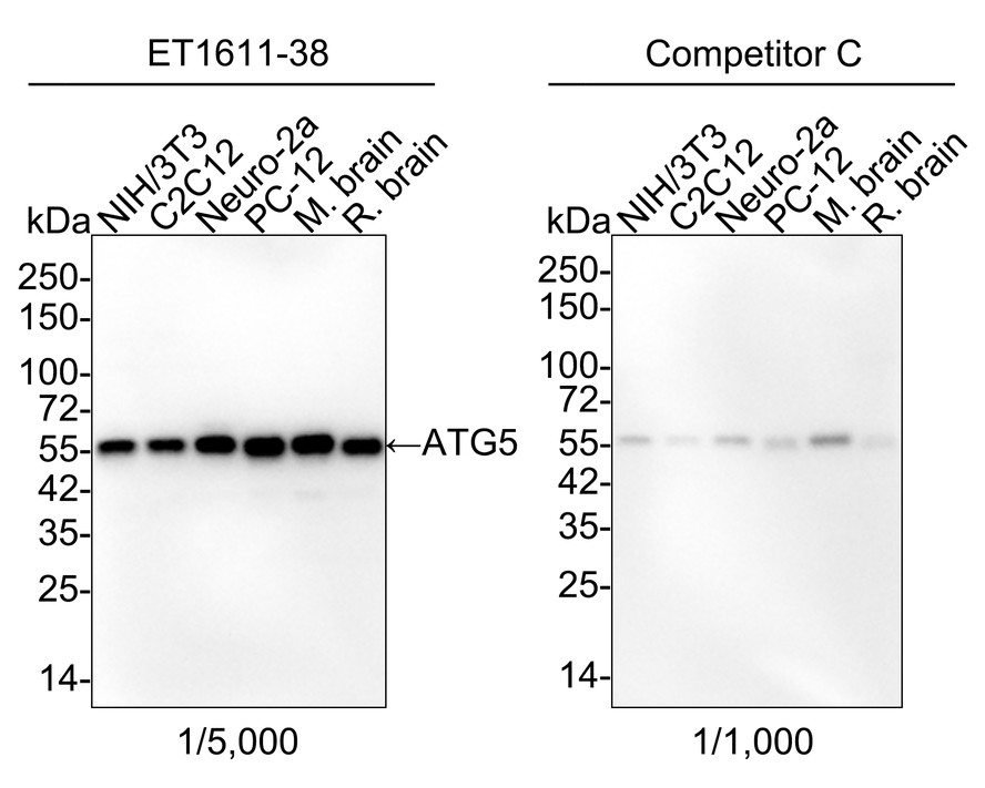
Western blot analysis of ATG5 on different lysates with Rabbit anti-ATG5 antibody (ET1611-38) at 1/5,000 dilution and competitor's antibody at 1/1,000 dilution.
Lane 1: NIH/3T3 cell lysate (15 µg/Lane)
Lane 2: C2C12 cell lysate (15 µg/Lane)
Lane 3: Neuro-2a cell lysate (15 µg/Lane)
Lane 4: PC-12 cell lysate (15 µg/Lane)
Lane 5: Mouse brain tissue lysate (15 µg/Lane)
Lane 6: Rat brain tissue lysate (15 µg/Lane)
Predicted band size: 32 kDa
Observed band size: 55 kDa
Exposure time: 35 seconds; ECL: K1802;
4-20% SDS-PAGE gel.
Proteins were transferred to a PVDF membrane and blocked with 5% NFDM/TBST for 1 hour at room temperature. The primary antibody (ET1611-38) at 1/5,000 dilution and competitor's antibody at 1/1,000 dilution were used in 5% NFDM/TBST at 4℃ overnight. Goat Anti-Rabbit IgG - HRP Secondary Antibody (HA1001) at 1/50,000 dilution was used for 1 hour at room temperature. -
Western blot analysis of ATG5 on different lysates with Rabbit anti-ATG5 antibody (ET1611-38) at 1/1,000 dilution.
Lane 1: HeLa cell lysate (20 µg/Lane)
Lane 2: A431 cell lysate (20 µg/Lane)
Lane 3: PC-12 cell lysate (20 µg/Lane)
Lane 4: NIH/3T3 cell lysate (20 µg/Lane)
Lane 5: Neuro-2a cell lysate (20 µg/Lane)
Predicted band size: 32 kDa
Observed band size: 55 kDa
Exposure time: 1 minute; ECL: K1801;
4-20% SDS-PAGE gel.
Proteins were transferred to a PVDF membrane and blocked with 5% NFDM/TBST for 1 hour at room temperature. The primary antibody (ET1611-38) at 1/1,000 dilution was used in 5% NFDM/TBST at room temperature for 2 hours. Goat Anti-Rabbit IgG - HRP Secondary Antibody (HA1001) at 1:100,000 dilution was used for 1 hour at room temperature. -
☑ Knockout (KO)
All lanes: Western blot analysis of ATG5 with anti-ATG5 antibody [SN73-07] (ET1611-38) at 1/1,000 dilution.
Lane 1: Wild-type Huh7 whole cell lysate.
Lane 2: ATG5 knockout Huh7 whole cell lysate.
ET1611-38 was shown to specifically react with ATG5 in wild-type Huh7 cells. No band was observed when ATG5 knockout sample was tested. Wild-type and ATG5 knockout samples were subjected to SDS-PAGE. Proteins were transferred to a PVDF membrane and blocked with 5% NFDM in TBST for 1 hour at room temperature. The primary Anti-ATG5 antibody (ET1611-38, 1/1,000) and Anti-GAPDH antibody (ET1601-4, 1/10,000) were used in 5% BSA at room temperature for 2 hours. Goat Anti-Rabbit IgG H&L (HRP) Secondary Antibody (HA1001) at 1:200,000 dilution was used for 1 hour at room temperature.
Cell lysate was provided by Ubigene Biosciences (Ubigene Biosciences Co., Ltd., Guangzhou, China). -
☑ Knockout (KO)
All lanes: Western blot analysis of ATG5 with anti-ATG5 antibody [SN73-07] (ET1611-38) at 1/1,000 dilution.
Lane 1: Wild-type Vero-E6 whole cell lysate.
Lane 2: ATG5 knockout Vero-E6 whole cell lysate.
ET1611-38 was shown to specifically react with ATG5 in wild-type Vero-E6 cells. No band was observed when ATG5 knockout sample was tested. Wild-type and ATG5 knockout samples were subjected to SDS-PAGE. Proteins were transferred to a PVDF membrane and blocked with 5% NFDM in TBST for 1 hour at room temperature. The primary Anti-ATG5 antibody (ET1611-38, 1/1,000) and Anti-GAPDH antibody (ET1601-4, 1/10,000) were used in 5% BSA at room temperature for 2 hours. Goat Anti-Rabbit IgG H&L (HRP) Secondary Antibody (HA1001) at 1:200,000 dilution was used for 1 hour at room temperature.
Cell lysate was provided by Ubigene Biosciences (Ubigene Biosciences Co., Ltd., Guangzhou, China). -
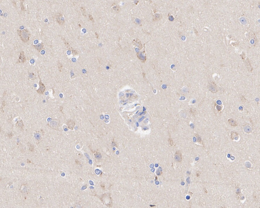
Immunohistochemical analysis of paraffin-embedded human brain tissue with Rabbit anti-ATG5 antibody (ET1611-38) at 1/2,000 dilution.
The section was pre-treated using heat mediated antigen retrieval with Tris-EDTA buffer (pH 9.0) for 20 minutes. The tissues were blocked in 1% BSA for 20 minutes at room temperature, washed with ddH2O and PBS, and then probed with the primary antibody (ET1611-38) at 1/2,000 dilution for 1 hour at room temperature. The detection was performed using an HRP conjugated compact polymer system. DAB was used as the chromogen. Tissues were counterstained with hematoxylin and mounted with DPX. -
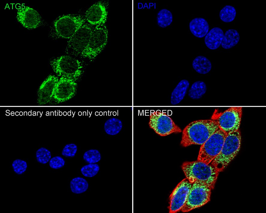
Immunocytochemistry analysis of NIH/3T3 cells labeling ATG5 with Rabbit anti-ATG5 antibody (ET1611-38) at 1/250 dilution.
Cells were fixed in 4% paraformaldehyde for 20 minutes at room temperature, permeabilized with 0.1% Triton X-100 in PBS for 5 minutes at room temperature, then blocked with 1% BSA in 10% negative goat serum for 1 hour at room temperature. Cells were then incubated with Rabbit anti-ATG5 antibody (ET1611-38) at 1/250 dilution in 1% BSA in PBST overnight at 4 ℃. Goat Anti-Rabbit IgG H&L (iFluor™ 488, HA1121) was used as the secondary antibody at 1/1,000 dilution. PBS instead of the primary antibody was used as the secondary antibody only control. Nuclear DNA was labelled in blue with DAPI. Beta tubulin (M1305-2, red) was stained at 1/100 dilution overnight at +4℃. Goat Anti-Mouse IgG H&L (iFluor™ 594, HA1126) was used as the secondary antibody at 1/1,000 dilution. -
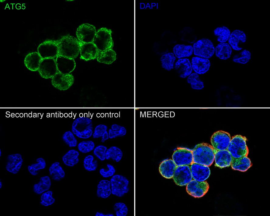
Immunocytochemistry analysis of PC-12 cells labeling ATG5 with Rabbit anti-ATG5 antibody (ET1611-38) at 1/100 dilution.
Cells were fixed in 4% paraformaldehyde for 20 minutes at room temperature, permeabilized with 0.1% Triton X-100 in PBS for 5 minutes at room temperature, then blocked with 1% BSA in 10% negative goat serum for 1 hour at room temperature. Cells were then incubated with Rabbit anti-ATG5 antibody (ET1611-38) at 1/100 dilution in 1% BSA in PBST overnight at 4 ℃. Goat Anti-Rabbit IgG H&L (iFluor™ 488, HA1121) was used as the secondary antibody at 1/1,000 dilution. PBS instead of the primary antibody was used as the secondary antibody only control. Nuclear DNA was labelled in blue with DAPI. Beta tubulin (M1305-2, red) was stained at 1/100 dilution overnight at +4℃. Goat Anti-Mouse IgG H&L (iFluor™ 594, HA1126) was used as the secondary antibody at 1/1,000 dilution. -
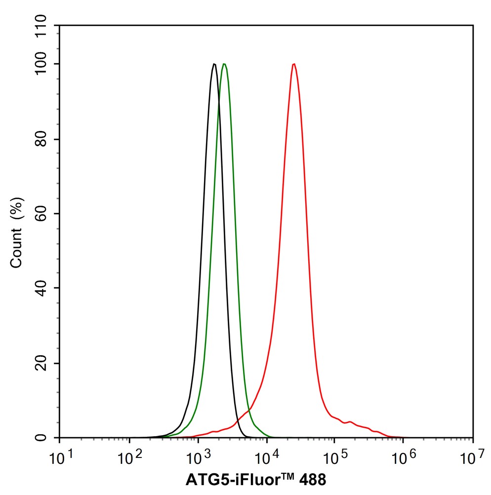
Flow cytometric analysis of NIH/3T3 cells labeling ATG5.
Cells were fixed and permeabilized. Then stained with the primary antibody (ET1611-38, 1/1,000) (red) compared with Rabbit IgG Isotype Control (green). After incubation of the primary antibody at +4℃ for an hour, the cells were stained with a iFluor™ 488 conjugate-Goat anti-Rabbit IgG Secondary antibody (HA1121) at 1/1,000 dilution for 30 minutes at +4℃. Unlabelled sample was used as a control (cells without incubation with primary antibody; black). -
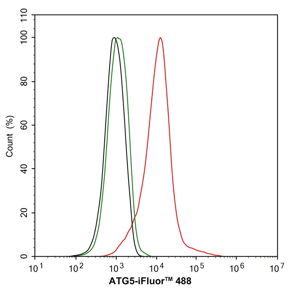
Flow cytometric analysis of PC-12 cells labeling ATG5.
Cells were fixed and permeabilized. Then stained with the primary antibody (ET1611-38, 1/1,000) (red) compared with Rabbit IgG Isotype Control (green). After incubation of the primary antibody at +4℃ for an hour, the cells were stained with a iFluor™ 488 conjugate-Goat anti-Rabbit IgG Secondary antibody (HA1121) at 1/1,000 dilution for 30 minutes at +4℃. Unlabelled sample was used as a control (cells without incubation with primary antibody; black). -
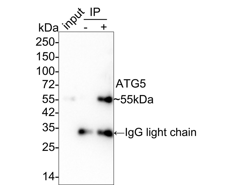
ATG5 was immunoprecipitated in 0.2mg HeLa cell lysate with ET1611-38 at 2 µg/25 µl agarose. Western blot was performed from the immunoprecipitate using ET1611-38 at 1/2,000 dilution. Anti-Rabbit IgG for IP Nano-secondary antibody (NBI01H) at 1/5,000 dilution was used for 1 hour at room temperature.
Lane 1: HeLa cell lysate (input)
Lane 2: Rabbit IgG instead of ET1611-38 in HeLa cell lysate
Lane 3: ET1611-38 IP in HeLa cell lysate
Blocking/Dilution buffer: 5% NFDM/TBST
Exposure time: 1 minute 21 seconds; ECL: K1802
请注意: All products are "FOR RESEARCH USE ONLY AND ARE NOT INTENDED FOR DIAGNOSTIC OR THERAPEUTIC USE"
引文
-
LAPTM5 Confers the Resistance to Venetoclax via Promoting the Autophagosome-Lysosome Fusion in Multiple Myeloma
Author: Yuxiang Li, Jing Bai, Dan Liu, Jinxia Hao, Ruyu Yan, Hongjuan Guo, Yuzhi Huang, Hongtao Yu, Hao Leng, Kecheng Zhou, Minxia Liu
PMID: 39753521
应用: WB
反应种属: Human
发表时间: 2025 Jan
-
 Citation
-
Bmal1 upregulates ATG5 expression to promote autophagy in skin cutaneous melanoma
Author: Tao Lei,et al
PMID: 39343115
应用: WB
反应种属: Human
发表时间: 2024 Oct
-
Citation
-
RPGR is a guanine nucleotide exchange factor for the small GTPase RAB37 required for retinal function via autophagy regulation
Author: Ruhong Ying,et al
PMID: 38536817
应用: WB
反应种属: Human
发表时间: 2024 Mar
-
Citation
-
Impaired autophagic flux in the human brain after traumatic brain injury
Author: Lang, Jiadong,et al
PMID: 38526944
应用: WB,IHC
反应种属: Human
发表时间: 2024 Mar
-
Citation
-
Short-chain fatty acids mitigate Methamphetamine-induced hepatic injuries in a Sigma-1 receptor-dependent manner
Author: Zhang Kaikai,et al
PMID: 38833980

应用: WB
反应种属: Mouse
发表时间: 2024 Jun
-
Citation
-
Obesity induced caveolin-1 impairs osteogenesis via activating mitophagy and inhibiting Sirt1 signaling
Author: Liu Shuai,et al
PMID: NOPMID20240609

应用: WB
反应种属: Mouse
发表时间: 2024 Jun
-
Citation
-
Exosomal miR-4645-5p from hypoxic bone marrow mesenchymal stem cells facilitates diabetic wound healing by restoring keratinocyte autophagy
Author: Shi Yan,et al
PMID: 38250706
应用: WB,IF
反应种属: Mouse
发表时间: 2024 Jan
-
Citation
-
Hyperuricemia Induces Reproductive Damage in Male Rats through Zinc Homeostasis Imbalance and Oxidative Stress
Author: Te Liger,et al
PMID: no pmid 240115
应用: WB
反应种属: Rat
发表时间: 2024 Jan
-
Citation
-
Hyperacetylated microtubules assist porcine deltacoronavirus nsp8 to degrade MDA5 via SQSTM1/p62-dependent selective autophagy
Author: Li Zhuang,et al
PMID: 38353538
应用: WB
反应种属: Pig
发表时间: 2024 Feb
-
Citation
-
Integrative transcriptomic and network pharmacology analysis reveals the neuroprotective role of BYHWD through enhancing autophagy by inhibiting Ctsb in intracerebral hemorrhage mice
Author: Cai Y, Yu Z, Yang X, et al
PMID: 37957754

应用: WB
反应种属: Mouse
发表时间: 2023 Nov
-
Citation
-
Wei-Tong-Xin ameliorated cisplatin-induced mitophagy and apoptosis in gastric antral mucosa by activating the Nrf2/HO-1 pathway
Author:
PMID: 36806345

应用: WB
反应种属: Mouse
发表时间: 2023 May
-
Citation
-
Chloroquine Intervenes Nephrotoxicity of Nilotinib through Deubiquitinase USP13‐Mediated Stabilization of Bcl‐XL
Author:
PMID: 37452432

应用: WB
反应种属: Human
发表时间: 2023 Jul
-
Citation
-
3-Bromopyruvate overcomes cetuximab resistance in human colorectal cancer cells by inducing autophagy-dependent ferroptosis
Author:
PMID: 37558749

应用: WB
反应种属: Human,Mouse
发表时间: 2023 Aug
-
Citation
-
TRAF6 autophagic degradation by avibirnavirus VP3 inhibits antiviral innate immunity via blocking NFKB/NF-κB activation
Author: Deng, T., Hu, B., Wang, X., Ding, S., Lin, L., Yan, Y., Peng, X., Zheng, X., Liao, M., Jin, Y., Dong, W., Gu, J., & Zhou, J.
PMID: 35266845

应用: WB
反应种属: Chicken
发表时间: 2022 Mar
-
Citation
-
Interleukin-17 promotes osteoclastogenesis and periodontal damage via autophagy in vitro and in vivo
Author:
PMID: 35219162

应用: WB
反应种属: Mouse
发表时间: 2022 Jun
-
Citation
-
Bone marrow mesenchymal stem cells facilitate diabetic wound healing through the restoration of epidermal cell autophagy via the HIF-1α/TGF-β1/SMAD pathway
Author: Shi, Y., Wang, S., Zhang, W., Zhu, Y., Fan, Z., Huang, Y., Li, F., & Yang, R.
PMID: 35841007

应用: WB,IF
反应种属: Human
发表时间: 2022 Jul
-
Citation
-
Fluid Shear Stress Regulates Osteogenic Differentiation via AnnexinA6-Mediated Autophagy in MC3T3-E1 Cells
Author: Pei, T., Su, G., Yang, J., Gao, W., Yang, X., Zhang, Y., Ren, J., Shen, Y., & Liu, X.
PMID: 36555344

应用: WB
反应种属: Human
发表时间: 2022 Dec
-
Citation
-
Methamphetamine Disturbs Gut Homeostasis and Reshapes Serum Metabolome, Inducing Neurotoxicity and Abnormal Behaviors in Mice
Author: Zhang, K. K., Chen, L. J., Li, J. H., Liu, J. L., Wang, L. B., Xu, L. L., Yang, J. Z., Li, X. W., Xie, X. L., & Wang, Q.
PMID: 35509309

应用: WB
反应种属: Mouse
发表时间: 2022 Apr
-
Citation
-
The Disulfiram/Copper Complex Induces Autophagic Cell Death in Colorectal Cancer by Targeting ULK1
Author: Hu, Yeting;Qian, Yucheng;Wei, Jingsun;Jin, Tian;Kong, Xiangxing;Cao, Hongfeng;Ding, Kefeng
PMID: 34887757
应用: WB
反应种属: Human
发表时间: 2021 Nov
-
Citation
-
Autophagy‐Sirt3 axis decelerates hematopoietic aging
Author: Jianrong Wang
PMID: 32951306
应用: WB
反应种属: Mouse
发表时间: 2020 Oct
-
Citation
-
Luteolin alleviates methamphetamine-induced neurotoxicity by suppressing PI3K/Akt pathway-modulated apoptosis and autophagy in rats
Author:
PMID: 32035215

应用: WB
反应种属: rat
发表时间: 2020 Mar
-
Citation
-
Autophagy negative-regulating Wnt signaling enhanced inflammatoryosteoclastogenesis from Pre-OCsin vitro
Author: Weilian Sun
PMID: 32199225
应用: WB
反应种属: Human
发表时间: 2020 Jun
-
Citation
-
Exopolysaccharide from Cryptococcus heimaeyensis S20 induces autophagic cell death in non-small cell lung cancer cells via ROS/p38 and ROS/ERK signalling
Author: Chao Shen
PMID: 32597573
应用: WB,IHC
反应种属: Human
发表时间: 2020 Aug
-
Citation
-
Sinomenine attenuates septic‐associated lung injury through the Nrf2‐Keap1 and autophagy
Author: Fengjie Huang
PMID: 31729764
应用: WB
反应种属: Mouse
发表时间: 2019 Feb
-
Citation
-
Water Extract of Sporoderm-Broken Spores of Ganoderma lucidum Induces Osteosarcoma Apoptosis and Restricts Autophagic Flux
Author: Weiqi Yan
PMID: 32021244
应用: WB,IHC
反应种属: Mouse
发表时间: 2019 Dec
-
Citation