概述
产品名称
beta Tubulin Mouse Monoclonal Antibody [A1-A4]
抗体类型
Mouse Monoclonal Antibody
免疫原
Synthetic peptide within Human Beta tubulin aa 151-200 / 444.
种属反应性
Human, Mouse, Rat
验证应用
WB, IF-Cell, IHC-P, FC
分子量
Predicted band size: 50 kDa
阳性对照
Hela cell lysates, NIH/3T3 cell lysates, PC-12 cell lysates, SKOV-3 cells, HeLa, 293, human tonsil tissue, human spleen tissue, human colon tissue, rat brain tissue, NIH/3T3.
偶联
unconjugated
克隆号
A1-A4
RRID
产品特性
形态
Liquid
浓度
2ug/ul
存放说明
Store at +4℃ after thawing. Aliquot store at -20℃ or -80℃. Avoid repeated freeze / thaw cycles.
存储缓冲液
1*PBS (pH7.4), 0.2% BSA, 40% Glycerol. Preservative: 0.05% Sodium Azide.
亚型
IgG3
纯化方式
Protein A affinity purified.
应用稀释度
-
WB
-
1:5,000-1:20,000
-
IF-Cell
-
1:100-1:500
-
IHC-P
-
1:50-1:200
-
FC
-
1:100-1:1,000
发表文章中的应用
发表文章中的种属
| Human | See 13 publications below |
| Mouse | See 7 publications below |
| human | See 4 publications below |
靶点
功能
Tubulins is one of several members of a small family of globular proteins. The most common members of the tubulins family are α-tubulins and β-tubulins. The beta-tubulins (relative molecular weight about 50 kDa) is counterpart of alpha-tubulin in tubulins heterodimer, it is coded by multiple tubulins genes and it is also posttranslationally modified. Heterogeneity of subunit is concentrated in C-terminal structural domain. Beta-Tubulins may have bound GTP or GDP. Under certain conditions β-tubulin can hydrolyze its bound GTP to GDP plus Pi, release the Pi, and exchange the GDP for GTP.
背景文献
1. "Tumoral and tissue-specific expression of the major human beta-tubulin isotypes."Leandro-Garcia L.J., Leskela S., Landa I., Montero-Conde C., Lopez-Jimenez E., Leton R., .Cytoskeleton 67:214-223(2010)
2. "Five mouse tubulin isotypes and their regulated expression during development."Lewis S.A., Lee M.G.-S., Cowan N.J.J. Cell Biol. 101:852-861(1985)
序列相似性
Belongs to the tubulin family.
组织特异性
Ubiquitously expressed with highest levels in spleen, thymus and immature brain.
翻译后修饰
Some glutamate residues at the C-terminus are polyglutamylated, resulting in polyglutamate chains on the gamma-carboxyl group. Polyglutamylation plays a key role in microtubule severing by spastin (SPAST). SPAST preferentially recognizes and acts on microtubules decorated with short polyglutamate tails: severing activity by SPAST increases as the number of glutamates per tubulin rises from one to eight, but decreases beyond this glutamylation threshold.; Some glutamate residues at the C-terminus are monoglycylated but not polyglycylated due to the absence of functional TTLL10 in human. Monoglycylation is mainly limited to tubulin incorporated into axonemes (cilia and flagella). Both polyglutamylation and monoglycylation can coexist on the same protein on adjacent residues, and lowering glycylation levels increases polyglutamylation, and reciprocally. The precise function of monoglycylation is still unclear (Probable).; Phosphorylated on Ser-172 by CDK1 during the cell cycle, from metaphase to telophase, but not in interphase. This phosphorylation inhibits tubulin incorporation into microtubules.
亚细胞定位
Cytoplasm, Cytoskeleton, Microtubule.
别名
beta 3 tubulin antibody
beta-4 antibody
CDCBM antibody
CDCBM1 antibody
CFEOM3 antibody
CFEOM3A antibody
FEOM3 antibody
M(beta)3 antibody
M(beta)6 antibody
MC1R antibody
展开beta 3 tubulin antibody
beta-4 antibody
CDCBM antibody
CDCBM1 antibody
CFEOM3 antibody
CFEOM3A antibody
FEOM3 antibody
M(beta)3 antibody
M(beta)6 antibody
MC1R antibody
Neuron specific beta III Tubulin antibody
Neuron-specific class III beta-tubulin antibody
QccE-11995 antibody
QccE-15186 antibody
TBB3_HUMAN antibody
Tubb 3 antibody
TUBB3 antibody
TUBB4 antibody
Tubulin beta 3 antibody
Tubulin beta 3 chain antibody
Tubulin beta 4 antibody
Tubulin beta III antibody
Tubulin beta-3 chain antibody
Tubulin beta-4 chain antibody
Tubulin beta-III antibody
折叠图片
-
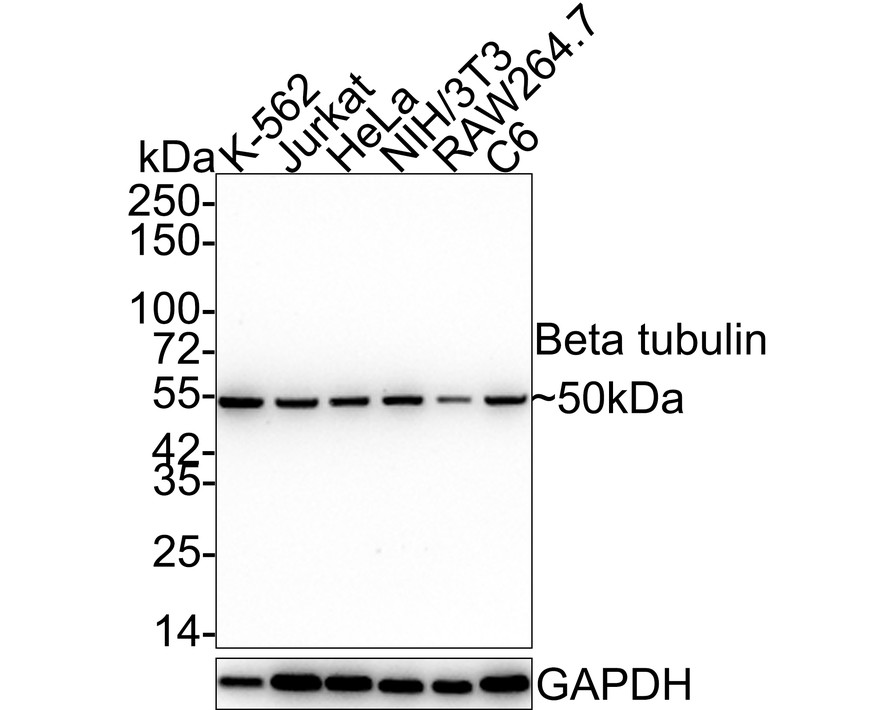
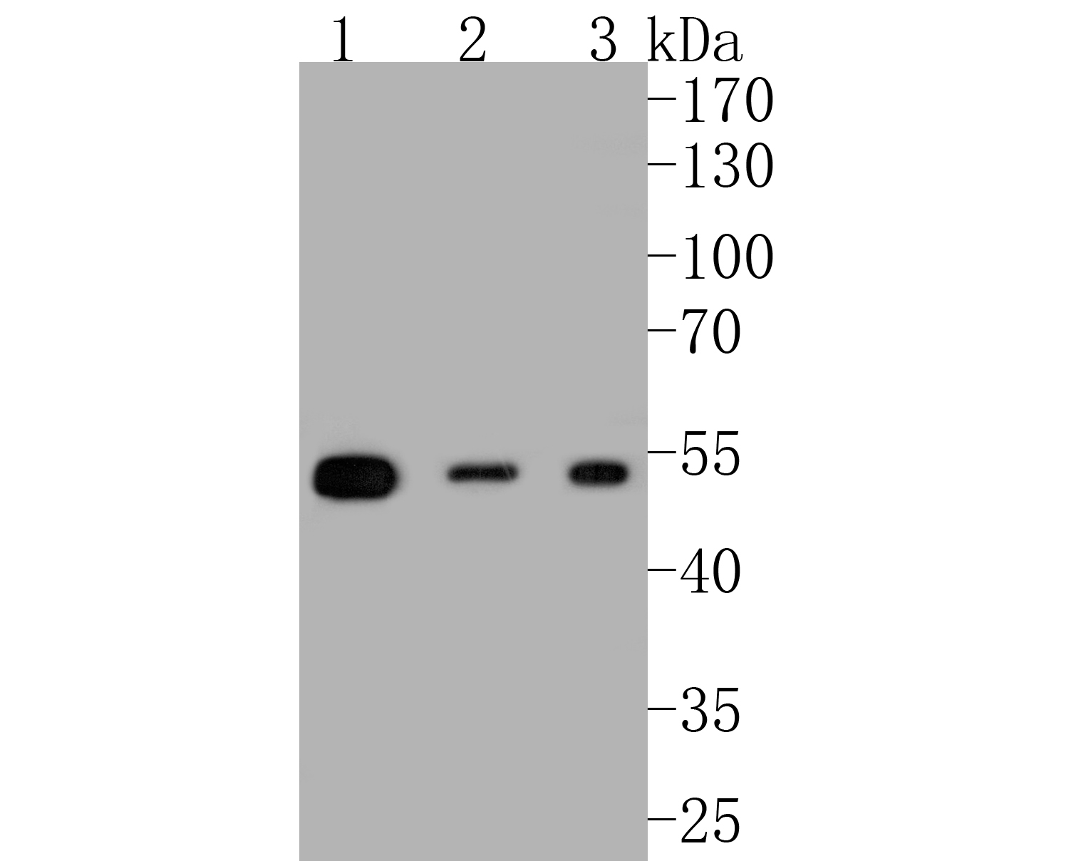
Western blot analysis of beta Tubulin on Hela cell lysates with Mouse anti-beta Tubulin antibody (M1305-2).
Hela cell lysates at 10 µg/Lane.
Predicted band size: 50 kDa
Observed band size: 50 kDa
8% SDS-PAGE gel.
Proteins were transferred to a PVDF membrane and blocked with 5% NFDM/TBST for 1 hour at room temperature. The primary antibody (M1305-2) at serial dilution was used in 5% NFDM/TBST at room temperature for 2 hours. Goat Anti-Mouse IgG - HRP Secondary Antibody (HA1006) at 1:100,000 dilution was used for 1 hour at room temperature. -
Western blot analysis of beta Tubulin on NIH/3T3 cell lysates with Mouse anti-beta Tubulin antibody (M1305-2).
NIH/3T3 cell lysates at 10 µg/Lane.
Predicted band size: 50 kDa
Observed band size: 50 kDa
8% SDS-PAGE gel.
Proteins were transferred to a PVDF membrane and blocked with 5% NFDM/TBST for 1 hour at room temperature. The primary antibody (M1305-2) at serial dilution was used in 5% NFDM/TBST at room temperature for 2 hours. Goat Anti-Mouse IgG - HRP Secondary Antibody (HA1006) at 1:100,000 dilution was used for 1 hour at room temperature. -
Western blot analysis of beta Tubulin on PC-12 cell lysates with Mouse anti-beta Tubulin antibody (M1305-2).
PC-12 cell lysates at 10 µg/Lane.
Predicted band size: 50 kDa
Observed band size: 50 kDa
8% SDS-PAGE gel.
Proteins were transferred to a PVDF membrane and blocked with 5% NFDM/TBST for 1 hour at room temperature. The primary antibody (M1305-2) at serial dilution was used in 5% NFDM/TBST at room temperature for 2 hours. Goat Anti-Mouse IgG - HRP Secondary Antibody (HA1006) at 1:100,000 dilution was used for 1 hour at room temperature. -

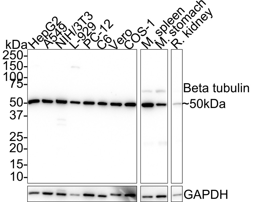
Western blot analysis of beta Tubulin on different lysates with Rabbit anti-beta Tubulin antibody (M1305-2) at 1/1,000 dilution.
Lane 1: HeLa cell lysate
Lane 2: HepG2 cell lysate
Lane 3: NIH/3T3 cell lysate
Lane 4: PC-12 cell lysate
Lane 5: C6 cell lysate
Lane 6: Vero cell lysate
Lane 7: COS-1 cell lysate
Lysates/proteins at 20 µg/Lane.
Predicted band size: 52 kDa
Observed band size: 52 kDa
Exposure time: 2 seconds;
4-20% SDS-PAGE gel.
Proteins were transferred to a PVDF membrane and blocked with 5% NFDM/TBST for 1 hour at room temperature. The primary antibody (M1305-2) at 1/1,000 dilution was used in 5% NFDM/TBST at 4℃ overnight. Goat Anti-Rabbit IgG - HRP Secondary Antibody (HA1001) at 1/50,000 dilution was used for 1 hour at room temperature. -
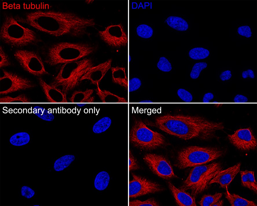
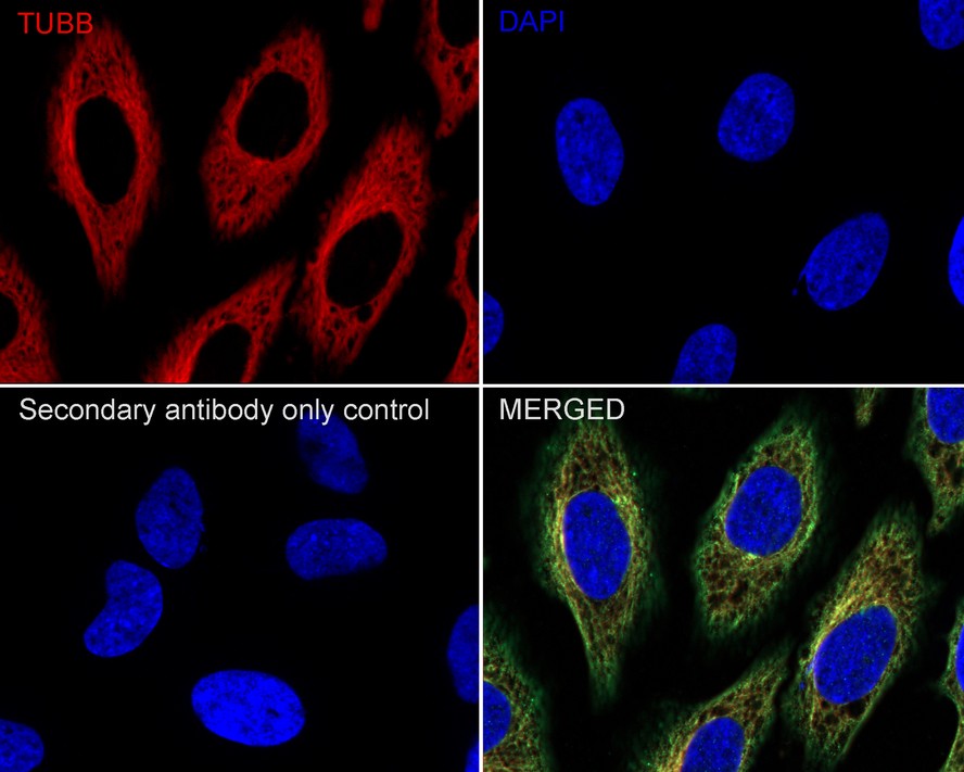
Immunocytochemistry analysis of HeLa cells labeling beta Tubulin with Mouse anti-beta Tubulin antibody (M1305-2) at 1/100 dilution.
Cells were fixed in 100% precooled methanol for 5 minutes at room temperature, then blocked with 1% BSA in 10% negative goat serum for 1 hour at room temperature. Cells were then incubated with Mouse anti-beta Tubulin antibody (M1305-2) at 1/100 dilution in 1% BSA in PBST overnight at 4 ℃. Goat Anti-Mouse IgG H&L (iFluor™ 594, HA1126) was used as the secondary antibody at 1/1,000 dilution. PBS instead of the primary antibody was used as the secondary antibody only control. Nuclear DNA was labelled in blue with DAPI. -
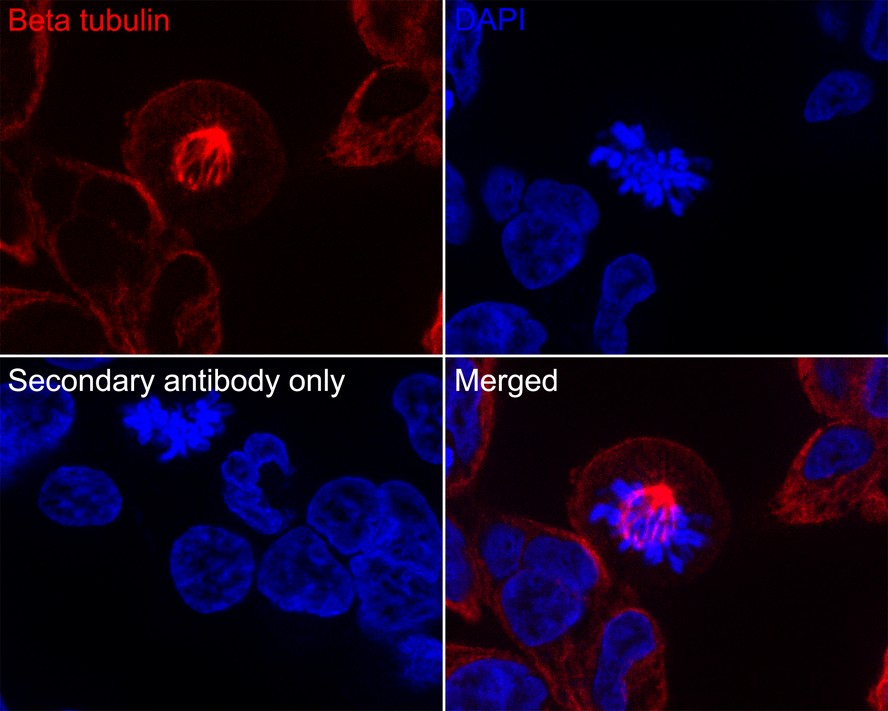
Immunocytochemistry analysis of 293 cells labeling beta Tubulin with Mouse anti-beta Tubulin antibody (M1305-2) at 1/100 dilution.
Cells were fixed in 4% paraformaldehyde for 20 minutes at room temperature, permeabilized with 0.1% Triton X-100 in PBS for 5 minutes at room temperature, then blocked with 1% BSA in 10% negative goat serum for 1 hour at room temperature. Cells were then incubated with Mouse anti-beta Tubulin antibody (M1305-2) at 1/100 dilution in 1% BSA in PBST overnight at 4 ℃. Goat Anti-Mouse IgG H&L (iFluor™ 594, HA1126) was used as the secondary antibody at 1/1,000 dilution. PBS instead of the primary antibody was used as the secondary antibody only control. Nuclear DNA was labelled in blue with DAPI. -

Immunohistochemical analysis of paraffin-embedded human tonsil tissue using anti-beta tubulin antibody. Counter stained with hematoxylin.
-
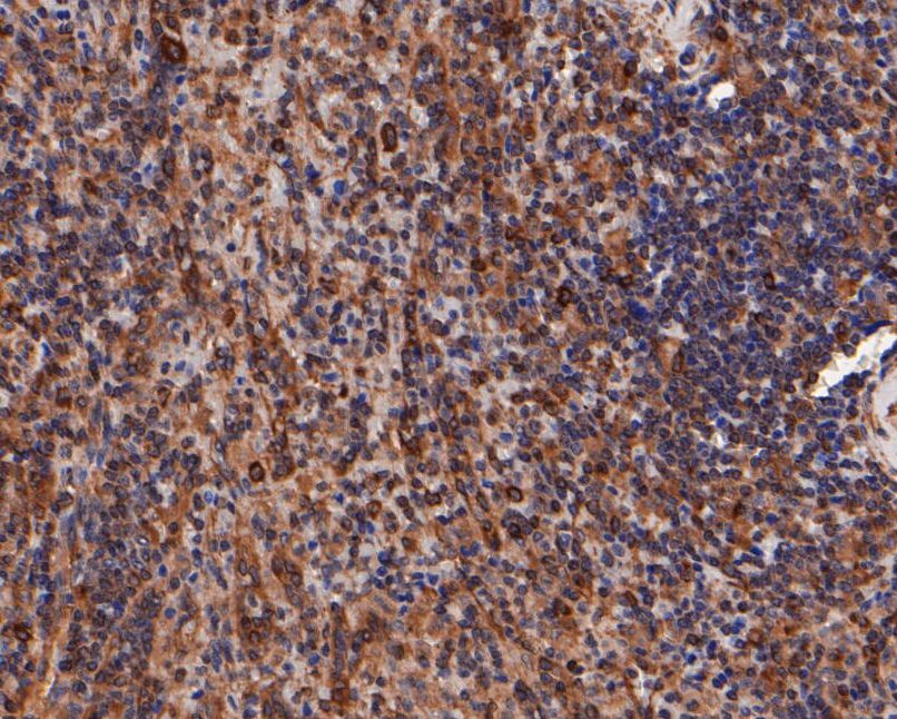
Immunohistochemical analysis of paraffin-embedded human spleen tissue using anti-beta tubulin antibody. Counter stained with hematoxylin.
-
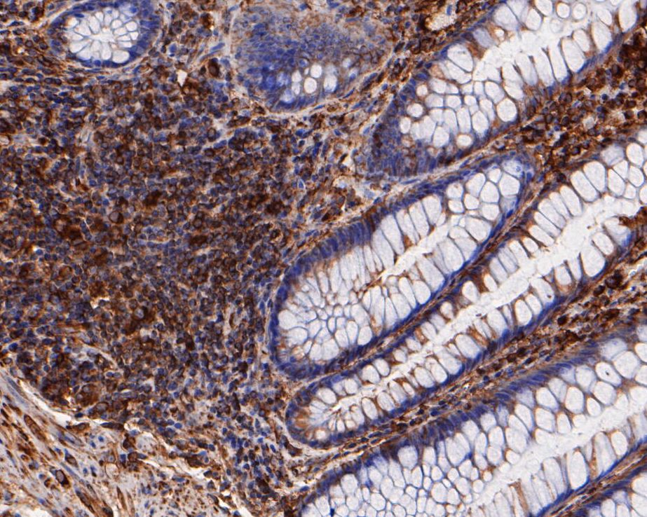
Immunohistochemical analysis of paraffin-embedded human colon tissue using anti-beta tubulin antibody. Counter stained with hematoxylin.
-
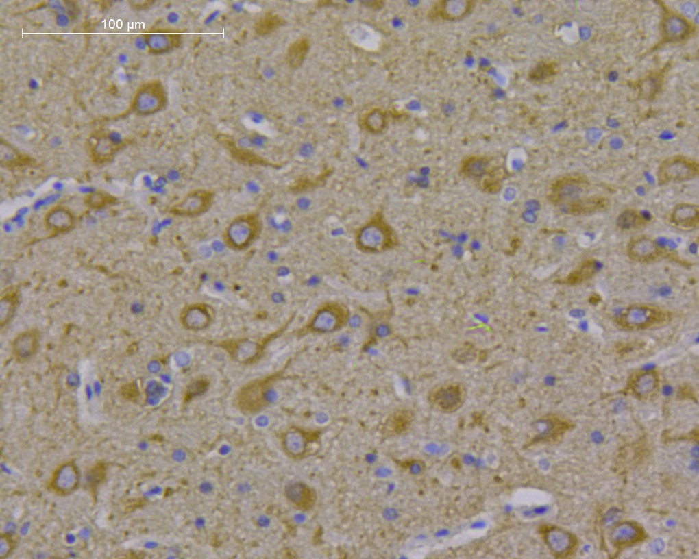
Immunohistochemical analysis of paraffin-embedded rat brain tissue using anti-beta tubulin antibody. Counter stained with hematoxylin.
-
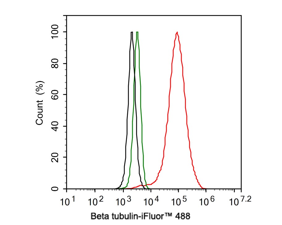
Flow cytometric analysis of HeLa cells labeling beta Tubulin.
Cells were fixed and permeabilized. Then stained with the primary antibody (M1305-2, 1μg/mL) (red) compared with Mouse IgG1 Isotype Control (green). After incubation of the primary antibody at +4℃ for an hour, the cells were stained with a iFluor™ 488 conjugate-Goat anti-Mouse IgG Secondary antibody (HA1125) at 1/1,000 dilution for 30 minutes at +4℃. Unlabelled sample was used as a control (cells without incubation with primary antibody; black). -

Flow cytometric analysis of NIH/3T3 cells labeling beta Tubulin.
Cells were fixed and permeabilized. Then stained with the primary antibody (M1305-2, 1μg/mL) (red) compared with Mouse IgG1 Isotype Control (green). After incubation of the primary antibody at +4℃ for an hour, the cells were stained with a iFluor™ 488 conjugate-Goat anti-Mouse IgG Secondary antibody (HA1125) at 1/1,000 dilution for 30 minutes at +4℃. Unlabelled sample was used as a control (cells without incubation with primary antibody; black). -
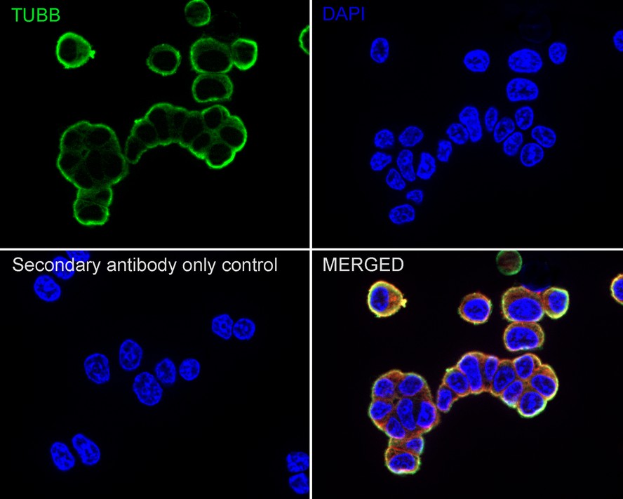
Immunocytochemistry analysis of PC-12 cells labeling beta Tubulin with Rabbit anti-beta Tubulin antibody (M1305-2) at 1/100 dilution.
Cells were fixed in 4% paraformaldehyde for 20 minutes at room temperature, permeabilized with 0.1% Triton X-100 in PBS for 5 minutes at room temperature, then blocked with 1% BSA in 10% negative goat serum for 1 hour at room temperature. Cells were then incubated with Rabbit anti-beta Tubulin antibody (M1305-2) at 1/100 dilution in 1% BSA in PBST overnight at 4 ℃. Goat Anti-Rabbit IgG H&L (iFluor™ 488, HA1121) was used as the secondary antibody at 1/1,000 dilution. PBS instead of the primary antibody was used as the secondary antibody only control. Nuclear DNA was labelled in blue with DAPI.
Beta tubulin (M1305-2, red) was stained at 1/100 dilution overnight at +4℃. Goat Anti-Mouse IgG H&L (iFluor™ 594, HA1126) was used as the secondary antibody at 1/1,000 dilution. -
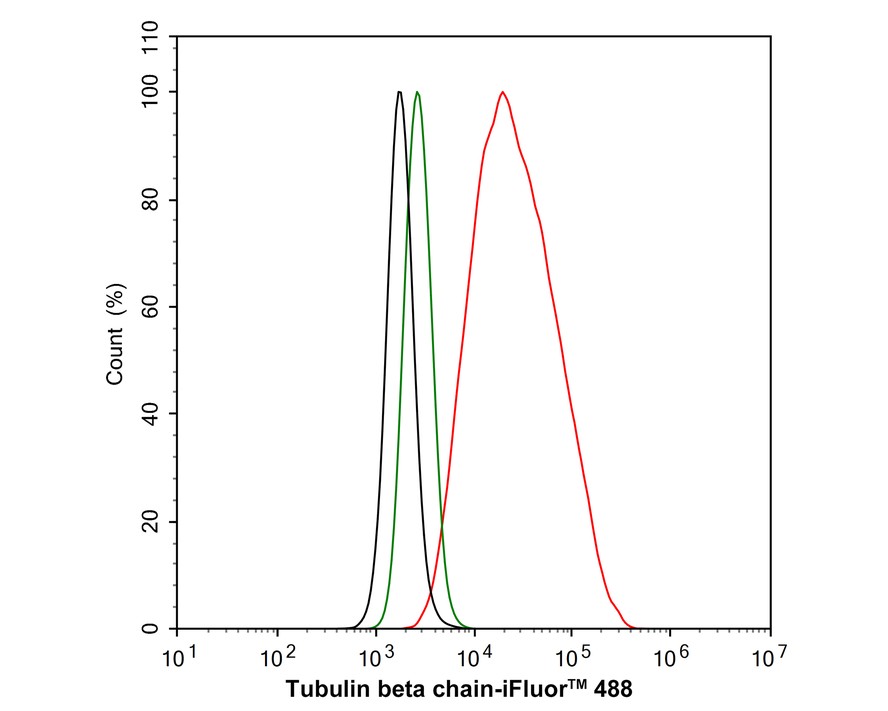
Flow cytometric analysis of HeLa cells labeling beta Tubulin.
Cells were fixed and permeabilized. Then stained with the primary antibody (M1305-2, 1μg/mL) (red) compared with Rabbit IgG Isotype Control (green). After incubation of the primary antibody at +4℃ for an hour, the cells were stained with a iFluor™ 488 conjugate-Goat anti-Rabbit IgG Secondary antibody (HA1121) at 1/1,000 dilution for 30 minutes at +4℃. Unlabelled sample was used as a control (cells without incubation with primary antibody; black). -
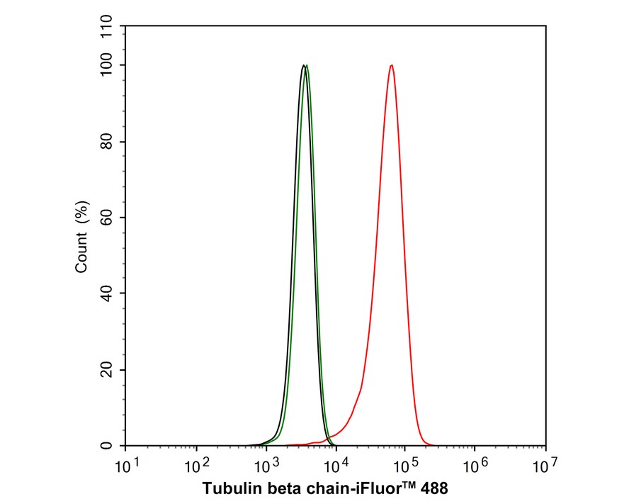
Flow cytometric analysis of NIH/3T3 cells labeling beta Tubulin.
Cells were fixed and permeabilized. Then stained with the primary antibody (M1305-2, 1μg/mL) (red) compared with Rabbit IgG Isotype Control (green). After incubation of the primary antibody at +4℃ for an hour, the cells were stained with a iFluor™ 488 conjugate-Goat anti-Rabbit IgG Secondary antibody (HA1121) at 1/1,000 dilution for 30 minutes at +4℃. Unlabelled sample was used as a control (cells without incubation with primary antibody; black). -
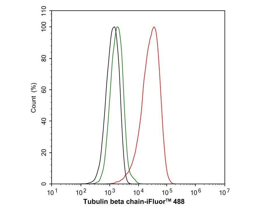
Flow cytometric analysis of PC-12 HeLa cells labeling beta Tubulin.
Cells were fixed and permeabilized. Then stained with the primary antibody (M1305-2, 1μg/mL) (red) compared with Rabbit IgG Isotype Control (green). After incubation of the primary antibody at +4℃ for an hour, the cells were stained with a iFluor™ 488 conjugate-Goat anti-Rabbit IgG Secondary antibody (HA1121) at 1/1,000 dilution for 30 minutes at +4℃. Unlabelled sample was used as a control (cells without incubation with primary antibody; black).
Please note: All products are "FOR RESEARCH USE ONLY AND ARE NOT INTENDED FOR DIAGNOSTIC OR THERAPEUTIC USE"
引文
-
Engineering Entomopathogenic Fungi Using Thermal-Responsive Polymer to Boost Their Resilience against Abiotic Stresses
Author: Yang Guang,et al
PMID: 39225683
应用: WB
反应种属:
发表时间: 2024 Sep
-
Citation
-
Dihydrotanshinone I promotes LDL uptake by HepG2 cells through increasing LDLR level
Author: Dan-Dan Hu,et al
PMID: NO PMID 2024101804
应用: WB
反应种属: Human
发表时间: 2024 Oct
-
Citation
-
Hsc70 promotes anti-tumor immunity by targeting PD-L1 for lysosomal degradation
Author: Xu Xiaoyan,et al
PMID: 38762492
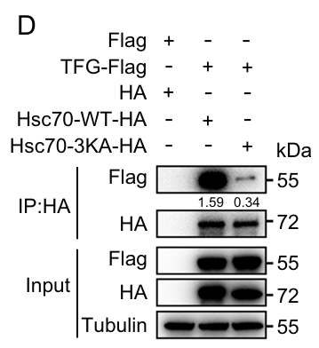
应用: WB
反应种属: Human
发表时间: 2024 May
-
Citation
-
Cigarette smoking inhibits myoblast regeneration by promoting proteasomal degradation of NPAT protein and hindering cell cycle progression
Author: Wang Jianfeng,et al
PMID: 38496008
应用: WB
反应种属: Mouse
发表时间: 2024 Mar
-
Citation
-
Subtle structural alteration in indisulam switches the molecular mechanisms for the inhibitory effect on the migration of gastric cancer cells
Author: Hou Changxu,et al
PMID: 38359488
应用: WB
反应种属: Human
发表时间: 2024 Feb
-
Citation
-
Gastrodin protects porcine sertoli cells from zearalenone-induced abnormal secretion of glial cell line-derived neurotrophic factor through the NOTCH signaling pathway.
Author:
PMID: 37285694
应用: WB
反应种属: Pig
发表时间: 2023 Sept
-
Citation
-
Increased Expression of SRSF1 Predicts Poor Prognosis in Multiple Myeloma
Author: Zhang, J., Wang, Z., Wang, K., Xin, D., Wang, L., Fan, Y., & Xu, Y.
PMID: 37206090
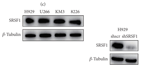
应用: WB
反应种属: Human
发表时间: 2023 May
-
Citation
-
A regulatory circuit comprising the CBP and SIRT7 regulates FAM134B-mediated ER-phagy
Author: Wang X, Jiang X, Li B, et al
PMID: 37043189
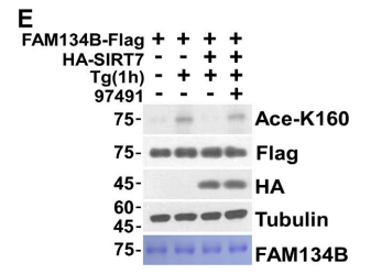
应用: WB
反应种属:
发表时间: 2023 May
-
Citation
-
TOPK promotes the growth of esophageal cancer in vitro and in vivo by enhancing YB1/eEF1A1 signal pathway
Author: Wu, W., Xu, J., Gao, D., Xie, Z., Chen, W., Li, W., Yuan, Q., Duan, L., Zhang, Y., Yang, X., Chen, Y., Dong, Z., Liu, K., & Jiang, Y.
PMID: 37328464
应用:
反应种属:
发表时间: 2023 Jun
-
Citation
-
ARIH1 activates STING-mediated T-cell activation and sensitizes tumors to immune checkpoint blockade
Author:
PMID: 37429863
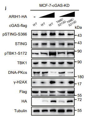
应用: WB
反应种属: Human
发表时间: 2023 Jul
-
Citation
-
VAPB-mediated ER-targeting stabilizes IRS-1 signalosomes to regulate insulin/IGF signaling
Author: Gao XK, Sheng ZK, Lu YH, et al
PMID: 37528084
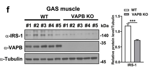
应用: WB
反应种属: Mouse
发表时间: 2023 Aug
-
Citation
-
STING promotes ferroptosis through NCOA4-dependent ferritinophagy in acute kidney injury
Author: Jin, L., Yu, B., Wang, H., Shi, L., Yang, J., Wu, L., Gao, C., Pan, H., Han, F., Lin, W., Lai, E. Y., Wang, Y. F., & Yang, Y.
PMID: 37634745
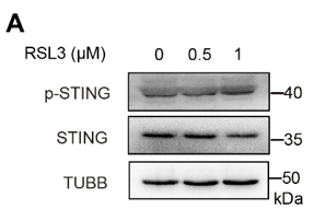
应用: WB
反应种属: Mouse
发表时间: 2023 Aug
-
Citation
-
The aryl sulfonamide indisulam inhibits gastric cancer cell migration by promoting the ubiquitination and degradation of the transcription factor ZEB1
Author: Lu, J., Li, D., Jiang, H., Li, Y., Lu, C., Chen, T., Wang, Y., Wang, X., Sun, W., Pu, Z., Qiao, C., Ma, J., & Xu, G.
PMID: 36805336

应用: WB
反应种属: Human
发表时间: 2023 Apr
-
Citation
-
Restoring nuclear entry of Sirtuin 2 in oligodendrocyte progenitor cells promotes remyelination during ageing
Author: Ma, X. R., Zhu, X., Xiao, Y., Gu, H. M., Zheng, S. S., Li, L., Wang, F., Dong, Z. J., Wang, D. X., Wu, Y., Yang, C., Jiang, W., Yao, K., Yin, Y., Zhang, Y., Peng, C., Gao, L., Meng, Z., Hu, Z., Liu, C., … Zhao, J. W.
PMID: 35264567
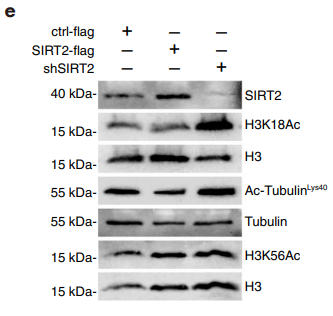
应用: WB
反应种属: Mouse
发表时间: 2022 Mar
-
Citation
-
Endothelial Shp2 deficiency controls alternative activation of macrophage preventing radiation-induced lung injury through notch signaling
Author: Liu, P., Li, Y., Li, M., Zhou, H., Zhang, H., Zhang, Y., Xu, J., Xu, Y., Zhang, J., Xia, B., Cheng, H., Ke, Y., & Zhang, X.
PMID: 35243230
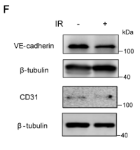
应用: WB
反应种属: Human
发表时间: 2022 Feb
-
Citation
-
Atg9A-mediated mitophagy is required for decidual differentiation of endometrial stromal cells
Author:
PMID: 36343573

应用: WB
反应种属: Human
发表时间: 2022 Dec
-
Citation
-
Inhibition of RIPK1 by ZJU-37 promotes oligodendrocyte progenitor proliferation and remyelination via NF-κB pathway
Author: Ma, X. R., Yang, S. Y., Zheng, S. S., Yan, H. H., Gu, H. M., Wang, F., Wu, Y., Dong, Z. J., Wang, D. X., Wang, Y., Meng, X., Sun, J., Xia, H. G., & Zhao, J. W.
PMID: 35365618
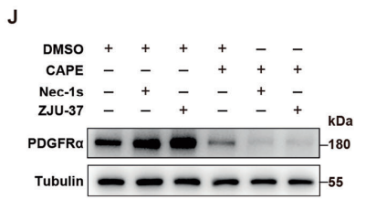
应用: WB
反应种属: Rat
发表时间: 2022 Apr
-
Citation
-
Neutralization of Hv1/HVCN1 With Antibody Enhances Microglia/Macrophages Myelin Clearance by Promoting Their Migration in the Brain
Author: Wang, F., Ma, X. R., Wu, Y., Xu, Y. C., Gu, H. M., Wang, D. X., Dong, Z. J., Li, H. L., Wang, L. B., & Zhao, J. W.
PMID: 34744634
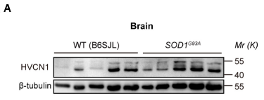
应用: WB
反应种属: Mouse
发表时间: 2021 Oct
-
Citation
-
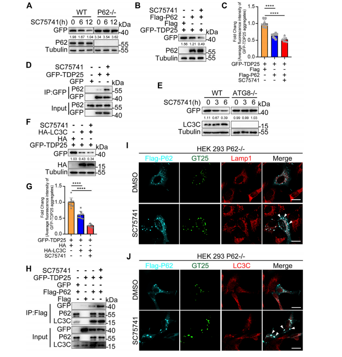
SC75741, A Novel c-Abl Inhibitor, Promotes the Clearance of TDP25 Aggregates via ATG5-Dependent Autophagy Pathway
Author: Zhou, D., Yan, H., Yang, S., Zhang, Y., Xu, X., Cen, X., Lei, K., & Xia, H.
PMID: 34776962
应用: WB
反应种属: Human
发表时间: 2021 Oct
-
Citation
-
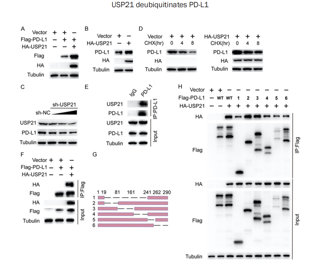
Deubiquitination and Stabilization of PD-L1 by USP21
Author: Yang, S., Yan, H., Wu, Y., Shan, B., Zhou, D., Liu, X., Mao, X., Zhou, S., Zhao, Q., & Xia, H.
PMID: 34956491
应用: WB
反应种属: Human
发表时间: 2021 Nov
-
Citation
-
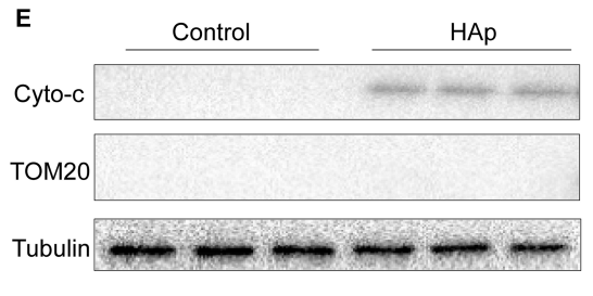
Nano-Sized Hydroxyapatite Induces Apoptosis and Osteogenic Differentiation of Vascular Smooth Muscle Cells via JNK/c-JUN Pathway. International journal of nanomedicine, 16, 3633–3648.
Author: Liu, Q., Xiang, P., Chen, M., Luo, Y., Zhao, Y., Zhu, J., Jing, W., & Yu, H.
PMID: 34079254
应用: WB
反应种属: Human
发表时间: 2021 May
-
Citation
-
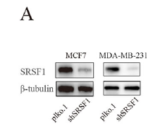
Splicing factor SRSF1 promotes breast cancer progression via oncogenic splice switching of PTPMT1. Journal of experimental & clinical cancer research : CR, 40(1), 171.
Author: Du, J. X., Luo, Y. H., Zhang, S. J., Wang, B., Chen, C., Zhu, G. Q., Zhu, P., Cai, C. Z., Wan, J. L., Cai, J. L., Chen, S. P., Dai, Z., & Zhu, W.
PMID: 33992102
应用: WB,IHC
反应种属: Human
发表时间: 2021 May
-
Citation
-
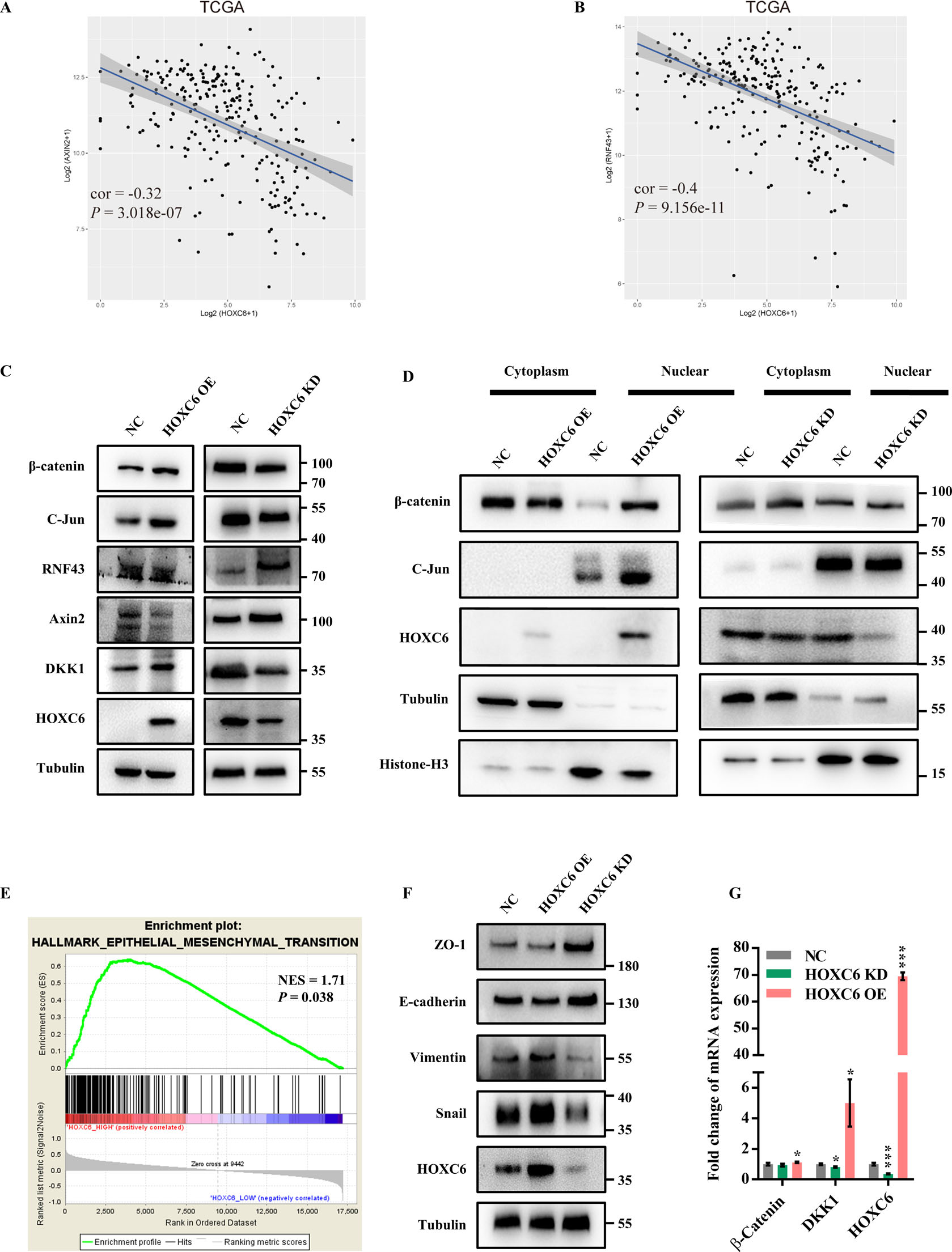
HomeoboxC6 promotes metastasis by orchestrating the DKK1/Wnt/β-catenin axis in right-sided colon cancer. Cell death & disease, 12(4), 337.
Author: Qi, L., Chen, J., Zhou, B., Xu, K., Wang, K., Fang, Z., Shao, Y., Yuan, Y., Zheng, S., & Hu, W.
PMID: 33795652
应用: WB
反应种属: Mouse
发表时间: 2021 Apr
-
Citation
-
Pharmacological targeting of MCL-1 promotes mitophagy and improves disease pathologies in an Alzheimer’s disease mouse model
Author: Jie Wu ;Ayaz Najafov;Hongguang Xia
PMID: 33184293
应用: WB,COIP
反应种属: human
发表时间: 2020 Nov
-
Citation
-
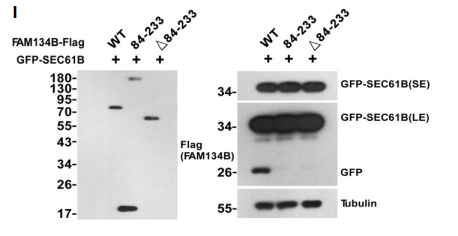
FAM134B oligomerization drives endoplasmic reticulum membrane scission for ER-phagy. The EMBO journal, 39(5), e102608.
Author: Jiang, X., Wang, X., Ding, X., Du, M., Li, B., Weng, X., Zhang, J., Li, L., Tian, R., Zhu, Q., Chen, S., Wang, L., Liu, W., Fang, L., Neculai, D., & Sun, Q.
PMID: 31930741
应用: WB
反应种属: Human
发表时间: 2020 Mar
-
Citation
-
Triptolide induces atrophy of myotubes by triggering IRS-1 degradation and activating the FoxO3 pathway. Toxicology in vitro : an international journal published in association with BIBRA, 65, 104793.
Author: Zhang Bao;Jianying Zhou
PMID: 32061799
应用: WB
反应种属: human
发表时间: 2020 Jun
-
Citation
-
Glucose-6-Phosphate Upregulates Txnip Expression by Interacting With MondoA
Author: Xiang Gao;Yan Luo
PMID: 31993438
应用: WB
反应种属: human
发表时间: 2020 Jan
-
Citation
-
P2Y12 regulates microglia activation and excitatory synaptic transmission in spinal lamina II neurons during neuropathic pain in rodents
Author: Dongping Du
PMID: 30778044
应用: WB
反应种属: rat
发表时间: 2019 Feb
-
Citation
-
Shp2 in myocytes is essential for cardiovascular and neointima development.
Author: Hongqiang Cheng;
PMID: 31634485
应用: WB
反应种属: Mouse
发表时间: 2019 Dec
-
Citation
-
JWA regulates TRAIL-induced apoptosis via MARCH8-mediated DR4 ubiquitination in cisplatin-resistant gastric cancer cells
Author: J Zhou
PMID: 28671676
应用: WB
反应种属: human
发表时间: 2017 Jul
-
Citation


