概述
产品名称
Phospho-AKT1 (T450) Recombinant Rabbit Monoclonal Antibody [SD08-12]
抗体类型
Recombinant Rabbit monoclonal Antibody
免疫原
Synthetic phospho-peptide corresponding to residues surrounding Thr450 of human AKT1.
种属反应性
Human, Mouse, Rat
验证应用
WB, IHC-P, IP, IF-Cell, FC
分子量
Predicted band size: 56 kDa
阳性对照
A549 cell lysate, MCF7 cell lysate, HeLa cell lysate, Jurkat cell lysate, C2C12 cell lysate, NIH/3T3 cell lysate, C6 cell lysate, PC-12 cell lysate, human breast cancer tissue, mouse kidney tissue, rat kidney tissue, human colon carcinoma tissue, mouse lung tissue, MCF7, C2C12, C6.
偶联
unconjugated
克隆号
SD08-12
RRID
产品特性
形态
Liquid
浓度
1ug/ul
存放说明
Store at +4℃ after thawing. Aliquot store at -20℃ or -80℃. Avoid repeated freeze / thaw cycles.
存储缓冲液
1*TBS (pH7.4), 0.05% BSA, 40% Glycerol. Preservative: 0.05% Sodium Azide.
亚型
IgG
纯化方式
Protein A affinity purified.
应用稀释度
-
WB
-
1:500-1:2,000
-
IHC-P
-
1:50-1:200
-
IP
-
Use at an assay dependent concentration.
-
IF-Cell
-
1:100
-
FC
-
1:1,000
发表文章中的应用
发表文章中的种属
靶点
功能
The serine/threonine kinase Akt family contains several members, including Akt1 (also designated PKB or RacPK), Akt2 and Akt 3, which exhibit sequence homology with the protein kinase A and C families and are encoded by the c-Akt proto-oncogene. All members of the Akt family have a pleckstrin homology domain. Akt1 and Akt2 are activated by PDGF stimulation. This activation is dependent on PDGFR-β tyrosine residues 740 and 751, which bind the subunit of the phosphatidylinositol 3-kinase (PI 3-kinase) complex. Activation of Akt1 by insulin or insulin-growth factor-1(IGF-1) results in phosphorylation of both Thr 308 and Ser 473. Phosphorylation of both residues is important to generate a high level of Akt1 activity, and the phosphorylation of Thr 308 is not dependent on phosphorylation of Ser 473 in vivo. Thus, Akt proteins become phosphorylated and activated in insulin/IGF-1-stimulated cells by an upstream kinase(s). The activation of Akt1 and Akt2 is inhibited by the PI kinase inhibitor wortmannin, suggesting that the protein signals downstream of the PI kinases.
背景文献
1. Liang D et al. Therapeutic efficacy of apelin on transplanted mesenchymal stem cells in hindlimb ischemic mice via regulation of autophagy. Sci Rep 6:21914 (2016).
2. Ma J et al. microRNA-22 attenuates neuronal cell apoptosis in a cell model of traumatic brain injury. Am J Transl Res 8:1895-902 (2016).
序列相似性
Belongs to the protein kinase superfamily. AGC Ser/Thr protein kinase family. RAC subfamily.
组织特异性
Expressed in prostate cancer and levels increase from the normal to the malignant state (at protein level). Expressed in all human cell types so far analyzed. The Tyr-176 phosphorylated form shows a significant increase in expression in breast cancers during the progressive stages i.e. normal to hyperplasia (ADH), ductal carcinoma in situ (DCIS), invasive ductal carcinoma (IDC) and lymph node metastatic (LNMM) stages.
翻译后修饰
O-GlcNAcylation at Thr-305 and Thr-312 inhibits activating phosphorylation at Thr-308 via disrupting the interaction between AKT1 and PDPK1. O-GlcNAcylation at Ser-473 also probably interferes with phosphorylation at this site.; Phosphorylation on Thr-308, Ser-473 and Tyr-474 is required for full activity. Activated TNK2 phosphorylates it on Tyr-176 resulting in its binding to the anionic plasma membrane phospholipid PA. This phosphorylated form localizes to the cell membrane, where it is targeted by PDPK1 and PDPK2 for further phosphorylations on Thr-308 and Ser-473 leading to its activation. Ser-473 phosphorylation by mTORC2 favors Thr-308 phosphorylation by PDPK1. Phosphorylated at Thr-308 and Ser-473 by IKBKE and TBK1. Ser-473 phosphorylation is enhanced by interaction with AGAP2 isoform 2 (PIKE-A). Ser-473 phosphorylation is enhanced in focal cortical dysplasias with Taylor-type balloon cells. Ser-473 phosphorylation is enhanced by signaling through activated FLT3 (By similarity). Ser-473 is dephosphorylated by PHLPP. Dephosphorylated at Thr-308 and Ser-473 by PP2A phosphatase. The phosphorylated form of PPP2R5B is required for bridging AKT1 with PP2A phosphatase. Ser-473 is dephosphorylated by CPPED1, leading to termination of signaling.; Ubiquitinated via 'Lys-48'-linked polyubiquitination by ZNRF1, leading to its degradation by the proteasome (By similarity). Ubiquitinated; undergoes both 'Lys-48'- and 'Lys-63'-linked polyubiquitination. TRAF6-induced 'Lys-63'-linked AKT1 ubiquitination is critical for phosphorylation and activation. When ubiquitinated, it translocates to the plasma membrane, where it becomes phosphorylated. When fully phosphorylated and translocated into the nucleus, undergoes 'Lys-48'-polyubiquitination catalyzed by TTC3, leading to its degradation by the proteasome. Also ubiquitinated by TRIM13 leading to its proteasomal degradation. Phosphorylated, undergoes 'Lys-48'-linked polyubiquitination preferentially at Lys-284 catalyzed by MUL1, leading to its proteasomal degradation.; Acetylated on Lys-14 and Lys-20 by the histone acetyltransferases EP300 and KAT2B. Acetylation results in reduced phosphorylation and inhibition of activity. Deacetylated at Lys-14 and Lys-20 by SIRT1. SIRT1-mediated deacetylation relieves the inhibition.
亚细胞定位
Cytoplasm, Nucleus, Cell membrane.
别名
AKT 1 antibody
AKT antibody
AKT1 antibody
AKT1_HUMAN antibody
MGC99656 antibody
PKB antibody
PKB-ALPHA antibody
PRKBA antibody
Protein Kinase B Alpha antibody
Protein kinase B antibody
展开AKT 1 antibody
AKT antibody
AKT1 antibody
AKT1_HUMAN antibody
MGC99656 antibody
PKB antibody
PKB-ALPHA antibody
PRKBA antibody
Protein Kinase B Alpha antibody
Protein kinase B antibody
Proto-oncogene c-Akt antibody
RAC Alpha antibody
RAC antibody
RAC-alpha serine/threonine-protein kinase antibody
RAC-PK-alpha antibody
折叠图片
-

☑ Cell treatment (CT)
Western blot analysis of Phospho-AKT1 (T450) on different lysates with Rabbit anti-Phospho-AKT1 (T450) antibody (ET1612-73) at 1/1,000 dilution.
Lane 1: A549 cell lysate
Lane 2: MCF7 cell lysate
Lane 3: HeLa cell lysate
Lane 4: Jurkat cell lysate
Lane 5: C2C12 cell lysate
Lane 6: NIH/3T3 cell lysate
Lane 7: C6 cell lysate
Lane 8: PC-12 cell lysate
Lane 9: MCF7 cell lysate, the membrane treated with λpp for 1 hour
Lane 10: C2C12 cell lysate, the membrane treated with λpp for 1 hour
Lane 11: C6 cell lysate, the membrane treated with λpp for 1 hour
Lysates/proteins at 20 µg/Lane.
Predicted band size: 54 kDa
Observed band size: 54 kDa
Exposure time: 3 minutes; ECL: K1802;
4-20% SDS-PAGE gel.
Proteins were transferred to a PVDF membrane and blocked with 5% NFDM/TBST for 1 hour at room temperature. The primary antibody (ET1612-73) at 1/1,000 dilution was used in 5% NFDM/TBST at 4℃ overnight. Goat Anti-Rabbit IgG - HRP Secondary Antibody (HA1001) at 1/50,000 dilution was used for 1 hour at room temperature. -

Immunohistochemical analysis of paraffin-embedded human breast cancer tissue with Rabbit anti-Phospho-AKT1 (T450) antibody (ET1612-73) at 1/200 dilution.
The section was pre-treated using heat mediated antigen retrieval with sodium citrate buffer (pH 6.0) for 2 minutes. The tissues were blocked in 1% BSA for 20 minutes at room temperature, washed with ddH2O and PBS, and then probed with the primary antibody (ET1612-73) at 1/200 dilution for 1 hour at room temperature. The detection was performed using an HRP conjugated compact polymer system. DAB was used as the chromogen. Tissues were counterstained with hematoxylin and mounted with DPX. -

Immunohistochemical analysis of paraffin-embedded mouse kidney tissue with Rabbit anti-Phospho-AKT1 (T450) antibody (ET1612-73) at 1/200 dilution.
The section was pre-treated using heat mediated antigen retrieval with sodium citrate buffer (pH 6.0) for 2 minutes. The tissues were blocked in 1% BSA for 20 minutes at room temperature, washed with ddH2O and PBS, and then probed with the primary antibody (ET1612-73) at 1/200 dilution for 1 hour at room temperature. The detection was performed using an HRP conjugated compact polymer system. DAB was used as the chromogen. Tissues were counterstained with hematoxylin and mounted with DPX. -

Immunohistochemical analysis of paraffin-embedded rat kidney tissue with Rabbit anti-Phospho-AKT1 (T450) antibody (ET1612-73) at 1/200 dilution.
The section was pre-treated using heat mediated antigen retrieval with sodium citrate buffer (pH 6.0) for 2 minutes. The tissues were blocked in 1% BSA for 20 minutes at room temperature, washed with ddH2O and PBS, and then probed with the primary antibody (ET1612-73) at 1/200 dilution for 1 hour at room temperature. The detection was performed using an HRP conjugated compact polymer system. DAB was used as the chromogen. Tissues were counterstained with hematoxylin and mounted with DPX. -

Immunohistochemical analysis of paraffin-embedded human colon carcinoma tissue with Rabbit anti-Phospho-AKT1 (T450) antibody (ET1612-73) at 1/50 dilution.
The section was pre-treated using heat mediated antigen retrieval with Tris-EDTA buffer (pH 9.0) for 20 minutes. The tissues were blocked in 1% BSA for 20 minutes at room temperature, washed with ddH2O and PBS, and then probed with the primary antibody (ET1612-73) at 1/50 dilution for 1 hour at room temperature. The detection was performed using an HRP conjugated compact polymer system. DAB was used as the chromogen. Tissues were counterstained with hematoxylin and mounted with DPX. -

Immunohistochemical analysis of paraffin-embedded mouse lung tissue with Rabbit anti-Phospho-AKT1 (T450) antibody (ET1612-73) at 1/200 dilution.
The section was pre-treated using heat mediated antigen retrieval with Tris-EDTA buffer (pH 9.0) for 20 minutes. The tissues were blocked in 1% BSA for 20 minutes at room temperature, washed with ddH2O and PBS, and then probed with the primary antibody (ET1612-73) at 1/200 dilution for 1 hour at room temperature. The detection was performed using an HRP conjugated compact polymer system. DAB was used as the chromogen. Tissues were counterstained with hematoxylin and mounted with DPX. -

Immunocytochemistry analysis of MCF7 cells labeling Phospho-AKT1 (T450) with Rabbit anti-Phospho-AKT1 (T450) antibody (ET1612-73) at 1/100 dilution.
Cells were fixed in 4% paraformaldehyde for 20 minutes at room temperature, permeabilized with 0.1% Triton X-100 in PBS for 5 minutes at room temperature, then blocked with 1% BSA in 10% negative goat serum for 1 hour at room temperature. Cells were then incubated with Rabbit anti-Phospho-AKT1 (T450) antibody (ET1612-73) at 1/100 dilution in 1% BSA in PBST overnight at 4 ℃. Goat Anti-Rabbit IgG H&L (iFluor™ 488, HA1121) was used as the secondary antibody at 1/1,000 dilution. PBS instead of the primary antibody was used as the secondary antibody only control. Nuclear DNA was labelled in blue with DAPI.
Beta tubulin (M1305-2, red) was stained at 1/100 dilution overnight at +4℃. Goat Anti-Mouse IgG H&L (iFluor™ 594, HA1126) was used as the secondary antibody at 1/1,000 dilution. -

Flow cytometric analysis of MCF7 cells labeling Phospho-AKT1 (T450).
Cells were fixed and permeabilized. Then stained with the primary antibody (ET1612-73, 1/1,000) (red) compared with Rabbit IgG Isotype Control (green). After incubation of the primary antibody at +4℃ for an hour, the cells were stained with a iFluor™ 488 conjugate-Goat anti-Rabbit IgG Secondary antibody (HA1121) at 1/1,000 dilution for 30 minutes at +4℃. Unlabelled sample was used as a control (cells without incubation with primary antibody; black). -

Immunocytochemistry analysis of C2C12 cells labeling Phospho-AKT1 (T450) with Rabbit anti-Phospho-AKT1 (T450) antibody (ET1612-73) at 1/100 dilution.
Cells were fixed in 4% paraformaldehyde for 20 minutes at room temperature, permeabilized with 0.1% Triton X-100 in PBS for 5 minutes at room temperature, then blocked with 1% BSA in 10% negative goat serum for 1 hour at room temperature. Cells were then incubated with Rabbit anti-Phospho-AKT1 (T450) antibody (ET1612-73) at 1/100 dilution in 1% BSA in PBST overnight at 4 ℃. Goat Anti-Rabbit IgG H&L (iFluor™ 488, HA1121) was used as the secondary antibody at 1/1,000 dilution. PBS instead of the primary antibody was used as the secondary antibody only control. Nuclear DNA was labelled in blue with DAPI.
Beta tubulin (M1305-2, red) was stained at 1/100 dilution overnight at +4℃. Goat Anti-Mouse IgG H&L (iFluor™ 594, HA1126) was used as the secondary antibody at 1/1,000 dilution. -

Flow cytometric analysis of C2C12 cells labeling Phospho-AKT1 (T450).
Cells were fixed and permeabilized. Then stained with the primary antibody (ET1612-73, 1/1,000) (red) compared with Rabbit IgG Isotype Control (green). After incubation of the primary antibody at +4℃ for an hour, the cells were stained with a iFluor™ 488 conjugate-Goat anti-Rabbit IgG Secondary antibody (HA1121) at 1/1,000 dilution for 30 minutes at +4℃. Unlabelled sample was used as a control (cells without incubation with primary antibody; black). -

Immunocytochemistry analysis of C6 cells labeling Phospho-AKT1 (T450) with Rabbit anti-Phospho-AKT1 (T450) antibody (ET1612-73) at 1/100 dilution.
Cells were fixed in 4% paraformaldehyde for 20 minutes at room temperature, permeabilized with 0.1% Triton X-100 in PBS for 5 minutes at room temperature, then blocked with 1% BSA in 10% negative goat serum for 1 hour at room temperature. Cells were then incubated with Rabbit anti-Phospho-AKT1 (T450) antibody (ET1612-73) at 1/100 dilution in 1% BSA in PBST overnight at 4 ℃. Goat Anti-Rabbit IgG H&L (iFluor™ 488, HA1121) was used as the secondary antibody at 1/1,000 dilution. PBS instead of the primary antibody was used as the secondary antibody only control. Nuclear DNA was labelled in blue with DAPI.
Beta tubulin (M1305-2, red) was stained at 1/100 dilution overnight at +4℃. Goat Anti-Mouse IgG H&L (iFluor™ 594, HA1126) was used as the secondary antibody at 1/1,000 dilution. -

Flow cytometric analysis of C6 cells labeling Phospho-AKT1 (T450).
Cells were fixed and permeabilized. Then stained with the primary antibody (ET1612-73, 1/1,000) (red) compared with Rabbit IgG Isotype Control (green). After incubation of the primary antibody at +4℃ for an hour, the cells were stained with a iFluor™ 488 conjugate-Goat anti-Rabbit IgG Secondary antibody (HA1121) at 1/1,000 dilution for 30 minutes at +4℃. Unlabelled sample was used as a control (cells without incubation with primary antibody; black).
Please note: All products are "FOR RESEARCH USE ONLY AND ARE NOT INTENDED FOR DIAGNOSTIC OR THERAPEUTIC USE"
引文
-
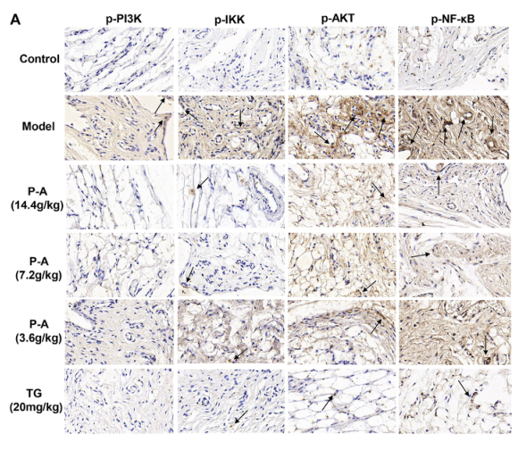
The mechanism of action of paeoniae radix rubra-angelicae sinensis radix drug pair in the treatment of rheumatoid arthritis through PI3K/AKT/NF-κB signaling pathway
Author: Li, J., Zhang, X., Guo, D., Shi, Y., Zhang, S., Yang, R., & Cheng, J.
PMID: 36992829

应用: WB,IHC
反应种属: Rat
发表时间: 2023 Mar
-
Citation
同靶点&同通路的产品
AKT1 Recombinant Rabbit Monoclonal Antibody [ST05-09]
Application: WB,IHC-P,IP,FC,IHC-Fr,IF-Cell
Reactivity: Human,Mouse,Rat
Conjugate: unconjugated
AKT1 Mouse Monoclonal Antibody [D9-9-C9]
Application: WB,IHC-P
Reactivity: Human,Mouse,Rat
Conjugate: unconjugated
AKT1 Recombinant Rabbit Monoclonal Antibody [PSH0-50]
Application: WB,IF-Cell,FC
Reactivity: Human,Mouse,Rat,Monkey
Conjugate: unconjugated
Phospho-AKT1 (S124) Recombinant Rabbit Monoclonal Antibody [JJ08-46]
Application: WB,IF-Cell,IF-Tissue,IHC-P,IP
Reactivity: Human,Mouse,Rat
Conjugate: unconjugated
Phospho-AKT1 (S129) Recombinant Rabbit Monoclonal Antibody [JE59-50]
Application: WB
Reactivity: Human,Mouse,Rat
Conjugate: unconjugated


