概述
产品名称
Beta III Tubulin Mouse Monoclonal Antibody [A8-D10]
抗体类型
Mouse Monoclonal Antibody
免疫原
Synthetic peptide (KLH-coupled) within human Tubulin beta-3 chain aa 401-450.
种属反应性
Human, Mouse, Rat
验证应用
WB, IF-Cell, IHC-P, FC, IF-Tissue
分子量
Predicted band size: 50 kDa
阳性对照
SH-SY5Y cell lysate, U-87 MG cell lysate, A-172 cell lysate, Neuro-2a cell lysate, PC-12 cell lysate, mouse brain tissue lysate, rat brain tissue lysate, HEK-293, SH-SY5Y, Neuro-2a, human brain tissue, mouse brain tissue, rat brain tissue, mouse hippocampus tissue, MCF7.
偶联
unconjugated
克隆号
A8-D10
RRID
产品特性
形态
Liquid
浓度
2ug/ul
存放说明
Store at +4℃ after thawing. Aliquot store at -20℃. Avoid repeated freeze / thaw cycles.
存储缓冲液
1*PBS (pH7.4), 0.2% BSA, 40% Glycerol. Preservative: 0.05% Sodium Azide.
亚型
IgG2a
纯化方式
Immunogen affinity purified.
应用稀释度
-
WB
-
1:2,000-1:5,000
-
IF-Cell
-
1:500-1:1,000
-
IHC-P
-
1:2,000
-
FC
-
1:1,000
-
IF-Tissue
-
1:200
发表文章中的应用
发表文章中的种属
靶点
功能
Tubulin is a compound of subunits of A tubulin and B tubulin. Class III beta tubulin (beta III-tubulin) is a vertebrate tubulin isotype specific to the neurons and mammalian testis cells, making it an ideal neuronal marker. Overexpression of class III beta tubulin is associated with the resistances of microtubule-targeted cancer drugs in lung cancer cell lines, breast cancer cell lines, and ovarian tumors.
背景文献
1. Tischfield M A et al. Human TUBB3 mutations perturb microtubule dynamics, kinesin interactions, and axon guidance. Cell 140:74-87 (2010).
2. Fourest-Lieuvin A et al. Microtubule regulation in mitosis: tubulin phosphorylation by the cyclin-dependent kinase Cdk1. Mol Biol Cell 17:1041-1050 (2006).
序列相似性
Belongs to the tubulin family.
组织特异性
Expression is primarily restricted to central and peripheral nervous system. Greatly increased expression in most cancerous tissues.
翻译后修饰
Some glutamate residues at the C-terminus are polyglutamylated, resulting in polyglutamate chains on the gamma-carboxyl group. Polyglutamylation plays a key role in microtubule severing by spastin (SPAST). SPAST preferentially recognizes and acts on microtubules decorated with short polyglutamate tails: severing activity by SPAST increases as the number of glutamates per tubulin rises from one to eight, but decreases beyond this glutamylation threshold.; Some glutamate residues at the C-terminus are monoglycylated but not polyglycylated due to the absence of functional TTLL10 in human. Monoglycylation is mainly limited to tubulin incorporated into axonemes (cilia and flagella). Both polyglutamylation and monoglycylation can coexist on the same protein on adjacent residues, and lowering glycylation levels increases polyglutamylation, and reciprocally. The precise function of monoglycylation is still unclear (Probable).; Phosphorylated on Ser-172 by CDK1 during the cell cycle, from metaphase to telophase, but not in interphase. This phosphorylation inhibits tubulin incorporation into microtubules.
亚细胞定位
Cytoplasm. Cytoskeleton. Microtubule.
别名
beta 3 tubulin antibody
beta 4 antibody
beta-4 antibody
CDCBM antibody
CDCBM1 antibody
CFEOM3 antibody
CFEOM3A antibody
FEOM3 antibody
M(beta)3 antibody
M(beta)6 antibody
展开beta 3 tubulin antibody
beta 4 antibody
beta-4 antibody
CDCBM antibody
CDCBM1 antibody
CFEOM3 antibody
CFEOM3A antibody
FEOM3 antibody
M(beta)3 antibody
M(beta)6 antibody
MC1R antibody
Neuron specific beta III Tubulin antibody
Neuron-specific class III beta-tubulin antibody
QccE-11995 antibody
QccE-15186 antibody
TBB3_HUMAN antibody
Tubb 3 antibody
TUBB3 antibody
TUBB4 antibody
Tubulin beta 3 antibody
Tubulin beta 3 chain antibody
Tubulin beta 4 antibody
Tubulin beta III antibody
Tubulin beta-3 chain antibody
Tubulin beta-4 chain antibody
Tubulin beta-III antibody
tuj 1 antibody
tuj1 antibody
折叠图片
-

Western blot analysis of Beta III Tubulin on different lysates with Mouse anti-Beta III Tubulin antibody (M0805-8) at 1/2,000 dilution and competitor's antibody at 1/2,000 dilution.
Lane 1: SH-SY5Y cell lysate
Lane 2: U-87 MG cell lysate
Lane 3: A-172 cell lysate
Lane 4: Neuro-2a cell lysate
Lane 5: PC-12 cell lysate
Lane 6: Mouse brain tissue lysate
Lane 7: Rat brain tissue lysate
Lysates/proteins at 10 µg/Lane.
Predicted band size: 50 kDa
Observed band size: 50 kDa
Exposure time: 11 seconds;
4-20% SDS-PAGE gel.
Proteins were transferred to a PVDF membrane and blocked with 5% NFDM/TBST for 1 hour at room temperature. The primary antibody (M0805-8) at 1/2,000 dilution and competitor's antibody at 1/2,000 dilution were used in 5% BSA at 4℃ overnight. Goat Anti-Mouse IgG - HRP Secondary Antibody (HA1006) at 1/50,000 dilution was used for 1 hour at room temperature. -

Immunocytochemistry analysis of HEK-293 cells labeling Beta III Tubulin with Mouse anti-Beta III Tubulin antibody (M0805-8) at 1/500 dilution.
Cells were fixed in 4% paraformaldehyde for 20 minutes at room temperature, permeabilized with 0.1% Triton X-100 in PBS for 5 minutes at room temperature, then blocked with 1% BSA in 10% negative goat serum for 1 hour at room temperature. Cells were then incubated with Mouse anti-Beta III Tubulin antibody (M0805-8) at 1/500 dilution in 1% BSA in PBST overnight at 4 ℃. Goat Anti-Mouse IgG H&L (iFluor™ 488, HA1125) was used as the secondary antibody at 1/1,000 dilution. PBS instead of the primary antibody was used as the secondary antibody only control. Nuclear DNA was labelled in blue with DAPI.
beta Tubulin (ET1602-4, red) was stained at 1/100 dilution overnight at +4℃. Goat Anti-Rabbit IgG H&L (iFluor™ 594, HA1122) were used as the secondary antibody at 1/1,000 dilution. -

Western blot analysis of Beta III Tubulin on different lysates with Mouse anti-Beta III Tubulin antibody (M0805-8) at 1/2,000 dilution.
Lane 1: SH-SY5Y cell lysate
Lane 2: U-87 MG cell lysate
Lane 3: A-172 cell lysate
Lane 4: Neuro-2a cell lysate
Lane 5: PC-12 cell lysate
Lane 6: Mouse brain tissue lysate
Lane 7: Rat brain tissue lysate
Lysates/proteins at 10 µg/Lane.
Predicted band size: 50 kDa
Observed band size: 50 kDa
Exposure time: 11 seconds;
4-20% SDS-PAGE gel.
Proteins were transferred to a PVDF membrane and blocked with 5% NFDM/TBST for 1 hour at room temperature. The primary antibody (M0805-8) at 1/2,000 dilution was used in 5% NFDM/TBST at 4℃ overnight. Goat Anti-Mouse IgG - HRP Secondary Antibody (HA1006) at 1/50,000 dilution was used for 1 hour at room temperature. -

Immunocytochemistry analysis of SH-SY5Y cells labeling Beta III Tubulin with Mouse anti-Beta III Tubulin antibody (M0805-8) at 1/500 dilution.
Cells were fixed in 4% paraformaldehyde for 20 minutes at room temperature, permeabilized with 0.1% Triton X-100 in PBS for 5 minutes at room temperature, then blocked with 1% BSA in 10% negative goat serum for 1 hour at room temperature. Cells were then incubated with Mouse anti-Beta III Tubulin antibody (M0805-8) at 1/500 dilution in 1% BSA in PBST overnight at 4 ℃. Goat Anti-Mouse IgG H&L (iFluor™ 488, HA1125) was used as the secondary antibody at 1/1,000 dilution. PBS instead of the primary antibody was used as the secondary antibody only control. Nuclear DNA was labelled in blue with DAPI.
beta Tubulin (ET1602-4, red) was stained at 1/100 dilution overnight at +4℃. Goat Anti-Rabbit IgG H&L (iFluor™ 594, HA1122) were used as the secondary antibody at 1/1,000 dilution. -

Immunocytochemistry analysis of Neuro-2a cells labeling Beta III Tubulin with Mouse anti-Beta III Tubulin antibody (M0805-8) at 1/500 dilution.
Cells were fixed in 4% paraformaldehyde for 20 minutes at room temperature, permeabilized with 0.1% Triton X-100 in PBS for 5 minutes at room temperature, then blocked with 1% BSA in 10% negative goat serum for 1 hour at room temperature. Cells were then incubated with Mouse anti-Beta III Tubulin antibody (M0805-8) at 1/500 dilution in 1% BSA in PBST overnight at 4 ℃. Goat Anti-Mouse IgG H&L (iFluor™ 488, HA1125) was used as the secondary antibody at 1/1,000 dilution. PBS instead of the primary antibody was used as the secondary antibody only control. Nuclear DNA was labelled in blue with DAPI. -

Immunohistochemical analysis of paraffin-embedded human brain tissue with Mouse anti-Beta III Tubulin antibody (M0805-8) at 1/2,000 dilution.
The section was pre-treated using heat mediated antigen retrieval with Tris-EDTA buffer (pH 9.0) for 20 minutes. The tissues were blocked in 1% BSA for 20 minutes at room temperature, washed with ddH2O and PBS, and then probed with the primary antibody (M0805-8) at 1/2,000 dilution for 1 hour at room temperature. The detection was performed using an HRP conjugated compact polymer system. DAB was used as the chromogen. Tissues were counterstained with hematoxylin and mounted with DPX. -

Immunohistochemical analysis of paraffin-embedded mouse brain tissue with Mouse anti-Beta III Tubulin antibody (M0805-8) at 1/2,000 dilution.
The section was pre-treated using heat mediated antigen retrieval with Tris-EDTA buffer (pH 9.0) for 20 minutes. The tissues were blocked in 1% BSA for 20 minutes at room temperature, washed with ddH2O and PBS, and then probed with the primary antibody (M0805-8) at 1/2,000 dilution for 1 hour at room temperature. The detection was performed using an HRP conjugated compact polymer system. DAB was used as the chromogen. Tissues were counterstained with hematoxylin and mounted with DPX. -

Immunohistochemical analysis of paraffin-embedded rat brain tissue with Mouse anti-Beta III Tubulin antibody (M0805-8) at 1/2,000 dilution.
The section was pre-treated using heat mediated antigen retrieval with Tris-EDTA buffer (pH 9.0) for 20 minutes. The tissues were blocked in 1% BSA for 20 minutes at room temperature, washed with ddH2O and PBS, and then probed with the primary antibody (M0805-8) at 1/2,000 dilution for 1 hour at room temperature. The detection was performed using an HRP conjugated compact polymer system. DAB was used as the chromogen. Tissues were counterstained with hematoxylin and mounted with DPX. -

Immunofluorescence analysis of paraffin-embedded mouse brain tissue labeling Beta III Tubulin (M0805-8) and Synaptophysin (ET1606-56).
The section was pre-treated using heat mediated antigen retrieval with Tris-EDTA buffer (pH 9.0) for 20 minutes. The tissues were blocked in 10% negative goat serum for 1 hour at room temperature, washed with PBS. And then probed with the primary antibodies Beta III Tubulin (M0805-8, green) at 1/200 dilution and Synaptophysin (ET1606-56, red) at 1/200 dilution overnight at 4 ℃, washed with PBS.
Alexa Fluor® 488 conjugate-Goat anti-Mouse IgG and Alexa Fluor® 594 conjugate-Goat anti-Rabbit IgG were used as the secondary antibodies at 1/500 dilution. DAPI was used as nuclear counterstain. -

Immunofluorescence analysis of paraffin-embedded mouse hippocampus tissue labeling Beta III Tubulin (M0805-8) and Synaptophysin (ET1606-56).
The section was pre-treated using heat mediated antigen retrieval with Tris-EDTA buffer (pH 9.0) for 20 minutes. The tissues were blocked in 10% negative goat serum for 1 hour at room temperature, washed with PBS. And then probed with the primary antibodies Beta III Tubulin (M0805-8, green) at 1/200 dilution and Synaptophysin (ET1606-56, red) at 1/200 dilution overnight at 4 ℃, washed with PBS.
Alexa Fluor® 488 conjugate-Goat anti-Mouse IgG and Alexa Fluor® 594 conjugate-Goat anti-Rabbit IgG were used as the secondary antibodies at 1/500 dilution. DAPI was used as nuclear counterstain. -

Immunofluorescence analysis of paraffin-embedded rat brain tissue labeling Beta III Tubulin (M0805-8) and Synaptophysin (ET1606-56).
The section was pre-treated using heat mediated antigen retrieval with Tris-EDTA buffer (pH 9.0) for 20 minutes. The tissues were blocked in 10% negative goat serum for 1 hour at room temperature, washed with PBS. And then probed with the primary antibodies Beta III Tubulin (M0805-8, green) at 1/200 dilution and Synaptophysin (ET1606-56, red) at 1/200 dilution overnight at 4 ℃, washed with PBS.
Alexa Fluor® 488 conjugate-Goat anti-Mouse IgG and Alexa Fluor® 594 conjugate-Goat anti-Rabbit IgG were used as the secondary antibodies at 1/500 dilution. DAPI was used as nuclear counterstain. -

Flow cytometric analysis of MCF7 cells labeling Beta III Tubulin.
Cells were fixed and permeabilized. Then stained with the primary antibody (M0805-8, 1/1,000) (red) compared with Mouse IgG1 Isotype Control (green). After incubation of the primary antibody at +4℃ for an hour, the cells were stained with a iFluor™ 488 conjugate-Goat anti-Mouse IgG Secondary antibody (HA1125) at 1/1,000 dilution for 30 minutes at +4℃. Unlabelled sample was used as a control (cells without incubation with primary antibody; black).
Please note: All products are "FOR RESEARCH USE ONLY AND ARE NOT INTENDED FOR DIAGNOSTIC OR THERAPEUTIC USE"
引文
-
Injectable Bombyx mori (B. mori) silk fibroin/MXene conductive hydrogel for electrically stimulating neural stem cells into neurons for treating brain damage
Author: Yang Zhangze,et al
PMID: 38486273
应用: IF
反应种属: Rat
发表时间: 2024 Mar
-
Citation
-
Promotion effect of TGF-β-Zfp423-ApoD pathway on lip sensory recovery after nerve sacrifice caused by nerve collateral compensation
Author: Ma, P., Zhang, G., Chen, S., Miao, C., Cao, Y., Wang, M., Liu, W., Shen, J., Tang, P. M., Men, Y., Ye, L., & Li, C.
PMID: 37286538
应用: WB
反应种属:
发表时间: 2023 Jun
-
Citation
-
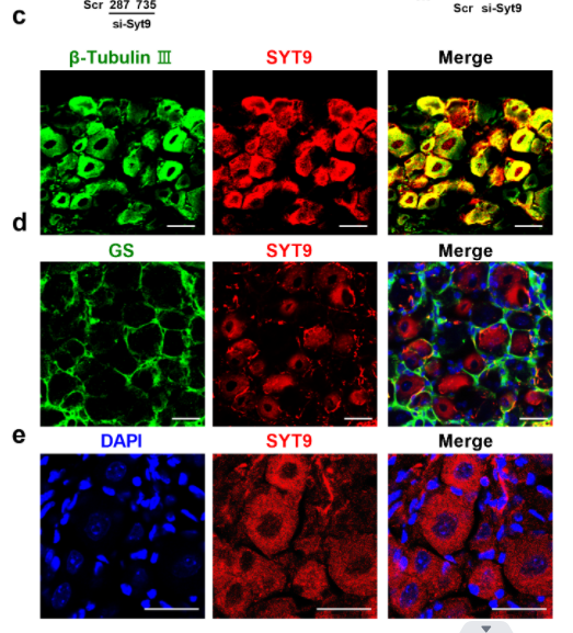
The Cytidine N-Acetyltransferase NAT10 Participates in Peripheral Nerve Injury-Induced Neuropathic Pain by Stabilizing SYT9 Expression in Primary Sensory Neurons
Author:
PMID: 36898834

应用: IF
反应种属: Mouse
发表时间: 2023 Apr
-
Citation
-
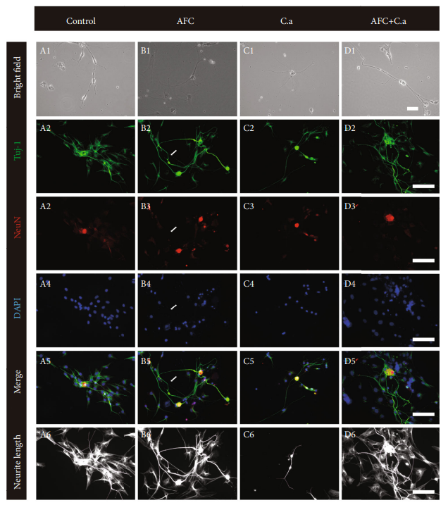
The Effect of Cutibacterium acnes Infection on Nerve Penetration in the Annulus Fibrosus of Lumbar Intervertebral Discs via Suppressing Oxidative Stress
Author: Shan, Z., Wang, X., Zong, W., Li, J., Zheng, B., Huang, B., Zhang, X., Chen, J., & Huang, Y.
PMID: 35265268

应用: IF
反应种属: Rat
发表时间: 2022 Feb
-
Citation
同靶点&同通路的产品
Beta III Tubulin Rabbit Polyclonal Antibody
Application: WB,IF-Cell,IHC-P,FC
Reactivity: Human,Mouse,Rat
Conjugate: unconjugated
Beta III Tubulin Rabbit Polyclonal Antibody
Application: WB,IF-Cell,IHC-P,FC
Reactivity: Human,Mouse,Rat
Conjugate: unconjugated
Beta III Tubulin Rabbit Polyclonal Antibody
Application: WB
Reactivity: Human,Mouse,Rat
Conjugate: unconjugated
Beta III Tubulin Recombinant Rabbit Monoclonal Antibody [SP06-00]
Application: WB,IHC-P,IP,FC,IHC-Fr,IF-Cell
Reactivity: Human,Mouse,Rat
Conjugate: unconjugated
iFluor™ 488 Conjugated Beta III Tubulin Mouse Monoclonal Antibody [A8-D10]
Application: IF-Cell,FC,IF-Tissue
Reactivity: Human,Mouse,Rat
Conjugate: iFluor™ 488


