概述
产品名称
Cytokeratin 8 Recombinant Rabbit Monoclonal Antibody [SU0338]
抗体类型
Recombinant Rabbit monoclonal Antibody
免疫原
Synthetic peptide within Human Cytokeratin 8 aa 321-370 / 483.
种属反应性
Human, Mouse
验证应用
WB, IF-Cell, IF-Tissue, IHC-P, IP, FC
分子量
Predicted band size: 54 kDa
阳性对照
Hela cell lysate, A431 cell lysate, Hela, MCF-7, A431, human liver tissue, human breast carcinoma tissue, human breast tissue, SK-Br-3, HepG2.
偶联
unconjugated
克隆号
SU0338
RRID
产品特性
形态
Liquid
浓度
1ug/ul
存放说明
Store at +4℃ after thawing. Aliquot store at -20℃. Avoid repeated freeze / thaw cycles.
存储缓冲液
1*TBS (pH7.4), 0.05% BSA, 40% Glycerol. Preservative: 0.05% Sodium Azide.
亚型
IgG
纯化方式
Protein A affinity purified.
应用稀释度
-
WB
-
1:1,000-1:2,000
-
IF-Cell
-
1:400-1:800
-
IF-Tissue
-
1:400-1:800
-
IHC-P
-
1:50-1:1,500
-
FC
-
1:500-1:1,000
-
IP
-
Use at an assay dependent concentration.
靶点
功能
Keratin, type II cytoskeletal 8 also known as cytokeratin-8 (CK-8) or keratin-8 (K8) is a keratin protein that is encoded in humans by the KRT8 gene. It is often paired with keratin 18. Antibodies to CK8 can be used to differentiate lobular carcinoma of the breast from ductal carcinoma of the breast. In normal tissue, it reacts mainly with secretory epithelia, but not with squamous epithelium, such as that found in the skin, cervix, and esophagus. However, it also reacts with a range of malignant cells, including those derived from secretory epithelia, but also some squamous carcinomata, such as spindle cell carcinoma. It is considered useful in identifying microscopic metastases of breast carcinoma in lymph nodes, and in distinguishing Paget's disease from malignant melanoma. It also reacts with neuroendocrine tumors. Keratin 8 is often used together with keratin 18 and keratin 19 to differentiate cells of epithelial origin from hematopoietic cells in tests that enumerate circulating tumor cells in blood.
背景文献
1. Ruiz A et al. Effect of hydroxychloroquine and characterization of autophagy in a mouse model of endometriosis. Cell Death Dis 7:e2059 (2016).
2. Xiao, L. et al. Three-dimensional epithelial and mesenchymal cell co-cultures form early tooth epithelium invagination-like structures: expression patterns of relevant molecules. J. Cell. Biochem. 113: 1875-1885(2012).
序列相似性
Belongs to the intermediate filament family.
组织特异性
Expressed in colon, placenta, liver and very weakly in exocervix. Increased expression observed in lymph nodes of breast carcinoma.
翻译后修饰
Phosphorylation at Ser-34 increases during mitosis. Hyperphosphorylated at Ser-53 in diseased cirrhosis liver. Phosphorylation increases by IL-6.; Proteolytically cleaved by caspases during epithelial cell apoptosis. Cleavage occurs at Asp-238 by either caspase-3, caspase-6 or caspase-7.; O-GlcNAcylation increases solubility, and decreases stability by inducing proteasomal degradation.
亚细胞定位
Nucleoplasm, Nucleus matrix, Cytoplasm.
别名
CARD2 antibody
Cell proliferation inducing gene 46 protein antibody
Cell proliferation inducing protein 46 antibody
CK 8 antibody
CK-8 antibody
CK8 antibody
CYK8 antibody
Cytokeratin 8 antibody
Cytokeratin-8 antibody
K2C8 antibody
展开CARD2 antibody
Cell proliferation inducing gene 46 protein antibody
Cell proliferation inducing protein 46 antibody
CK 8 antibody
CK-8 antibody
CK8 antibody
CYK8 antibody
Cytokeratin 8 antibody
Cytokeratin-8 antibody
K2C8 antibody
K2C8_HUMAN antibody
K8 antibody
Keratin 8 antibody
Keratin antibody
keratin type II cytoskeletal 8 antibody
Keratin-8 antibody
KRT8 antibody
type II cytoskeletal 8 antibody
Type-II keratin Kb8 antibody
折叠图片
-

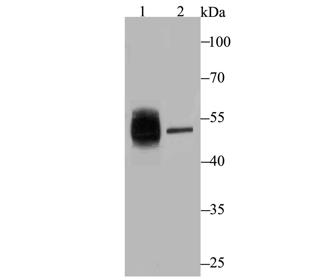
Western blot analysis of Cytokeratin 8 on different lysates with Rabbit anti-Cytokeratin 8 antibody (ET1608-32) at 1/1,000 dilution.
Lane 1: Hela cell lysate
Lane 2: A431 cell lysate
Lysates/proteins at 10 µg/Lane.
Predicted band size: 54 kDa
Observed band size: 54 kDa
Exposure time: 30 seconds;
10% SDS-PAGE gel.
Proteins were transferred to a PVDF membrane and blocked with 5% NFDM/TBST for 1 hour at room temperature. The primary antibody (ET1608-32) at 1/1,000 dilution was used in 5% NFDM/TBST at room temperature for 2 hours. Goat Anti-Rabbit IgG - HRP Secondary Antibody (HA1001) at 1:300,000 dilution was used for 1 hour at room temperature. -

☑ Knockdown (KD)
Western blot analysis of Cytokeratin 8 on different lysates with Rabbit anti-Cytokeratin 8 antibody (ET1608-32) at 1/2,000 dilution.
Lane 1: Hela-si NT cell lysate
Lane 2: Hela-si Cytokeratin 8 cell lysate
Lysates/proteins at 10 µg/Lane.
Predicted band size: 54 kDa
Observed band size: 54 kDa
Exposure time: 24 seconds;
4-20% SDS-PAGE gel.
ET1608-32 was shown to specifically react with Cytokeratin 8 in Hela-si NT cells. Weakened band was observed when Hela-si Cytokeratin 8 sample was tested. Hela-si NT and Hela-si Cytokeratin 8 samples were subjected to SDS-PAGE. Proteins were transferred to a PVDF membrane and blocked with 5% NFDM in TBST for 1 hour at room temperature. The primary antibody (ET1608-32, 1/2,000) and Loading control antibody (Rabbit anti-GAPDH, ET1601-4, 1/10,000) were used in 5% NFDM/TBST at 4℃ overnight. Goat Anti-rabbit IgG-HRP Secondary Antibody (HA1001) at 1:50,000 dilution was used for 1 hour at room temperature. -

ICC staining of Cytokeratin 8 in Hela cells (green). Formalin fixed cells were permeabilized with 0.1% Triton X-100 in TBS for 10 minutes at room temperature and blocked with 1% Blocker BSA for 15 minutes at room temperature. Cells were probed with the primary antibody (ET1608-32, 1/50) for 1 hour at room temperature, washed with PBS. Alexa Fluor®488 Goat anti-Rabbit IgG was used as the secondary antibody at 1/1,000 dilution. The nuclear counter stain is DAPI (blue).
-
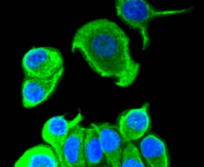
ICC staining of Cytokeratin 8 in MCF-7 cells (green). Formalin fixed cells were permeabilized with 0.1% Triton X-100 in TBS for 10 minutes at room temperature and blocked with 1% Blocker BSA for 15 minutes at room temperature. Cells were probed with the primary antibody (ET1608-32, 1/50) for 1 hour at room temperature, washed with PBS. Alexa Fluor®488 Goat anti-Rabbit IgG was used as the secondary antibody at 1/1,000 dilution. The nuclear counter stain is DAPI (blue).
-
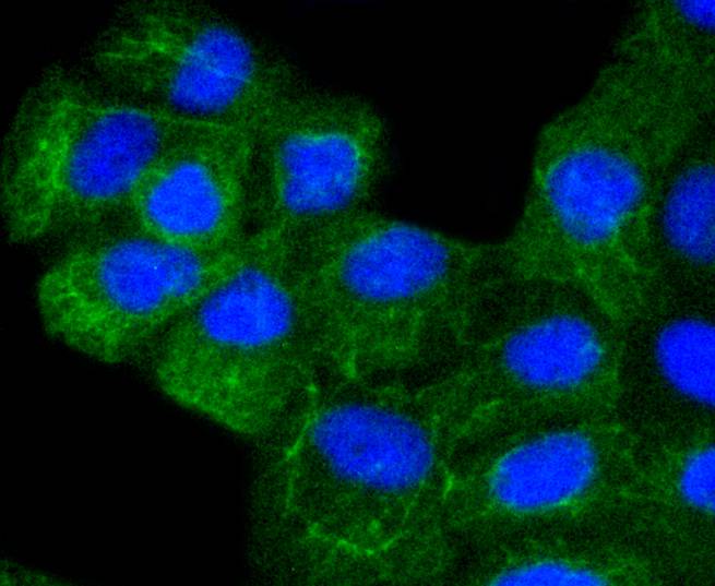
ICC staining of Cytokeratin 8 in A431 cells (green). Formalin fixed cells were permeabilized with 0.1% Triton X-100 in TBS for 10 minutes at room temperature and blocked with 1% Blocker BSA for 15 minutes at room temperature. Cells were probed with the primary antibody (ET1608-32, 1/50) for 1 hour at room temperature, washed with PBS. Alexa Fluor®488 Goat anti-Rabbit IgG was used as the secondary antibody at 1/1,000 dilution. The nuclear counter stain is DAPI (blue).
-
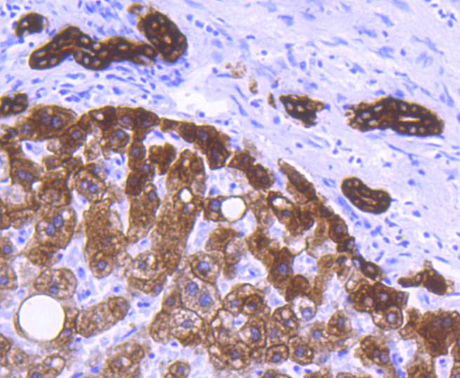
Immunohistochemical analysis of paraffin-embedded human liver tissue using anti-Cytokeratin 8 antibody. The section was pre-treated using heat mediated antigen retrieval with Tris-EDTA buffer (pH 8.0-8.4) for 20 minutes.The tissues were blocked in 5% BSA for 30 minutes at room temperature, washed with ddH2O and PBS, and then probed with the primary antibody (ET1608-32, 1/50) for 30 minutes at room temperature. The detection was performed using an HRP conjugated compact polymer system. DAB was used as the chromogen. Tissues were counterstained with hematoxylin and mounted with DPX.
-
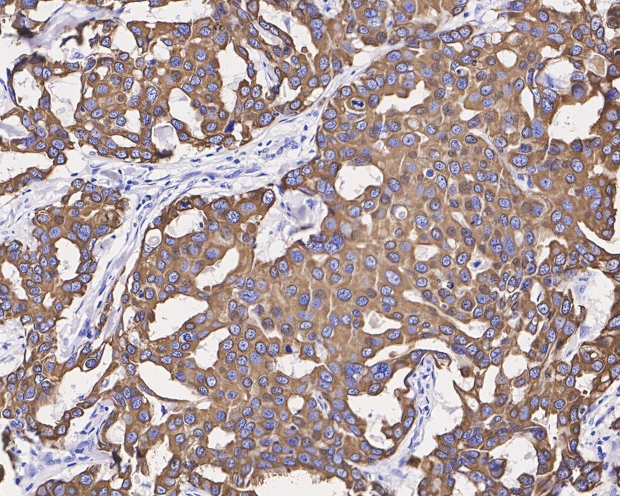
Immunohistochemical analysis of paraffin-embedded human breast carcinoma tissue with Rabbit anti-Cytokeratin 8 antibody (ET1608-32) at 1/1,500 dilution.
The section was pre-treated using heat mediated antigen retrieval with Tris-EDTA buffer (pH 9.0) for 20 minutes. The tissues were blocked in 1% BSA for 20 minutes at room temperature, washed with ddH2O and PBS, and then probed with the primary antibody (ET1608-32) at 1/1,500 dilution for 1 hour at room temperature. The detection was performed using an HRP conjugated compact polymer system. DAB was used as the chromogen. Tissues were counterstained with hematoxylin and mounted with DPX. -
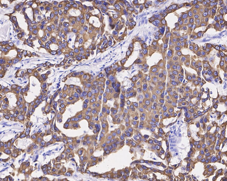
Immunohistochemical analysis of paraffin-embedded human breast carcinoma tissue with Rabbit anti-Cytokeratin 8 antibody (ET1608-32) at 1/1,500 dilution.
The section was not undergone antigen retrieval. The tissues were blocked in 1% BSA for 20 minutes at room temperature, washed with ddH2O and PBS, and then probed with the primary antibody (ET1608-32) at 1/1,500 dilution for 1 hour at room temperature. The detection was performed using an HRP conjugated compact polymer system. DAB was used as the chromogen. Tissues were counterstained with hematoxylin and mounted with DPX. -
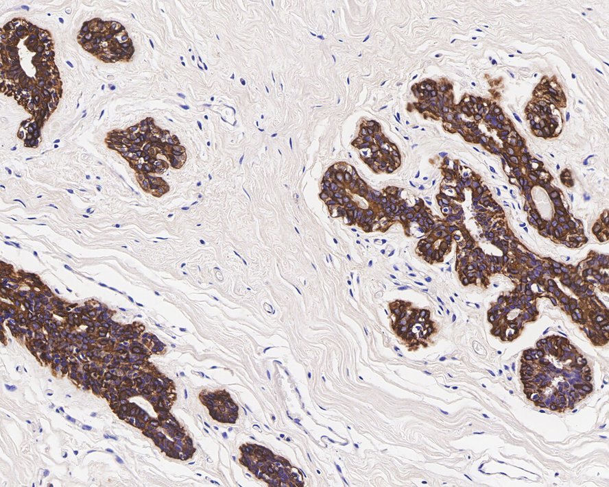
Immunohistochemical analysis of paraffin-embedded human breast tissue with Rabbit anti-Cytokeratin 8 antibody (ET1608-32) at 1/1,500 dilution.
The section was not undergone antigen retrieval. The tissues were blocked in 1% BSA for 20 minutes at room temperature, washed with ddH2O and PBS, and then probed with the primary antibody (ET1608-32) at 1/1,500 dilution for 1 hour at room temperature. The detection was performed using an HRP conjugated compact polymer system. DAB was used as the chromogen. Tissues were counterstained with hematoxylin and mounted with DPX. -

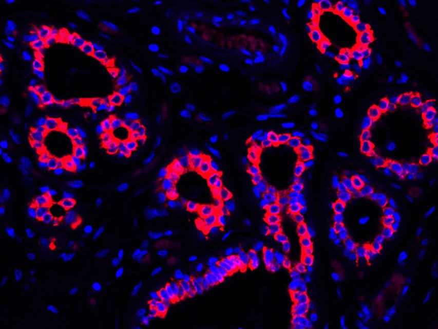
Immunofluorescence analysis of paraffin-embedded human breast tissue labeling Cytokeratin 8 (ET1608-32).
The section was pre-treated using heat mediated antigen retrieval with Tris-EDTA buffer (pH 9.0) for 20 minutes. The tissues were blocked in 10% negative goat serum for 1 hour at room temperature, washed with PBS. And then probed with the primary antibodies Cytokeratin 8 (ET1608-32, red) at 1/400 dilution at +4℃ overnight, washed with PBS.
Goat Anti-Rabbit IgG H&L (iFluor™ 594, HA1122) was used as the secondary antibodies at 1/1,000 dilution. Nuclei were counterstained with DAPI (blue). -
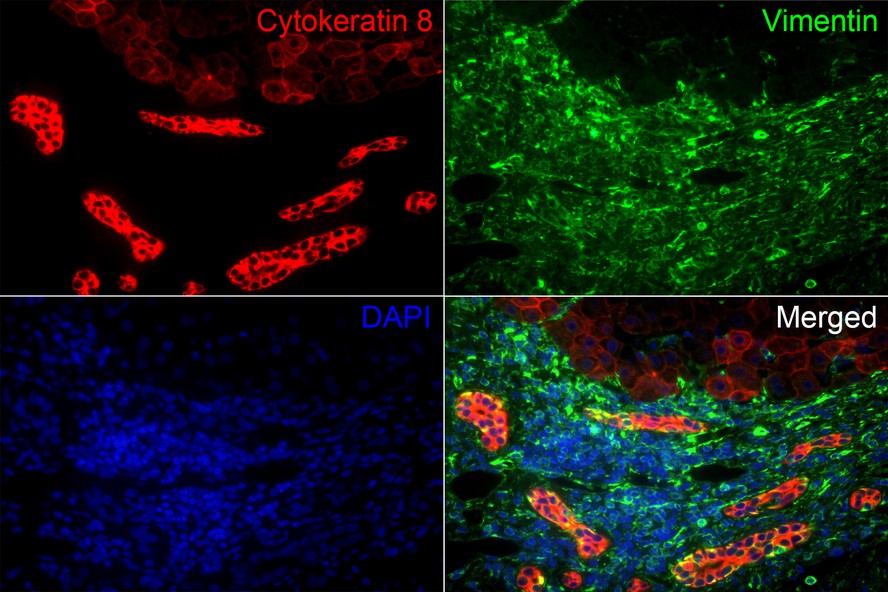
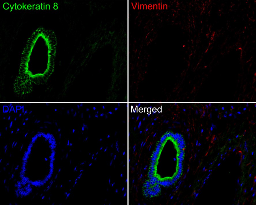
Immunofluorescence analysis of paraffin-embedded human liver tissue labeling Cytokeratin 8 (ET1608-32) and Vimentin (EM0401).
The section was pre-treated using heat mediated antigen retrieval with Tris-EDTA buffer (pH 9.0) for 20 minutes. The tissues were blocked in 10% negative goat serum for 1 hour at room temperature, washed with PBS. And then probed with the primary antibodies Cytokeratin 8 (ET1608-32, red) at 1/50 dilution and Vimentin (EM0401, green) at 1/500 dilution at +4℃ overnight, washed with PBS.
Goat Anti-Rabbit IgG H&L (iFluor™ 594, HA1122) and Goat Anti-Mouse IgG H&L (iFluor™ 488, HA1125) were used as the secondary antibodies at 1/1,000 dilution. Nuclei were counterstained with DAPI (blue). -
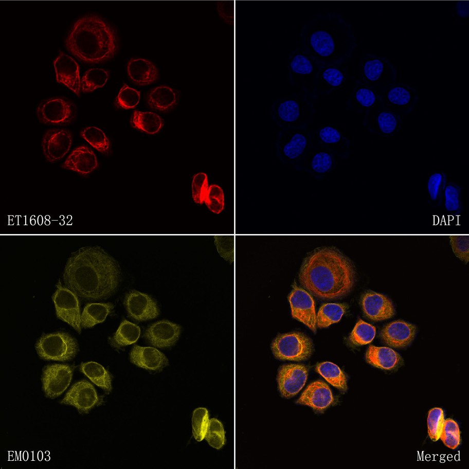
Immunocytochemistry analysis of SK-Br-3 cells labeling Cytokeratin 8 (ET1608-32).
Cells were fixed in 4% paraformaldehyde and permeabilized with 0.05% Triton X-100 in PBS for 10 minutes, and then blocked with 2% negative goat serum for 15 minutes at room temperature. Cells were probed with the primary antibody Cytokeratin 8 (ET1608-32, red) at 1/50 dilution and Beta-tubulin (EM0103, yellow) at 1/500 dilution at +4℃ overnight.
Goat Anti-Rabbit IgG H&L (iFluor™ 594, HA1122) and Goat Anti-Mouse IgG H&L (iFluor™ 488, HA1125) were used as the secondary antibodies at 1/1,000 dilution. Nuclei were counterstained with DAPI (blue). -
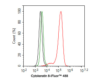
Flow cytometric analysis of HepG2 cells labeling Cytokeratin 8.
Cells were fixed and permeabilized. Then stained with the primary antibody (ET1608-32, 1ug/ml) (red) compared with Rabbit IgG Isotype Control (green). After incubation of the primary antibody at +4℃ for an hour, the cells were stained with a iFluor™ 488 conjugate-Goat anti-Rabbit IgG Secondary antibody (HA1121) at 1/1,000 dilution for 30 minutes at +4℃. Unlabelled sample was used as a control (cells without incubation with primary antibody; black). -

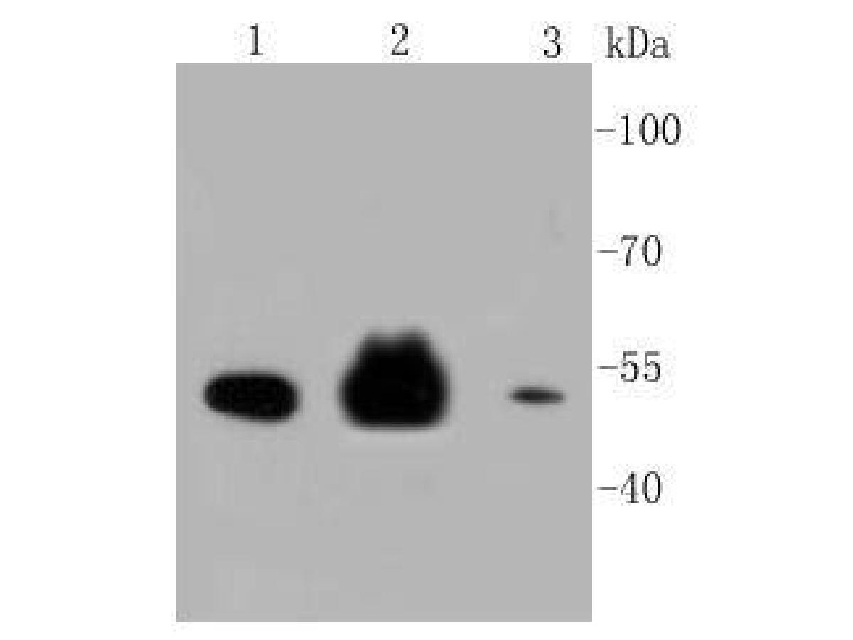
Cytokeratin 8 was immunoprecipitated in 0.2mg HeLa cell lysate with ET1608-32 at 2 µg/25 µl agarose. Western blot was performed from the immunoprecipitate using ET1608-32 at 1/2,000 dilution. Anti-Rabbit IgG for IP Nano-secondary antibody (NBI01H) at 1/5,000 dilution was used for 1 hour at room temperature.
Lane 1: HeLa cell lysate (input)
Lane 2: Rabbit IgG instead of ET1608-32 in HeLa cell lysate
Lane 3: ET1608-32 IP in HeLa cell lysate
Blocking/Dilution buffer: 5% NFDM/TBST
Exposure time: 43 seconds -
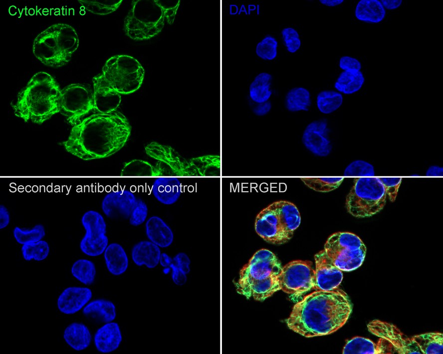
Immunocytochemistry analysis of HepG2 cells labeling Cytokeratin 8 with Rabbit anti-Cytokeratin 8 antibody (ET1608-32) at 1/100 dilution.
Cells were fixed in 4% paraformaldehyde for 20 minutes at room temperature, permeabilized with 0.1% Triton X-100 in PBS for 5 minutes at room temperature, then blocked with 1% BSA in 10% negative goat serum for 1 hour at room temperature. Cells were then incubated with Rabbit anti-Cytokeratin 8 antibody (ET1608-32) at 1/100 dilution in 1% BSA in PBST overnight at 4 ℃. Goat Anti-Rabbit IgG H&L (iFluor™ 488, HA1121) was used as the secondary antibody at 1/1,000 dilution. PBS instead of the primary antibody was used as the secondary antibody only control. Nuclear DNA was labelled in blue with DAPI.
Beta tubulin (M1305-2, red) was stained at 1/100 dilution overnight at +4℃. Goat Anti-Mouse IgG H&L (iFluor™ 594, HA1126) was used as the secondary antibody at 1/1,000 dilution.
Please note: All products are "FOR RESEARCH USE ONLY AND ARE NOT INTENDED FOR DIAGNOSTIC OR THERAPEUTIC USE"
同靶点&同通路的产品
iFluor™ 594 Conjugated Cytokeratin 8 Recombinant Rabbit Monoclonal Antibody [SU0338]
Application: IF-Cell,IF-Tissue,FC
Reactivity: Human,Mouse
Conjugate: iFluor™ 594
Cytokeratin 8 Rabbit Polyclonal Antibody
Application: WB,IF-Cell,IHC-P,FC
Reactivity: Human,Mouse,Rat
Conjugate: unconjugated
Cytokeratin 8 Rabbit Polyclonal Antibody
Application: WB,IF-Cell,IHC-P,FC
Reactivity: Human,Mouse,Rat
Conjugate: unconjugated
Cytokeratin 8
Application:
Reactivity:
Conjugate:
Cytokeratin 8 Mouse Monoclonal Antibody [A1-B11]
Application: WB,IF-Cell,IHC-P,FC
Reactivity: Human,Mouse,Rat
Conjugate: unconjugated
iFluor™ 488 Conjugated Cytokeratin 8 Recombinant Rabbit Monoclonal Antibody [SU0338]
Application: IF-Tissue,IF-Cell
Reactivity: Human
Conjugate: iFluor™ 488
iFluor™ 647 Conjugated Cytokeratin 8 Recombinant Rabbit Monoclonal Antibody [SU0338]
Application: IF-Cell,IF-Tissue
Reactivity: Human,Mouse
Conjugate: iFluor™ 647
Cytokeratin 8 Rabbit Polyclonal Antibody
Application: WB,IF-Cell,IHC-P,FC
Reactivity: Human
Conjugate: unconjugated


