ZO1 Recombinant Rabbit Monoclonal Antibody [PSH07-09]


Catalog# HA722797
ZO1 Recombinant Rabbit Monoclonal Antibody [PSH07-09]
-
WB
-
IF-Cell
-
mIHC
-
Human
-
Mouse
-
Monkey
-
HA751131
不含抗保成分
-
Cynomolgus monkey
-
unconjugated
概述
产品名称
ZO1 Recombinant Rabbit Monoclonal Antibody [PSH07-09]
抗体类型
Recombinant Rabbit monoclonal Antibody
免疫原
Recombinant protein within human ZO1 aa 1,401-1,748.
种属反应性
Human, Mouse, Monkey (Predicted: Cynomolgus monkey)
验证应用
WB, IF-Cell, mIHC
分子量
Predicted band size: 195 kDa
阳性对照
A431 cell lysate, 293T cell lysate, HepG2 cell lysate, HeLa cell lysate, U-2 OS cell lysate, NIH/3T3 cell lysate, C2C12 cell lysate, COS-1 cell lysate, Mouse testis tissue lysate, Rat testis tissue lysate, MCF7.
偶联
unconjugated
克隆号
PSH07-09
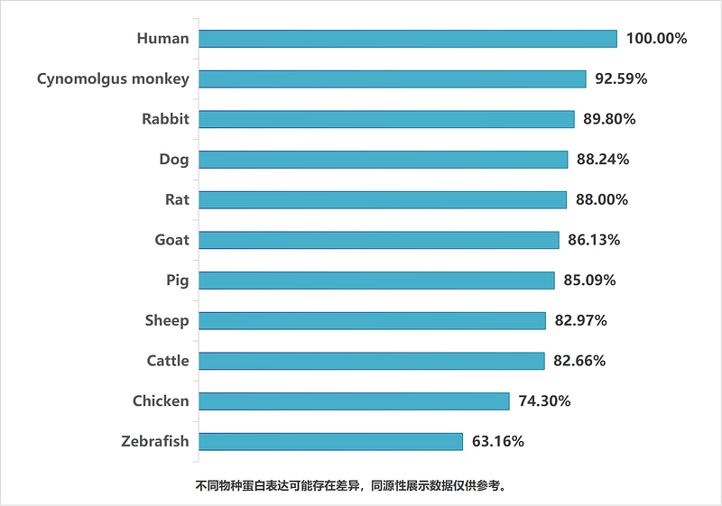
同源性数据
产品特性
形态
Liquid
浓度
存放说明
Shipped at 4℃. Store at +4℃ short term (1-2 weeks). It is recommended to aliquot into single-use upon delivery. Store at -20℃ long term.
存储缓冲液
PBS (pH7.4), 0.1% BSA, 40% Glycerol. Preservative: 0.05% Sodium Azide.
亚型
IgG
纯化方式
Protein A affinity purified.
应用稀释度
-
WB
-
1:1,000
-
IF-Cell
-
1:500-1:2,000
-
mIHC
-
1:100
靶点
功能
Zonula occludens-1 ZO-1, also known as Tight junction protein-1 is a 220-kD peripheral membrane protein that is encoded by the TJP1 gene in humans. It belongs to the family of zonula occludens proteins (ZO-1, ZO-2, and ZO-3), which are tight junction-associated proteins and of which, ZO-1 is the first to be cloned. It was first isolated in 1986 by Stevenson and Goodenough using a monoclonal antibody raised in rodent liver to recognise a 225-kD polypeptide in whole liver homogenates and in tight junction-enriched membrane fractions. It has a role as a scaffold protein which cross-links and anchors Tight Junction (TJ) strand proteins, which are fibril-like structures within the lipid bilayer, to the actin cytoskeleton. This gene encodes a protein located on a cytoplasmic membrane surface of intercellular tight junctions. The encoded protein may be involved in signal transduction at cell–cell junctions. Two transcript variants encoding distinct isoforms have been identified for this gene.
背景文献
1. Tsurudome Y et al. Decreased ZO1 expression causes loss of time-dependent tight junction function in the liver of ob/ob mice. Mol Biol Rep. 2022 Dec
2. Han F et al. GLTSCR1 coordinates alternative splicing and transcription elongation of ZO1 to regulate colorectal cancer progression. J Mol Cell Biol. 2022 Jun
亚细胞定位
Cell membrane, Cell junction, tight junction, gap junction, Cell projection, podosome.
UNIPROT
别名
Tight junction protein 1 antibody
Tight junction protein ZO-1 antibody
Tight junction protein ZO1 antibody
TJP1 antibody
zo-1 antibody
Zo1 antibody
ZO1_HUMAN antibody
Zona occludens 1 antibody
Zona occludens 1 protein antibody
Zona occludens protein 1 antibody
展开图片
-
Western blot analysis of ZO1 on different lysates with Rabbit anti-ZO1 antibody (HA722797) at 1/1,000 dilution.
Lane 1: A431 cell lysate (20 µg/Lane)
Lane 2: 293T cell lysate (20 µg/Lane)
Lane 3: HepG2 cell lysate (20 µg/Lane)
Lane 4: HeLa cell lysate (20 µg/Lane)
Lane 5: U-2 OS cell lysate (20 µg/Lane)
Lane 6: NIH/3T3 cell lysate (20 µg/Lane)
Lane 7: C2C12 cell lysate (20 µg/Lane)
Lane 8: COS-1 cell lysate (20 µg/Lane)
Lane 9: Mouse testis tissue lysate (40 µg/Lane)
Lane 10: Rat testis tissue lysate (40 µg/Lane)
Predicted band size: 195 kDa
Observed band size: 250 kDa
Exposure time: 3 minutes; ECL: K1802;
4-20% SDS-PAGE gel.
Proteins were transferred to a PVDF membrane and blocked with 5% NFDM/TBST for 1 hour at room temperature. The primary antibody (HA722797) at 1/1,000 dilution was used in 5% NFDM/TBST at 4℃ overnight. Goat Anti-Rabbit IgG - HRP Secondary Antibody (HA1001) at 1/50,000 dilution was used for 1 hour at room temperature. -
Immunocytochemistry analysis of MCF7 cells labeling ZO1 with Rabbit anti-ZO1 antibody (HA722797) at 1/2,000 dilution.
Cells were fixed in 4% paraformaldehyde for 20 minutes at room temperature, permeabilized with 0.1% Triton X-100 in PBS for 5 minutes at room temperature, then blocked with 1% BSA in 10% negative goat serum for 1 hour at room temperature. Cells were then incubated with Rabbit anti-ZO1 antibody (HA722797) at 1/2,000 dilution in 1% BSA in PBST overnight at 4 ℃. Goat Anti-Rabbit IgG H&L (iFluor™ 488, HA1121) was used as the secondary antibody at 1/1,000 dilution. PBS instead of the primary antibody was used as the secondary antibody only control. Nuclear DNA was labelled in blue with DAPI.
Beta tubulin (M1305-2, red) was stained at 1/100 dilution overnight at +4℃. Goat Anti-Mouse IgG H&L (iFluor™ 594, HA1126) was used as the secondary antibody at 1/1,000 dilution.
请注意: All products are "FOR RESEARCH USE ONLY AND ARE NOT INTENDED FOR DIAGNOSTIC OR THERAPEUTIC USE"
引文
-
Dietary goat milk extracellular vesicles remodel the microbiota–gut–brain axis to alleviate adolescent anxiety
期刊: Food Research International
DOI: 10.1016/j.foodres.2026.118537
IF: 8
应用: WB
反应种属: Mouse
发表时间: 2026 Feb
-
Assessment of microplastic toxicity on blood-testis barrier using 3D cell spheroids
期刊: Ecotoxicology And Environmental Safety
DOI: 10.1016/j.ecoenv.2025.119062
IF: 6.1
应用: IF
反应种属: Mouse
发表时间: 2025 Sept
-
Metallothionein 2A alleviates ulcerative colitis by inhibiting ferroptosis in intestinal epithelial cells with Tfrc downregulation
期刊: Journal Of Trace Elements In Medicine And Biology
DOI: 10.1016/j.jtemb.2025.127746
IF: 3.3
应用: WB
反应种属: Human
发表时间: 2025 Sept
-
Atractylenolide III Alleviates Inflammation in Cerebral Ischemia/Reperfusion Injury by Modulating the PI3K/Akt/NF-κB Signaling Pathway
期刊: Journal Of Ethnopharmacology
DOI: 10.1016/j.jep.2025.120644
IF: 5.4
应用: IHC,WB
反应种属: Rat
发表时间: 2025 Sept
-
Study on the unique effects of Wubi Shanyao Pills in improving postmenopausal osteoporosis via the “gut-bone” axis
期刊: Phytomedicine
DOI: 10.1016/j.phymed.2025.157447
IF: 8.3
应用: IHC,WB
反应种属: Mouse
发表时间: 2025 Oct
-
Nicotinamide mononucleotide rescues Di-n-butyl phthalate induced blood-brain barrier damage via NAD+/Sirt1/FOXO1a pathway activation
期刊: Ecotoxicology And Environmental Safety
DOI: 10.1016/j.ecoenv.2025.119290
IF: 6.1
应用: WB
反应种属: Mouse,Human
发表时间: 2025 Oct
-
Rhubarb polysaccharides alleviate hypertriglyceridemia-induced acute pancreatitis and regulate gut microbiota-mediated tryptophan metabolism
期刊: Journal Of Functional Foods
DOI: 10.1016/j.jff.2025.107104
IF: 4
应用: IHC
反应种属: Mouse
发表时间: 2025 Nov
-
Atractylenolide III Ameliorates Ulcerative Colitis By Targeting IL-17RA to Suppress Macrophage M1 Polarization
期刊: Journal Of Agricultural And Food Chemistry
DOI: 10.1021/acs.jafc.5c09397
IF: 6.2
应用: WB
反应种属: Mouse
发表时间: 2025 Nov
-
Activin A exacerbates neonatal necrotizing enterocolitis via ALK4-mediated apoptosis and barrier disruption
期刊: International Immunopharmacology
DOI: 10.1016/j.intimp.2025.115043
IF: 4.7
应用: IHC
反应种属: Mouse
发表时间: 2025 Jun
-
Astragalus membranaceus extract attenuates ulcerative colitis by integrating multiomics and the PI3K/AKT signaling pathway
期刊: Frontiers In Pharmacology
DOI: 10.3389/fphar.2025.1585748
IF: 4.8
应用: WB,IF
反应种属: Mouse
发表时间: 2025 Jun
-
Betulinic Acid Reduces Intestinal Inflammation and Enhances Intestinal Tight Junctions by Modulating the PPAR-γ/NF-κB Signaling Pathway in Intestinal Cells and Organoids
期刊: Nutrients
DOI: 10.3390/nu17132052
IF: 5
应用: IF,WB
反应种属: Mouse,Human
发表时间: 2025 Jun
-
Probiotic efficacy of Cetobacterium somerae (CGMCC No. 28843): promoting intestinal digestion, absorption, and structural integrity in juvenile grass carp (Ctenopharyngodon idella)
期刊: Journal of Animal Science and Biotechnology
DOI: 10.1186/s40104-025-01224-7
IF: 6.5
应用: IF
反应种属: Fish
发表时间: 2025 Jul
-
Sanzi Yangqin Decoction improved acute lung injury by regulating the TLR2-mediated NF-κB/NLRP3 signaling pathway and inhibiting the activation of NLRP3 inflammasome
期刊: Phytomedicine
DOI: 10.1016/j.phymed.2025.156438
IF: 8.3
应用: WB
反应种属: Mouse
发表时间: 2025 Jan
-
Sanzi Yangqin Decoction improved acute lung injury by regulating the TLR2-mediated NF-κB/NLRP3 signaling pathway and inhibiting the activation of NLRP3 inflammasome
期刊: Phytomedicine
DOI:
IF: 6.7
应用: WB
反应种属: Mouse
发表时间: 2025 Jan
-
Mesenchymal Stem Cells Armed with DNA Nanorobots as a Modality for Combination Therapy of Inflammatory Bowel Disease
期刊: ACS Applied Materials & Interfaces
DOI: 10.1021/acsami.4c08923
IF: 8.3
应用: WB
反应种属: Mouse
发表时间: 2025 Feb
-
Clostridium perfringens can promote the formation of fatty liver in cows
期刊: Veterinary Microbiology
DOI: 10.1016/j.vetmic.2025.110826
IF: 2.7
应用: WB
反应种属: Mouse
发表时间: 2025 Dec
-
Polystyrene nanoplastics-induced methuosis in brain microvascular endothelial cells: Rescue via ESCRT membrane repair system
期刊: Ecotoxicology And Environmental Safety
DOI: 10.1016/j.ecoenv.2025.118800
IF: 6.1
应用: IF
反应种属: Mouse
发表时间: 2025 Aug
 浙公网安备 33019202000643号