概述
产品名称
SIRT1 Rabbit Polyclonal Antibody
抗体类型
Rabbit Polyclonal Antibody
免疫原
Synthetic peptide within Human SIRT1 aa 698-747 / 747.
种属反应性
Human, Mouse
验证应用
WB, IHC-P, FC, IF-Cell
分子量
Predicted band size: 82 kDa
阳性对照
Hela cell lysate, Jurkat cell lysate, F9 cell lysate, HeLa, F9, human colon carcinoma tissue, human lung carcinoma tissue, mouse liver tissue, mouse testis tissue.
偶联
unconjugated
RRID
产品特性
形态
Liquid
存放说明
Shipped at 4℃. Store at +4℃ short term (1-2 weeks). It is recommended to aliquot into single-use upon delivery. Store at -20℃ long term.
存储缓冲液
1*PBS (pH7.4), 0.2% BSA, 40% Glycerol. Preservative: 0.05% Sodium Azide.
亚型
IgG
纯化方式
Immunogen affinity purified.
应用稀释度
-
WB
-
1:1,000-1:2,000
-
IHC-P
-
1:200
-
FC
-
1:1,000
-
IF-Cell
-
1:100
发表文章中的应用
| WB | 查看 8 篇文献如下 |
| IF | 查看 2 篇文献如下 |
| IHC-P | 查看 2 篇文献如下 |
发表文章中的种属
| Mouse | 查看 7 篇文献如下 |
| Human | 查看 2 篇文献如下 |
靶点
功能
SirT1, the mammalian ortholog of Sir2, is a nuclear protein implicated in the regulation of many cellular processes, including apoptosis, cellular senescence, endocrine signaling, glucose homeostasis, aging, and longevity. Targets of SirT1 include acetylated p53, p300, Ku70, forkhead (FoxO) transcription factors, PPARγ, and the PPARγ coactivator-1α (PGC-1α) protein. Deacetylation of p53 and FoxO transcription factors represses apoptosis and increases cell survival. SirT1 deacetylase activity is inhibited by nicotinamide and activated by resveratrol.
背景文献
1. "Human Sir2-related protein SIRT1 associates with the bHLH repressors HES1 and HEY2 and is involved in HES1- and HEY2-mediated transcriptional repression." Takata T., Ishikawa F. Biochem. Biophys. Res. Commun. 301:250-257(2003)
2. "Inhibition of silencing and accelerated aging by nicotinamide, a putative negative regulator of yeast sir2 and human SIRT1." Bitterman K.J., Anderson R.M., Cohen H.Y., Latorre-Esteves M., Sinclair D.A. J. Biol. Chem. 277:45099-45107(2002)
3. "Human SirT1 interacts with histone H1 and promotes formation of facultative heterochromatin." Vaquero A., Scher M., Lee D., Erdjument-Bromage H., Tempst P., Reinberg D.Mol. Cell 16:93-105(2004)
4. "Human immunodeficiency virus type 1 Tat protein inhibits the SIRT1 deacetylase and induces T cell hyperactivation." Kwon H.S., Brent M.M., Getachew R., Jayakumar P., Chen L.F., Schnolzer M., McBurney M.W., Marmorstein R., Greene W.C., Ott M. Cell Host Microbe 3:158-167(2008)
序列相似性
Belongs to the sirtuin family. Class I subfamily.
组织特异性
Widely expressed.
翻译后修饰
Methylated on multiple lysine residues; methylation is enhanced after DNA damage and is dispensable for deacetylase activity toward p53/TP53.; Phosphorylated. Phosphorylated by STK4/MST1, resulting in inhibition of SIRT1-mediated p53/TP53 deacetylation. Phosphorylation by MAPK8/JNK1 at Ser-27, Ser-47, and Thr-530 leads to increased nuclear localization and enzymatic activity. Phosphorylation at Thr-530 by DYRK1A and DYRK3 activates deacetylase activity and promotes cell survival. Phosphorylation by mammalian target of rapamycin complex 1 (mTORC1) at Ser-47 inhibits deacetylation activity. Phosphorylated by CaMK2, leading to increased p53/TP53 and NF-kappa-B p65/RELA deacetylation activity (By similarity). Phosphorylation at Ser-27 implicating MAPK9 is linked to protein stability. There is some ambiguity for some phosphosites: Ser-159/Ser-162 and Thr-544/Ser-545.; Proteolytically cleaved by cathepsin B upon TNF-alpha treatment to yield catalytic inactive but stable SirtT1 75 kDa fragment (75SirT1).; S-nitrosylated by GAPDH, leading to inhibit the NAD-dependent protein deacetylase activity.; Acetylated at various Lys residues. Deacetylated via an autocatalytic mechanism. Autodeacetylation at Lys-238 promotes its protein deacetylase activity.
亚细胞定位
Nucleus, cytoplasm, Mitochondrion.
UNIPROT
别名
75SirT1 antibody
hSIR2 antibody
hSIRT1 antibody
HST2, S. cerevisiae, homolog of antibody
NAD dependent deacetylase sirtuin 1 antibody
NAD dependent protein deacetylase sirtuin 1 antibody
OTTHUMP00000198111 antibody
OTTHUMP00000198112 antibody
Regulatory protein SIR2 homolog 1 antibody
SIR1_HUMAN antibody
展开75SirT1 antibody
hSIR2 antibody
hSIRT1 antibody
HST2, S. cerevisiae, homolog of antibody
NAD dependent deacetylase sirtuin 1 antibody
NAD dependent protein deacetylase sirtuin 1 antibody
OTTHUMP00000198111 antibody
OTTHUMP00000198112 antibody
Regulatory protein SIR2 homolog 1 antibody
SIR1_HUMAN antibody
SIR2 antibody
SIR2 like 1 antibody
SIR2 like protein 1 antibody
SIR2, S.cerevisiae, homolog-like 1 antibody
SIR2-like protein 1 antibody
SIR2ALPHA antibody
SIR2L1 antibody
Sirt1 antibody
SirtT1 75 kDa fragment antibody
Sirtuin (silent mating type information regulation 2 homolog) 1 (S. cerevisiae) antibody
Sirtuin 1 antibody
Sirtuin type 1 antibody
折叠图片
-

Western blot analysis of SIRT1 on different lysates with Rabbit anti-SIRT1 antibody (ER130811) at 1/500 dilution.
Lane 1: Hela cell lysate
Lane 2: Jurkat cell lysate
Lysates/proteins at 10 µg/Lane.
Predicted band size: 82 kDa
Observed band size: 110 kDa
Exposure time: 2 minutes;
6% SDS-PAGE gel.
Proteins were transferred to a PVDF membrane and blocked with 5% NFDM/TBST for 1 hour at room temperature. The primary antibody (ER130811) at 1/500 dilution was used in 5% NFDM/TBST at room temperature for 2 hours. Goat Anti-Rabbit IgG - HRP Secondary Antibody (HA1001) at 1:300,000 dilution was used for 1 hour at room temperature. -

Western blot analysis of SIRT1 on F9 cell lysates with Rabbit anti-SIRT1 antibody (ER130811) at 1/2,000 dilution.
Lysates/proteins at 10 µg/Lane.
Predicted band size: 82 kDa
Observed band size: 110 kDa
Exposure time: 1 minute;
8% SDS-PAGE gel.
Proteins were transferred to a PVDF membrane and blocked with 5% NFDM/TBST for 1 hour at room temperature. The primary antibody (ER130811) at 1/2,000 dilution was used in 5% NFDM/TBST at room temperature for 2 hours. Goat Anti-Rabbit IgG - HRP Secondary Antibody (HA1001) at 1:300,000 dilution was used for 1 hour at room temperature. -

Immunocytochemistry analysis of HeLa cells labeling SIRT1 with Rabbit anti-SIRT1 antibody (ER130811) at 1/100 dilution.
Cells were fixed in 4% paraformaldehyde for 20 minutes at room temperature, permeabilized with 0.1% Triton X-100 in PBS for 5 minutes at room temperature, then blocked with 1% BSA in 10% negative goat serum for 1 hour at room temperature. Cells were then incubated with Rabbit anti-SIRT1 antibody (ER130811) at 1/100 dilution in 1% BSA in PBST overnight at 4 ℃. Goat Anti-Rabbit IgG H&L (iFluor™ 488, HA1121) was used as the secondary antibody at 1/1,000 dilution. PBS instead of the primary antibody was used as the secondary antibody only control. Nuclear DNA was labelled in blue with DAPI.
Beta tubulin (M1305-2, red) was stained at 1/100 dilution overnight at +4℃. Goat Anti-Mouse IgG H&L (iFluor™ 594, HA1126) was used as the secondary antibody at 1/1,000 dilution. -

Immunocytochemistry analysis of F9 cells labeling SIRT1 with Rabbit anti-SIRT1 antibody (ER130811) at 1/100 dilution.
Cells were fixed in 4% paraformaldehyde for 20 minutes at room temperature, permeabilized with 0.1% Triton X-100 in PBS for 5 minutes at room temperature, then blocked with 1% BSA in 10% negative goat serum for 1 hour at room temperature. Cells were then incubated with Rabbit anti-SIRT1 antibody (ER130811) at 1/100 dilution in 1% BSA in PBST overnight at 4 ℃. Goat Anti-Rabbit IgG H&L (iFluor™ 488, HA1121) was used as the secondary antibody at 1/1,000 dilution. PBS instead of the primary antibody was used as the secondary antibody only control. Nuclear DNA was labelled in blue with DAPI.
Beta tubulin (M1305-2, red) was stained at 1/100 dilution overnight at +4℃. Goat Anti-Mouse IgG H&L (iFluor™ 594, HA1126) was used as the secondary antibody at 1/1,000 dilution. -

Immunohistochemical analysis of paraffin-embedded human colon carcinoma tissue using anti-SIRT1 antibody. Counter stained with hematoxylin.
-

Immunohistochemical analysis of paraffin-embedded human lung carcinoma tissue using anti-SIRT1 antibody. Counter stained with hematoxylin.
-

Immunohistochemical analysis of paraffin-embedded mouse liver tissue with Rabbit anti-SIRT1 antibody (ER130811) at 1/200 dilution.
The section was pre-treated using heat mediated antigen retrieval with sodium citrate buffer (pH 6.0) (high pressure) for 2 minutes. The tissues were blocked in 1% BSA for 20 minutes at room temperature, washed with ddH2O and PBS, and then probed with the primary antibody (ER130811) at 1/200 dilution for 1 hour at room temperature. The detection was performed using an HRP conjugated compact polymer system. DAB was used as the chromogen. Tissues were counterstained with hematoxylin and mounted with DPX. -

Immunohistochemical analysis of paraffin-embedded mouse testis tissue with Rabbit anti-SIRT1 antibody (ER130811) at 1/200 dilution.
The section was pre-treated using heat mediated antigen retrieval with sodium citrate buffer (pH 6.0) (high pressure) for 2 minutes. The tissues were blocked in 1% BSA for 20 minutes at room temperature, washed with ddH2O and PBS, and then probed with the primary antibody (ER130811) at 1/200 dilution for 1 hour at room temperature. The detection was performed using an HRP conjugated compact polymer system. DAB was used as the chromogen. Tissues were counterstained with hematoxylin and mounted with DPX. -

Flow cytometric analysis of F9 cells labeling SIRT1.
Cells were fixed and permeabilized. Then stained with the primary antibody (ER130811, 1/1,000) (red) compared with Rabbit IgG Isotype Control (green). After incubation of the primary antibody at +4℃ for an hour, the cells were stained with a iFluor™ 488 conjugate-Goat anti-Rabbit IgG Secondary antibody (HA1121) at 1/1,000 dilution for 30 minutes at +4℃. Unlabelled sample was used as a control (cells without incubation with primary antibody; black). -

☑ Knockdown (KD)
Western blot analysis of SIRT1 on different lysates with Rabbit anti-SIRT1 antibody (ER130811) at 1/20,000 dilution.
Lane 1: HEK-293-si NT cell lysate
Lane 2: HEK-293-si SIRT1 cell lysate
Lysates/proteins at 10 µg/Lane.
Predicted band size: 82 kDa
Observed band size: 110 kDa
Exposure time: 50 seconds;
4-20% SDS-PAGE gel.
Proteins were transferred to a PVDF membrane and blocked with 5% NFDM/TBST for 1 hour at room temperature. The primary antibody (ER130811) at 1/20,000 dilution was used in 5% NFDM/TBST at 4℃ overnight. Goat Anti-Rabbit IgG - HRP Secondary Antibody (HA1001) at 1/100,000 dilution was used for 1 hour at room temperature.
请注意: All products are "FOR RESEARCH USE ONLY AND ARE NOT INTENDED FOR DIAGNOSTIC OR THERAPEUTIC USE"
引文
-
Akkermansia muciniphila ameliorates olanzapine-induced metabolic dysfunction-associated steatotic liver disease via PGRMC1/SIRT1/FOXO1 signaling pathway
Author: Hui Chen, Ting Cao, ChenQuan Lin, ShiMeng Jiao, YiFang He, ZhenYu Zhu, QiuJin Guo, RenRong Wu, HuaLin Cai, BiKui Zhang
PMID: 40176900
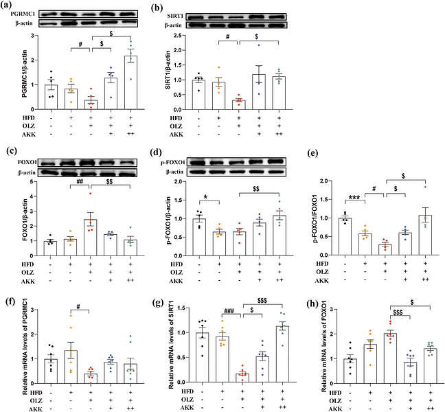
期刊: Frontiers In Pharmacology
应用: WB
反应种属: Mouse
发表时间: 2025 Mar
-
Citation
-
Salidroside alleviates bone cancer pain by inhibiting Th17/Treg imbalance through the AMPK/SIRT1 pathway
Author: Kesong Zheng, Chengwei Yang, Mingming Han, Fang Kang, Juan Li
PMID: 40609386
期刊: Phytomedicine
应用: WB
反应种属: Mouse
发表时间: 2025 Jun
-
Citation
-
ASIC1a Promotes nucleus pulposus derived stem cells apoptosis through modulation of SIRT3-dependent mitochondrial redox homeostasis in intervertebral disc degeneration
Author: Zhi-Gang Zhang, Liang Kang, Lu-Ping Zhou, Yan-Xin Wang, Chong-Yu Jia, Chen-Hao Zhao, Bo Zhang, Jia-Qi Wang, Hua-Qing Zhang, Ren-Jie Zhang, Cai-Liang Shen
PMID: 40625233
期刊: Redox Report
应用: WB
反应种属: Human
发表时间: 2025 Jul
-
Citation
-
Unconjugated bilirubin promotes uric acid restoration by activating hepatic AMPK pathway
Author: Yingqiong Zhang, Yujia Chen, Xiaojing Chen, Yue Gao, Jun Luo, Shuanghui Lu, Qi Li, Ping Li, Mengru Bai, Ting Jiang, Nanxin Zhang, Bichen Zhang, Binxin Chen, Hui Zhou, Huidi Jiang, Nengming Lin
PMID: 39299526
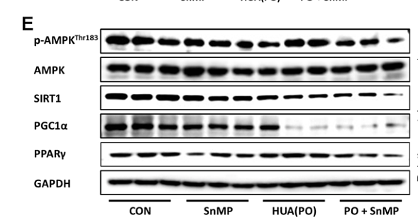
期刊: Free Radical Biology And Medicine
应用: WB
反应种属: Mouse
发表时间: 2024 Sept
-
Citation
-
Mangiferin and EGCG Compounds Fight Against Hyperlipidemia by Promoting FFA Oxidation via AMPK/PPARα
Author: Yahui Xu, Jie Zhang, Ting Zhang, Minghui Zi, Qiao Zhang
PMID: 39735726

期刊: Peroxisome Proliferator-Activated Receptor Research
应用:
反应种属:
发表时间: 2024 Dec
-
Citation
-
Exercise improved bone health in aging mice: a role of SIRT1 in regulating autophagy and osteogenic differentiation of BMSCs
Author:
PMID: 37476496
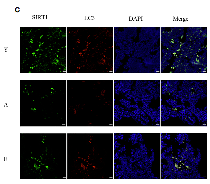
期刊: Frontiers In Endocrinology
应用: IF
反应种属: Mouse
发表时间: 2023 Jul
-
Citation
-
miR-146a impedes the anti-aging effect of AMPK via NAMPT suppression and NAD+/SIRT inactivation
Author: Gong, H., Chen, H., Xiao, P., Huang, N., Han, X., Zhang, J., Yang, Y., Li, T., Zhao, T., Tai, H., Xu, W., Zhang, G., Gong, C., Yang, M., Tang, X., & Xiao, H.
PMID: 35241643
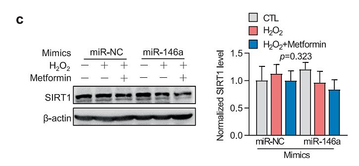
期刊: Signal Transduction And Targeted Therapy
应用: WB
反应种属: Mouse
发表时间: 2022 Mar
-
Citation
-
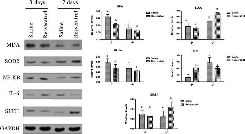
Effect of resveratrol on mouse ovarian vitrification and transplantation. Reproductive biology and endocrinology : RB&E, 19(1), 54.
Author: Wang, D., Geng, M., Gan, D., Han, G., Gao, G., Xing, A., Cui, Y., & Hu, Y.
PMID: 33836793
期刊:
应用: WB,IHC-P
反应种属: Mouse
发表时间: 2021 Apr
-
Citation
-
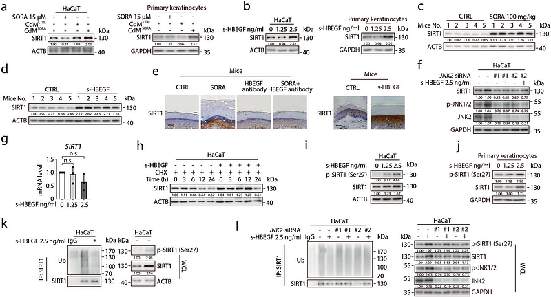
s-HBEGF/SIRT1 circuit-dictated crosstalk between vascular endothelial cells and keratinocytes mediates sorafenib-induced hand-foot skin reaction that can be reversed by nicotinamide. Cell research, 30(9), 779–793.
Author: Luo, P., Yan, H., Chen, X., Zhang, Y., Zhao, Z., Cao, J., Zhu, Y., Du, J., Xu, Z., Zhang, X., Zeng, S., Yang, B., Ma, S., & He, Q.
PMID: 32296111
期刊: Cell Research
应用: WB,IF,IHC-P
反应种属: Human
发表时间: 2020 Sep
-
Citation
-
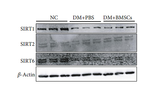
Mesenchymal Stem Cells Attenuate Diabetic Lung Fibrosis via Adjusting Sirt3-Mediated Stress Responses in Rats
Author: Yang Chen, Fuping Zhang, Di Wang, Lan Li, Haibo Si, Chengshi Wang, Jingping Liu, Younan Chen, Jingqiu Cheng, Yanrong Lu
PMID: 32089781
期刊: Oxidative Medicine And Cellular Longevity
应用: WB
反应种属: Mouse
发表时间: 2020 Feb
-
Citation
Alternative Products
同靶点 & 同通路的产品
Phospho-SIRT1 (S47) Recombinant Rabbit Monoclonal Antibody [SN06-40]
Application: WB,IF-Cell,IHC-P
Reactivity: Human
Conjugate: unconjugated
SIRT1 Mouse Monoclonal Antibody [7-C5-B2]
Application: WB,IF-Cell,IHC-P
Reactivity: Human,Mouse
Conjugate: unconjugated
SIRT1 Recombinant Rabbit Monoclonal Antibody [SZ04-01]
Application: WB,IF-Cell,IF-Tissue,IHC-P,ChIP
Reactivity: Human,Mouse,Rat
Conjugate: unconjugated
SIRT1 Recombinant Rabbit Monoclonal Antibody [SZ04-01] - BSA and Azide free
Application: WB,IF-Cell,IF-Tissue,IHC-P,ChIP
Reactivity: Human,Mouse,Rat
Conjugate: unconjugated
Phospho-SIRT1 (T530) Recombinant Rabbit Monoclonal Antibody [JJ206-6]
Application: WB,IF-Cell,IF-Tissue,IHC-P,FC
Reactivity: Human
Conjugate: unconjugated
SIRT1 Recombinant Rabbit Monoclonal Antibody [JE11-04]
Application: WB,IF-Cell
Reactivity: Human
Conjugate: unconjugated


