Histone H4 (acetyl K5) Recombinant Rabbit Monoclonal Antibody [SR31-07]


Catalog# ET1602-40
Histone H4 (acetyl K5) Recombinant Rabbit Monoclonal Antibody [SR31-07]
-
WB
-
IF-Cell
-
IF-Tissue
-
IHC-P
-
IP
-
ChIP
-
CUT&Tag-seq
-
Human
-
Mouse
-
Rat
-
HA750050
不含抗保成分
-
unconjugated
概述
产品名称
Histone H4 (acetyl K5) Recombinant Rabbit Monoclonal Antibody [SR31-07]
抗体类型
Recombinant Rabbit monoclonal Antibody
免疫原
Synthetic peptide within Human Histone H4 aa 1-50 / 103 (acetyl K5).
种属反应性
Human, Mouse, Rat
验证应用
WB, IF-Cell, IF-Tissue, IHC-P, IP, ChIP, CUT&Tag-seq
分子量
Predicted band size: 11 kDa
阳性对照
HeLa cell lysate, NIH/3T3 cell lysate, NIH/3T3 treated with 400nM TSA for 18 hours cell lysate, C6 cell lysate, C6 treated with 1μM TSA for 18 hours cell lysate, HeLa, NIH/3T3, PC-12, human testis tissue, human colon tissue, human tonsil tissue, human colon carcinoma tissue, mouse testis tissue, mouse colon tissue, mouse brain tissue.
偶联
unconjugated
克隆号
SR31-07
RRID
产品特性
形态
Liquid
浓度
存放说明
Shipped at 4℃. Store at +4℃ short term (1-2 weeks). It is recommended to aliquot into single-use upon delivery. Store at -20℃ long term.
存储缓冲液
1*TBS (pH7.4), 0.05% BSA, 40% Glycerol. Preservative: 0.05% Sodium Azide.
亚型
IgG
纯化方式
Protein A affinity purified.
应用稀释度
-
WB
-
1:1,000-1:5,000
-
IF-Cell
-
1:100-1:500
-
IF-Tissue
-
1:50-1:400
-
IHC-P
-
1:200-1:1,000
-
IP
-
Use at an assay dependent concentration.
-
ChIP
-
Use 0.5~2 μg for 25 μg of chromatin.
靶点
功能
Eukaryotic histones are basic and water soluble nuclear proteins that form hetero-octameric nucleosome particles by wrapping 146 base pairs of DNA in a left-handed super-helical turn sequentially to form chromosomal fiber. Two molecules of each of the four core histones (H2A, H2B, H3, and H4) form the octamer; formed of two H2A-H2B dimers and two H3-H4 dimers, forming two nearly symmetrical halves by tertiary structure. Over 80% of nucleosomes contain the linker Histone H1, derived from an intronless gene, that interacts with linker DNA between nucleosomes and mediates compaction into higher order chromatin. Histones are subject to posttranslational modification by enzymes primarily on their N-terminal tails, but also in their globular domains. Such modifications include methylation, citrullination, acetylation, phosphorylation, sumoylation, ubiquitination and ADP-ribosylation.
背景文献
1. Kim DW et al. A histone deacetylase inhibitor improves hypothyroidism caused by a TRa1 mutant. Hum Mol Genet 23:2651-64 (2014).
2. Ren Y et al. Potential of adipose-derived mesenchymal stem cells and skeletal muscle-derived satellite cells for somatic cell nuclear transfer mediated transgenesis in Arbas Cashmere goats. PLoS One 9:e93583 (2014).
序列相似性
Belongs to the histone H4 family.
翻译后修饰
Acetylation at Lys-6 (H4K5ac), Lys-9 (H4K8ac), Lys-13 (H4K12ac) and Lys-17 (H4K16ac) occurs in coding regions of the genome but not in heterochromatin.; Citrullination at Arg-4 (H4R3ci) by PADI4 impairs methylation.; Monomethylation and asymmetric dimethylation at Arg-4 (H4R3me1 and H4R3me2a, respectively) by PRMT1 favors acetylation at Lys-9 (H4K8ac) and Lys-13 (H4K12ac). Demethylation is performed by JMJD6. Symmetric dimethylation on Arg-4 (H4R3me2s) by the PRDM1/PRMT5 complex may play a crucial role in the germ-cell lineage.; Monomethylated, dimethylated or trimethylated at Lys-21 (H4K20me1, H4K20me2, H4K20me3). Monomethylation is performed by SET8. Dimethylation and trimethylation is performed by KMT5B and KMT5C and induces gene silencing (By similarity).; Phosphorylated by PAK2 at Ser-48 (H4S47ph). This phosphorylation increases the association of H3.3-H4 with the histone chaperone HIRA, thus promoting nucleosome assembly of H3.3-H4 and inhibiting nucleosome assembly of H3.1-H4.; Ubiquitinated by the CUL4-DDB-RBX1 complex in response to ultraviolet irradiation. This may weaken the interaction between histones and DNA and facilitate DNA accessibility to repair proteins. Monoubiquitinated at Lys-92 of histone H4 (H4K91ub1) in response to DNA damage. The exact role of H4K91ub1 in DNA damage response is still unclear but it may function as a licensing signal for additional histone H4 post-translational modifications such as H4 Lys-21 methylation (H4K20me).; Sumoylated, which is associated with transcriptional repression.; Crotonylation (Kcr) is specifically present in male germ cells and marks testis-specific genes in post-meiotic cells, including X-linked genes that escape sex chromosome inactivation in haploid cells. Crotonylation marks active promoters and enhancers and confers resistance to transcriptional repressors. It is also associated with post-meiotically activated genes on autosomes.; Butyrylation of histones marks active promoters and competes with histone acetylation.; Glutarylation at Lys-92 (H4K91glu) destabilizes nucleosomes by promoting dissociation of the H2A-H2B dimers from nucleosomes.
亚细胞定位
Nucleus, Chromosome.
别名
Histone gene cluster 1, H4A antibody
Histone gene cluster 2, H4 antibody
dJ160A22.1 antibody
dJ160A22.2 antibody
dJ221C16.1 antibody
dJ221C16.9 antibody
FO108 antibody
H4 antibody
H4 histone family, member A antibody
H4 histone family, member B antibody
展开图片
-
☑ Cell treatment (CT)
Western blot analysis of Histone H4 (acetyl K5) on different lysates with Rabbit anti-Histone H4 (acetyl K5) antibody (ET1602-40) at 1/1,000 dilution.
Lane 1: HeLa cell lysate
Lane 2: NIH/3T3 cell lysate
Lane 3: NIH/3T3 treated with 400nM TSA for 18 hours cell lysate
Lane 4: C6 cell lysate
Lane 5: C6 treated with 1μM TSA for 18 hours cell lysate
Lysates/proteins at 10 µg/Lane.
Predicted band size: 11 kDa
Observed band size: 11 kDa
Exposure time: 8 seconds; ECL: K1801;
4-20% SDS-PAGE gel.
Proteins were transferred to a PVDF membrane and blocked with 5% NFDM/TBST for 1 hour at room temperature. The primary antibody (ET1602-40) at 1/1,000 dilution was used in 5% NFDM/TBST at 4℃ overnight. Goat Anti-Rabbit IgG - HRP Secondary Antibody (HA1001) at 1/50,000 dilution was used for 1 hour at room temperature. -
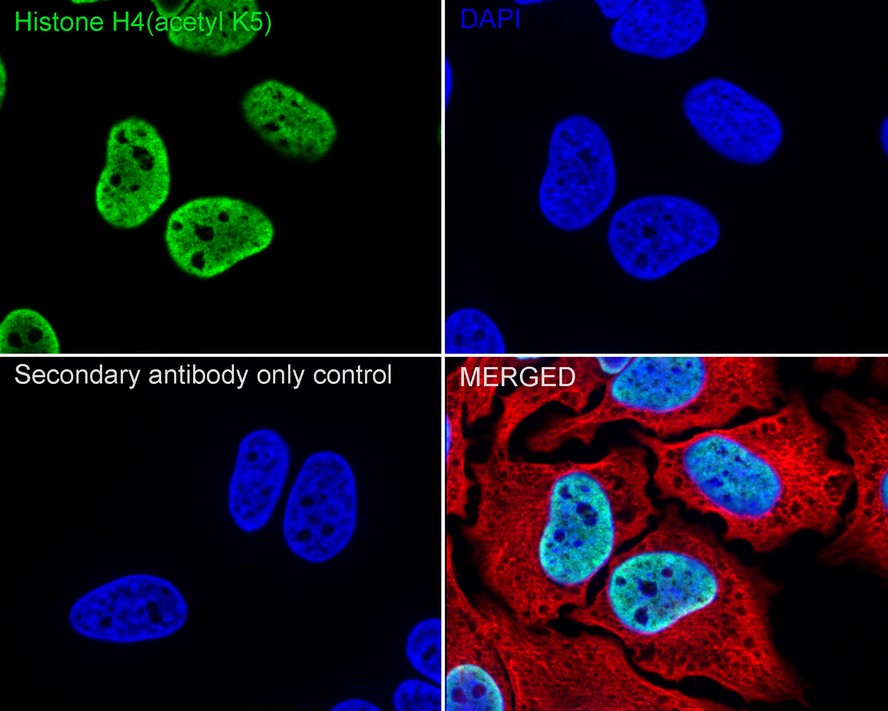
Immunocytochemistry analysis of HeLa cells labeling Histone H4 (acetyl K5) with Rabbit anti-Histone H4 (acetyl K5) antibody (ET1602-40) at 1/500 dilution.
Cells were fixed in 4% paraformaldehyde for 15 minutes at room temperature, permeabilized with 0.1% Triton X-100 in PBS for 15 minutes at room temperature, then blocked with 1% BSA in 10% negative goat serum for 1 hour at room temperature. Cells were then incubated with Rabbit anti-Histone H4 (acetyl K5) antibody (ET1602-40) at 1/500 dilution in 1% BSA in PBST overnight at 4 ℃. Goat Anti-Rabbit IgG H&L (iFluor™ 488, HA1121) was used as the secondary antibody at 1/1,000 dilution. PBS instead of the primary antibody was used as the secondary antibody only control. Nuclear DNA was labelled in blue with DAPI.
Beta tubulin (HA601187, red) was stained at 1/100 dilution overnight at +4℃. Goat Anti-Mouse IgG H&L (iFluor™ 594, HA1126) was used as the secondary antibody at 1/1,000 dilution. -
Immunocytochemistry analysis of NIH/3T3 cells labeling Histone H4 (acetyl K5) with Rabbit anti-Histone H4 (acetyl K5) antibody (ET1602-40) at 1/500 dilution.
Cells were fixed in 4% paraformaldehyde for 15 minutes at room temperature, permeabilized with 0.1% Triton X-100 in PBS for 15 minutes at room temperature, then blocked with 1% BSA in 10% negative goat serum for 1 hour at room temperature. Cells were then incubated with Rabbit anti-Histone H4 (acetyl K5) antibody (ET1602-40) at 1/500 dilution in 1% BSA in PBST overnight at 4 ℃. Goat Anti-Rabbit IgG H&L (iFluor™ 488, HA1121) was used as the secondary antibody at 1/1,000 dilution. PBS instead of the primary antibody was used as the secondary antibody only control. Nuclear DNA was labelled in blue with DAPI.
Beta tubulin (HA601187, red) was stained at 1/100 dilution overnight at +4℃. Goat Anti-Mouse IgG H&L (iFluor™ 594, HA1126) was used as the secondary antibody at 1/1,000 dilution. -
Immunocytochemistry analysis of PC-12 cells labeling Histone H4 (acetyl K5) with Rabbit anti-Histone H4 (acetyl K5) antibody (ET1602-40) at 1/100 dilution.
Cells were fixed in 4% paraformaldehyde for 15 minutes at room temperature, permeabilized with 0.1% Triton X-100 in PBS for 15 minutes at room temperature, then blocked with 1% BSA in 10% negative goat serum for 1 hour at room temperature. Cells were then incubated with Rabbit anti-Histone H4 (acetyl K5) antibody (ET1602-40) at 1/100 dilution in 1% BSA in PBST overnight at 4 ℃. Goat Anti-Rabbit IgG H&L (iFluor™ 488, HA1121) was used as the secondary antibody at 1/1,000 dilution. PBS instead of the primary antibody was used as the secondary antibody only control. Nuclear DNA was labelled in blue with DAPI.
Beta tubulin (HA601187, red) was stained at 1/100 dilution overnight at +4℃. Goat Anti-Mouse IgG H&L (iFluor™ 594, HA1126) was used as the secondary antibody at 1/1,000 dilution. -
Immunohistochemical analysis of paraffin-embedded human testis tissue with Rabbit anti-Histone H4 (acetyl K5) antibody (ET1602-40) at 1/1,000 dilution.
The section was pre-treated using heat mediated antigen retrieval with sodium citrate buffer (pH 6.0) (high pressure) for 2 minutes. The tissues were blocked in 1% BSA for 20 minutes at room temperature, washed with ddH2O and PBS, and then probed with the primary antibody (ET1602-40) at 1/1,000 dilution for 1 hour at room temperature. The detection was performed using an HRP conjugated compact polymer system. DAB was used as the chromogen. Tissues were counterstained with hematoxylin and mounted with DPX. -
Immunohistochemical analysis of paraffin-embedded human colon tissue with Rabbit anti-Histone H4 (acetyl K5) antibody (ET1602-40) at 1/1,000 dilution.
The section was pre-treated using heat mediated antigen retrieval with sodium citrate buffer (pH 6.0) (high pressure) for 2 minutes. The tissues were blocked in 1% BSA for 20 minutes at room temperature, washed with ddH2O and PBS, and then probed with the primary antibody (ET1602-40) at 1/1,000 dilution for 1 hour at room temperature. The detection was performed using an HRP conjugated compact polymer system. DAB was used as the chromogen. Tissues were counterstained with hematoxylin and mounted with DPX. -
Immunohistochemical analysis of paraffin-embedded human tonsil tissue using anti-Histone H4 (acetyl K5) antibody. The section was pre-treated using heat mediated antigen retrieval with Tris-EDTA buffer (pH 9.0) for 20 minutes.The tissues were blocked in 5% BSA for 30 minutes at room temperature, washed with ddH2O and PBS, and then probed with the primary antibody (ET1602-40, 1/1,000) for 30 minutes at room temperature. The detection was performed using an HRP conjugated compact polymer system. DAB was used as the chromogen. Tissues were counterstained with hematoxylin and mounted with DPX.
-
Immunohistochemical analysis of paraffin-embedded human colon carcinoma tissue using anti-Histone H4 (acetyl K5) antibody. The section was pre-treated using heat mediated antigen retrieval with Tris-EDTA buffer (pH 9.0) for 20 minutes.The tissues were blocked in 5% BSA for 30 minutes at room temperature, washed with ddH2O and PBS, and then probed with the primary antibody (ET1602-40, 1/200) for 30 minutes at room temperature. The detection was performed using an HRP conjugated compact polymer system. DAB was used as the chromogen. Tissues were counterstained with hematoxylin and mounted with DPX.
-
Immunohistochemical analysis of paraffin-embedded mouse testis tissue using anti-Histone H4 (acetyl K5) antibody. The section was pre-treated using heat mediated antigen retrieval with Tris-EDTA buffer (pH 9.0) for 20 minutes.The tissues were blocked in 5% BSA for 30 minutes at room temperature, washed with ddH2O and PBS, and then probed with the primary antibody (ET1602-40, 1/1,000) for 30 minutes at room temperature. The detection was performed using an HRP conjugated compact polymer system. DAB was used as the chromogen. Tissues were counterstained with hematoxylin and mounted with DPX.
-
Immunofluorescence staining of paraffin- embedded rat brain tissue using anti-Histone H4(acetyl K5) antibody.The section was pre-treated using heat mediated antigen retrieval with Tris-EDTA buffer (pH 9.0) for 20 minutes. The tissues were blocked in 10% negative goat serum for 1 hour at room temperature, washed with PBS, and then probed with ET1602-40 at 1/300 dilution for 10 hours at 4℃ and detected using Alexa Fluor® 488 conjugate-Goat anti-Rabbit IgG (H+L) Secondary Antibody at a dilution of 1:500 for 1 hour at room temperature.
-
Chromatin immunoprecipitations were performed with cross-linked chromatin from HeLa cells with Histone H4 (acetyl K5) (ET1602-40) or Normal Rabbit IgG according to the ChIP protocol. The enriched DNA was quantified by real-time PCR using indicated primers. The amount of immunoprecipitated DNA in each sample is represented as signal relative to the total amount of input chromatin, which is equivalent to one.
请注意: All products are "FOR RESEARCH USE ONLY AND ARE NOT INTENDED FOR DIAGNOSTIC OR THERAPEUTIC USE"
引文
-
Panobinostat (LBH589) inhibits Wnt/β-catenin signaling pathway via upregulating APCL expression in breast cancer
期刊: Cellular Signalling
DOI:
IF: 3.388
应用: WB
反应种属: Human
发表时间: 2019 Jul
 浙公网安备 33019202000643号