概述
产品名称
Caveolin-1 Recombinant Rabbit Monoclonal Antibody [SZ02-01]
抗体类型
Recombinant Rabbit monoclonal Antibody
免疫原
Synthetic peptide within Human Caveolin-1 aa 129-178 / 178.
种属反应性
Human, Mouse, Rat
验证应用
WB, IF-Cell, IF-Tissue, IHC-P
分子量
Predicted band size: 20 kDa
阳性对照
A549 cell lysate, A431 cell lysate, human lung tissue lysate, mouse lung tissue lysate, mouse heart tissue lysate, rat lung tissue lysate, rat heart tissue lysate, A549, MCF-7, NIH/3T3, human liver tissue, human liver carcinoma tissue, human uterus tissue, mouse heart tissue.
偶联
unconjugated
克隆号
SZ02-01
RRID
产品特性
形态
Liquid
存放说明
Shipped at 4℃. Store at +4℃ short term (1-2 weeks). It is recommended to aliquot into single-use upon delivery. Store at -20℃ long term.
存储缓冲液
1*TBS (pH7.4), 0.05% BSA, 40% Glycerol. Preservative: 0.05% Sodium Azide.
亚型
IgG
纯化方式
Protein A affinity purified.
应用稀释度
-
WB
-
1:500-1:5,000
-
IF-Cell
-
1:50-1:200
-
IF-Tissue
-
1:1,000
-
IHC-P
-
1:200-1:10,000
发表文章中的应用
| WB | 查看 3 篇文献如下 |
发表文章中的种属
| Human | 查看 2 篇文献如下 |
| Mouse | 查看 2 篇文献如下 |
靶点
功能
Caveolae (also known as plasmalemmal vesicles) are 50-100 nM flask-shaped membranes that represent a subcompartment of the plasma membrane. On the basis of morphological studies, caveolae have been implicated to function in the transcytosis of various macromolecules (including LDL) across capillary endothelial cells, uptake of small molecules via potocytosis and the compartmentalization of certain signaling molecules including G protein-coupled receptors. Three proteins, caveolin-1, caveolin-2 and caveolin-3, have been identified as principal components of caveolae. Two forms of caveolin-1, designated alpha and beta, share a distinct but overlapping cellular distribution and differ by an amino terminal 31 amino acid sequence which is absent from the beta isoform. Caveolin-1 shares 31% identity with caveolin-2 and 65% identity with caveolin-3 at the amino acid level. Functionally, the three proteins differ in their interactions with heterotrimeric G protein isoforms.
背景文献
1. Chen D et al. Glioma cell proliferation controlled by ERK activity-dependent surface expression of PDGFRA. PLoS One 9:e87281 (2014).
2. Olofsson A et al. Uptake of Helicobacter pylori vesicles is facilitated by clathrin-dependent and clathrin-independent endocytic pathways. MBio 5:e00979-14 (2014).
序列相似性
Belongs to the caveolin family.
组织特异性
Skeletal muscle, liver, stomach, lung, kidney and heart (at protein level). Expressed in the brain.
翻译后修饰
Ubiquitinated. Undergo monoubiquitination and multi- and/or polyubiquitination. Monoubiquitination of N-terminal lysines promotes integration in a ternary complex with UBXN6 and VCP which promotes oligomeric CAV1 targeting to lysosomes for degradation.; The initiator methionine for isoform 2 is removed during or just after translation. The new N-terminal amino acid is then N-acetylated.; Phosphorylated at Tyr-14 by ABL1 in response to oxidative stress.
亚细胞定位
Golgi apparatus membrane, Cell membrane, Membrane.
别名
BSCL3 antibody
CAV antibody
CAV1 antibody
CAV1_HUMAN antibody
caveolae protein, 22 kD antibody
caveolin 1 alpha isoform antibody
caveolin 1 beta isoform antibody
Caveolin 1 caveolae protein 22kDa antibody
Caveolin-1 antibody
Caveolin1 antibody
展开BSCL3 antibody
CAV antibody
CAV1 antibody
CAV1_HUMAN antibody
caveolae protein, 22 kD antibody
caveolin 1 alpha isoform antibody
caveolin 1 beta isoform antibody
Caveolin 1 caveolae protein 22kDa antibody
Caveolin-1 antibody
Caveolin1 antibody
cell growth-inhibiting protein 32 antibody
CGL3 antibody
LCCNS antibody
MSTP085 antibody
OTTHUMP00000025031 antibody
PPH3 antibody
VIP 21 antibody
VIP21 antibody
折叠图片
-

Western blot analysis of Caveolin-1 on different lysates with Rabbit anti-Caveolin-1 antibody (ET1603-1) at different dilutions.
Lane 1/2: A549 cell lysate
Lane 3/4: A431 cell lysate
Lysates/proteins at 10 µg/Lane.
Predicted band size: 20 kDa
Observed band size: 20 kDa
Exposure time: 2 minutes;
12% SDS-PAGE gel.
Proteins were transferred to a PVDF membrane and blocked with 5% NFDM/TBST for 1 hour at room temperature. The primary antibody (ET1603-1) at different dilutions were used in 5% NFDM/TBST at room temperature for 2 hours. Goat Anti-Rabbit IgG - HRP Secondary Antibody (HA1001) at 1:300,000 dilution was used for 1 hour at room temperature. -

Western blot analysis of Caveolin-1 on different lysates with Rabbit anti-Caveolin-1 antibody (ET1603-1) at 1/1,000 dilution.
Lane 1: A549 cell lysate (10 µg/Lane)
Lane 2: A431 cell lysate (10 µg/Lane)
Lane 3: Human lung tissue lysate (40 µg/Lane)
Lane 4: Mouse lung tissue lysate (40 µg/Lane)
Lane 5: Mouse heart tissue lysate (40 µg/Lane)
Lane 6: Rat lung tissue lysate (40 µg/Lane)
Lane 7: Rat heart tissue lysate (40 µg/Lane)
Predicted band size: 20 kDa
Observed band size: 20/18 kDa
Exposure time: Lane 1-4: 6 seconds; Lane 5-7: 30 seconds; ECL: K1801;
4-20% SDS-PAGE gel.
Proteins were transferred to a PVDF membrane and blocked with 5% NFDM/TBST for 1 hour at room temperature. The primary antibody (ET1603-1) at 1/1,000 dilution was used in 5% NFDM/TBST at 4℃ overnight. Goat Anti-Rabbit IgG - HRP Secondary Antibody (HA1001) at 1/50,000 dilution was used for 1 hour at room temperature. -

ICC staining of Caveolin-1 in A549 cells (green). Formalin fixed cells were permeabilized with 0.1% Triton X-100 in TBS for 10 minutes at room temperature and blocked with 1% Blocker BSA for 15 minutes at room temperature. Cells were probed with the primary antibody (ET1603-1, 1/50) for 1 hour at room temperature, washed with PBS. Alexa Fluor®488 Goat anti-Rabbit IgG was used as the secondary antibody at 1/1,000 dilution. The nuclear counter stain is DAPI (blue).
-

ICC staining of Caveolin-1 in MCF-7 cells (green). Formalin fixed cells were permeabilized with 0.1% Triton X-100 in TBS for 10 minutes at room temperature and blocked with 1% Blocker BSA for 15 minutes at room temperature. Cells were probed with the primary antibody (ET1603-1, 1/50) for 1 hour at room temperature, washed with PBS. Alexa Fluor®488 Goat anti-Rabbit IgG was used as the secondary antibody at 1/1,000 dilution. The nuclear counter stain is DAPI (blue).
-

ICC staining of Caveolin-1 in NIH/3T3 cells (green). Formalin fixed cells were permeabilized with 0.1% Triton X-100 in TBS for 10 minutes at room temperature and blocked with 1% Blocker BSA for 15 minutes at room temperature. Cells were probed with the primary antibody (ET1603-1, 1/50) for 1 hour at room temperature, washed with PBS. Alexa Fluor®488 Goat anti-Rabbit IgG was used as the secondary antibody at 1/1,000 dilution. The nuclear counter stain is DAPI (blue).
-

Immunohistochemical analysis of paraffin-embedded human liver tissue with Rabbit anti-Caveolin-1 antibody (ET1603-1) at 1/1,000 dilution.
The section was pre-treated using heat mediated antigen retrieval with Tris-EDTA buffer (pH 9.0) for 20 minutes. The tissues were blocked in 1% BSA for 20 minutes at room temperature, washed with ddH2O and PBS, and then probed with the primary antibody (ET1603-1) at 1/1,000 dilution for 1 hour at room temperature. The detection was performed using an HRP conjugated compact polymer system. DAB was used as the chromogen. Tissues were counterstained with hematoxylin and mounted with DPX. -

Immunohistochemical analysis of paraffin-embedded human liver carcinoma tissue with Rabbit anti-Caveolin-1 antibody (ET1603-1) at 1/200 dilution.
The section was pre-treated using heat mediated antigen retrieval with Tris-EDTA buffer (pH 9.0) for 20 minutes. The tissues were blocked in 1% BSA for 20 minutes at room temperature, washed with ddH2O and PBS, and then probed with the primary antibody (ET1603-1) at 1/200 dilution for 1 hour at room temperature. The detection was performed using an HRP conjugated compact polymer system. DAB was used as the chromogen. Tissues were counterstained with hematoxylin and mounted with DPX. -

Immunohistochemical analysis of paraffin-embedded human uterus tissue with Rabbit anti-Caveolin-1 antibody (ET1603-1) at 1/200 dilution.
The section was pre-treated using heat mediated antigen retrieval with Tris-EDTA buffer (pH 9.0) for 20 minutes. The tissues were blocked in 1% BSA for 20 minutes at room temperature, washed with ddH2O and PBS, and then probed with the primary antibody (ET1603-1) at 1/200 dilution for 1 hour at room temperature. The detection was performed using an HRP conjugated compact polymer system. DAB was used as the chromogen. Tissues were counterstained with hematoxylin and mounted with DPX. -

Immunohistochemical analysis of paraffin-embedded mouse heart tissue with Rabbit anti-Caveolin-1 antibody (ET1603-1) at 1/10,000 dilution.
The section was pre-treated using heat mediated antigen retrieval with Tris-EDTA buffer (pH 9.0) for 20 minutes. The tissues were blocked in 1% BSA for 20 minutes at room temperature, washed with ddH2O and PBS, and then probed with the primary antibody (ET1603-1) at 1/10,000 dilution for 1 hour at room temperature. The detection was performed using an HRP conjugated compact polymer system. DAB was used as the chromogen. Tissues were counterstained with hematoxylin and mounted with DPX. -

Application: IF-Tissue
Species: Mouse
Site: heart
Sample: Paraffin-embedded section
Antibody concentration: 1/1,000
请注意: All products are "FOR RESEARCH USE ONLY AND ARE NOT INTENDED FOR DIAGNOSTIC OR THERAPEUTIC USE"
引文
-
The novel role of Kallistatin in linking metabolic syndromes and cognitive memory deterioration by inducing amyloid-β plaques accumulation and tau protein hyperphosphorylation
Author: Weiwei Qi, Yanlan Long, Ziming Li, Zhen Zhao, Jinhui Shi, Wanting Xie, Laijian Wang, Yandan Tan, Ti Zhou, Minting Liang, Ping Jiang, Bin Jiang, Xia Yang, Guoquan Gao
PMID: 40762561
期刊: eLife
应用: WB
反应种属: Mouse
发表时间: 2025 Aug
-
Citation
-
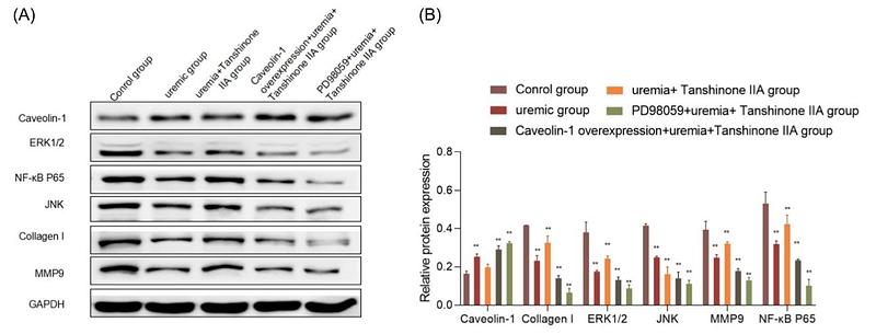
Effects of tanshinone IIA on endothelial cell dysfunction in uremic condition
Author: Li-hua Wang, Bo Yang, Zhe Wang, Lan Jia, Hai-yan Chen, Xue-qing Bi
PMID: 39051181

期刊: Journal Of Biochemical And Molecular Toxicology
应用: WB
反应种属: Human
发表时间: 2024 Jul
-
Citation
-
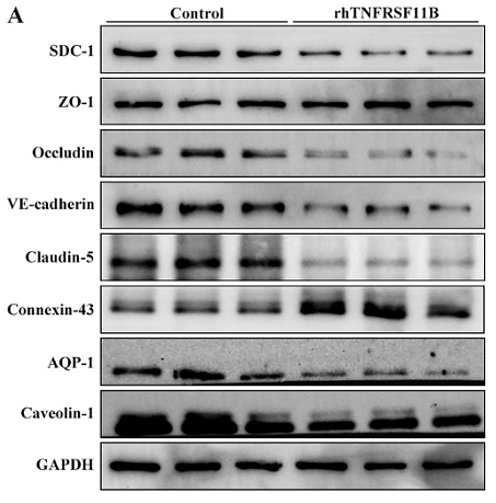
Plasma TNFRSF11B as a New Predictive Inflammatory Marker of Sepsis-ARDS with Endothelial Dysfunction
Author:
PMID: 37851947

期刊: Journal Of Proteome Research
应用: WB
反应种属: Human,Mouse
发表时间: 2023 Oct
-
Citation
同靶点 & 同通路的产品
Caveolin-1 Rabbit Polyclonal Antibody
Application: WB,IF-Cell,IHC-P,FC
Reactivity: Human,Mouse,Rat
Conjugate: unconjugated
Caveolin-1 Mouse Monoclonal Antibody [A3E6]
Application: WB,IHC-P
Reactivity: Human
Conjugate: unconjugated
Caveolin-1 Mouse Monoclonal Antibody [19-D6]
Application: WB,IHC-P
Reactivity: Human,Mouse,Rat
Conjugate: unconjugated
Caveolin-1 Mouse Monoclonal Antibody [A3E7]
Application: WB,IF-Cell,IHC-P,FC
Reactivity: Human
Conjugate: unconjugated
Caveolin-1 Mouse Monoclonal Antibody [A3E8]
Application: WB,IF-Cell,IHC-P,FC
Reactivity: Human
Conjugate: unconjugated
Caveolin-1 Mouse Monoclonal Antibody [A3E4]
Application: WB,IHC-P,FC
Reactivity: Human,Mouse
Conjugate: unconjugated


