p53 Mouse Monoclonal Antibody [7-5]


-
-
-
-
-
-
2+
Catalog# EM20603
p53 Mouse Monoclonal Antibody [7-5]
-
WB
-
IF-Cell
-
IHC-P
-
FC
-
Human
概述
产品名称
p53 Mouse Monoclonal Antibody [7-5]
抗体类型
Mouse Monoclonal Antibody
免疫原
Recombinant protein within human p53 aa 50-393.
种属反应性
Human
验证应用
WB, IF-Cell, IHC-P, FC
分子量
Predicted band size: 53 kDa
阳性对照
A431 cell lysate, 293 cell lysate, A431, human colonic carcinoma tissue, human gastric carcinoma tissue.
偶联
unconjugated
克隆号
7-5
RRID
产品特性
形态
Liquid
浓度
2ug/ul
存放说明
Store at +4℃ after thawing. Aliquot store at -20℃ or -80℃. Avoid repeated freeze / thaw cycles.
存储缓冲液
1*PBS (pH7.4), 0.2% BSA, 40% Glycerol. Preservative: 0.05% Sodium Azide.
亚型
IgG2b
纯化方式
Protein A affinity purified.
应用稀释度
-
WB
-
1:1,000-1:2,000
-
IHC-P
-
1:100-1:200
-
FC
-
1:100-1:200
-
IF-Cell
-
1:100-1:200
发表文章中的应用
| WB | 查看 3 篇文献如下 |
| IF | 查看 1 篇文献如下 |
发表文章中的种属
| Human | 查看 2 篇文献如下 |
| multiple myeloma cell | 查看 1 篇文献如下 |
靶点
功能
Tumor protein P53, also known as p53, cellular tumor antigen p53 (UniProt name), the Guardian of the Genome, phosphoprotein p53, tumor suppressor p53, antigen NY-CO-13, or transformation-related protein 53 (TRP53), is any isoform of a protein encoded by homologous genes in various organisms, such as TP53 (humans) and Trp53 (mice). The p53 proteins (originally thought to be, and often spoken of as, a single protein) are crucial in vertebrates, where they prevent cancer formation.[6] As such, p53 has been described as "the guardian of the genome" because of its role in conserving stability by preventing genome mutation. Hence TP53 is classified as a tumor suppressor gene. The name p53 was given in 1979 describing the apparent molecular mass; SDS-PAGE analysis indicates that it is a 53-kilodalton (kDa) protein. However, the actual mass of the full-length p53 protein (p53α) based on the sum of masses of the amino acid residues is only 43.7 kDa. This difference is due to the high number of proline residues in the protein, which slow its migration on SDS-PAGE, thus making it appear heavier than it actually is. In addition to the full-length protein, the human TP53 gene encodes at least 15 protein isoforms, ranging in size from 3.5 to 43.7 kDa. All these p53 proteins are called the p53 isoforms. The TP53 gene is the most frequently mutated gene (>50%) in human cancer, indicating that the TP53 gene plays a crucial role in preventing cancer formation. TP53 gene encodes proteins that bind to DNA and regulate gene expression to prevent mutations of the genome.
背景文献
1. SVH-B interacts directly with p53 and suppresses the transcriptional activity of p53."Zhou X., Yang G., Huang R., Chen X., Hu G.FEBS Lett. 581:4943-4948(2007)
2. Stabilization and activation of p53 induced by Cdk5 contributes to neuronal cell death."Lee J.-H., Kim H.-S., Lee S.-J., Kim K.-T.J. Cell Sci. 120:2259-2271(2007)
序列相似性
Belongs to the p53 family.
组织特异性
Ubiquitous. Isoforms are expressed in a wide range of normal tissues but in a tissue-dependent manner. Isoform 2 is expressed in most normal tissues but is not detected in brain, lung, prostate, muscle, fetal brain, spinal cord and fetal liver. Isoform 3 is expressed in most normal tissues but is not detected in lung, spleen, testis, fetal brain, spinal cord and fetal liver. Isoform 7 is expressed in most normal tissues but is not detected in prostate, uterus, skeletal muscle and breast. Isoform 8 is detected only in colon, bone marrow, testis, fetal brain and intestine. Isoform 9 is expressed in most normal tissues but is not detected in brain, heart, lung, fetal liver, salivary gland, breast or intestine.
翻译后修饰
Acetylated. Acetylation of Lys-382 by CREBBP enhances transcriptional activity. Deacetylation of Lys-382 by SIRT1 impairs its ability to induce proapoptotic program and modulate cell senescence. Deacetylation by SIRT2 impairs its ability to induce transcription activation in a AKT-dependent manner.; Phosphorylation on Ser residues mediates transcriptional activation. Phosphorylated by HIPK1 (By similarity). Phosphorylation at Ser-9 by HIPK4 increases repression activity on BIRC5 promoter. Phosphorylated on Thr-18 by VRK1. Phosphorylated on Ser-20 by CHEK2 in response to DNA damage, which prevents ubiquitination by MDM2. Phosphorylated on Ser-20 by PLK3 in response to reactive oxygen species (ROS), promoting p53/TP53-mediated apoptosis. Phosphorylated on Thr-55 by TAF1, which promotes MDM2-mediated degradation. Phosphorylated on Ser-33 by CDK7 in a CAK complex in response to DNA damage. Phosphorylated on Ser-46 by HIPK2 upon UV irradiation. Phosphorylation on Ser-46 is required for acetylation by CREBBP. Phosphorylated on Ser-392 following UV but not gamma irradiation. Phosphorylated on Ser-15 upon ultraviolet irradiation; which is enhanced by interaction with BANP. Phosphorylated by NUAK1 at Ser-15 and Ser-392; was initially thought to be mediated by STK11/LKB1 but it was later shown that it is indirect and that STK11/LKB1-dependent phosphorylation is probably mediated by downstream NUAK1. It is unclear whether AMP directly mediates phosphorylation at Ser-15. Phosphorylated on Thr-18 by isoform 1 and isoform 2 of VRK2. Phosphorylation on Thr-18 by isoform 2 of VRK2 results in a reduction in ubiquitination by MDM2 and an increase in acetylation by EP300. Stabilized by CDK5-mediated phosphorylation in response to genotoxic and oxidative stresses at Ser-15, Ser-33 and Ser-46, leading to accumulation of p53/TP53, particularly in the nucleus, thus inducing the transactivation of p53/TP53 target genes. Phosphorylated by DYRK2 at Ser-46 in response to genotoxic stress. Phosphorylated at Ser-315 and Ser-392 by CDK2 in response to DNA-damage. Phosphorylation at Ser-15 is required for interaction with DDX3X and gamma-tubulin.; Dephosphorylated by PP2A-PPP2R5C holoenzyme at Thr-55. SV40 small T antigen inhibits the dephosphorylation by the AC form of PP2A.; May be O-glycosylated in the C-terminal basic region. Studied in EB-1 cell line.; Ubiquitinated by MDM2 and SYVN1, which leads to proteasomal degradation. Ubiquitinated by RFWD3, which works in cooperation with MDM2 and may catalyze the formation of short polyubiquitin chains on p53/TP53 that are not targeted to the proteasome. Ubiquitinated by MKRN1 at Lys-291 and Lys-292, which leads to proteasomal degradation. Deubiquitinated by USP10, leading to its stabilization. Ubiquitinated by TRIM24, RFFL, RNF34 and RNF125, which leads to proteasomal degradation. Ubiquitination by TOPORS induces degradation. Deubiquitination by USP7, leading to stabilization. Isoform 4 is monoubiquitinated in an MDM2-independent manner. Ubiquitinated by COP1, which leads to proteasomal degradation. Ubiquitination and subsequent proteasomal degradation is negatively regulated by CCAR2. Polyubiquitinated by C10orf90/FATS, polyubiquitination is 'Lys-48'-linkage independent and non-proteolytic, leading to TP53 stabilization (By similarity).; Monomethylated at Lys-372 by SETD7, leading to stabilization and increased transcriptional activation. Monomethylated at Lys-370 by SMYD2, leading to decreased DNA-binding activity and subsequent transcriptional regulation activity. Lys-372 monomethylation prevents interaction with SMYD2 and subsequent monomethylation at Lys-370. Dimethylated at Lys-373 by EHMT1 and EHMT2. Monomethylated at Lys-382 by KMT5A, promoting interaction with L3MBTL1 and leading to repress transcriptional activity. Dimethylation at Lys-370 and Lys-382 diminishes p53 ubiquitination, through stabilizing association with the methyl reader PHF20. Demethylation of dimethylated Lys-370 by KDM1A prevents interaction with TP53BP1 and represses TP53-mediated transcriptional activation. Monomethylated at Arg-333 and dimethylated at Arg-335 and Arg-337 by PRMT5; methylation is increased after DNA damage and might possibly affect TP53 target gene specificity.; Sumoylated with SUMO1. Sumoylated at Lys-386 by UBC9.
亚细胞定位
Nucleus, Cytoplasm
UNIPROT
别名
Antigen NY-CO-13 antibody
BCC7 antibody
Cellular tumor antigen p53 antibody
FLJ92943 antibody
LFS1 antibody
Mutant tumor protein 53 antibody
p53 antibody
p53 tumor suppressor antibody
P53_HUMAN antibody
Phosphoprotein p53 antibody
展开图片
-
☑ Knockout (KO)
All lanes: Western blot analysis of p53 with anti-p53 antibody (EM20603) at 1:500 dilution.
Lane 1/2: Wild-type A431 whole cell lysate (10 µg).
Lane 3/4: p53 knockout A431 whole cell lysate (10 µg).
EM20603 was shown to specifically react with p53 in wild-type A431 cells. No band was observed when p53 knockout sample was tested. Wild-type and p53 knockout samples were subjected to SDS-PAGE. Proteins were transferred to a PVDF membrane and blocked with 5% NFDM in TBST for 1 hour at room temperature. The primary antibody (EM20603, 1/500) and Loading control antibody (Rabbit anti-GAPDH, ET1601-4, 1/10,000) was used in 5% BSA at room temperature for 2 hours. Goat Anti-Mouse IgG-HRP Secondary Antibody (HA1006) at 1:100,000 dilution was used for 1 hour at room temperature. -
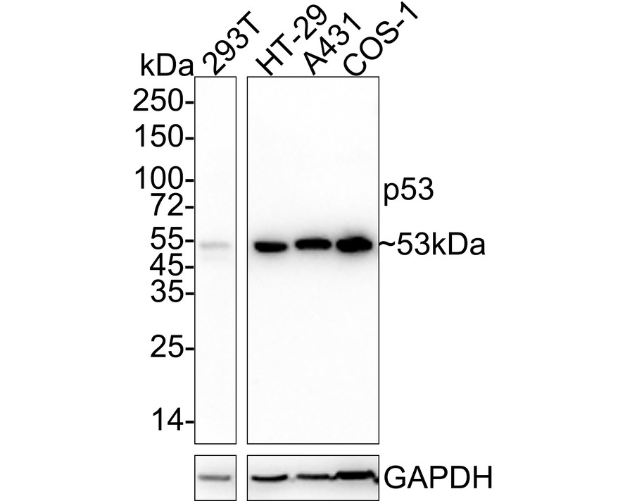
Western blot analysis of p53 on different lysates with Mouse anti-p53 antibody (EM20603) at 1/2,000 dilution.
Lane 1: A431 cell lysate
Lane 2: 293 cell lysate
Lysates/proteins at 10 µg/Lane.
Predicted band size: 53 kDa
Observed band size: 53 kDa
Exposure time: 2 minutes;
10% SDS-PAGE gel.
Proteins were transferred to a PVDF membrane and blocked with 5% NFDM/TBST for 1 hour at room temperature. The primary antibody (EM20603) at 1/2,000 dilution was used in 5% NFDM/TBST at room temperature for 2 hours. Goat Anti-Mouse IgG - HRP Secondary Antibody (HA1006) at 1:150,000 dilution was used for 1 hour at room temperature. -
ICC staining p53 in A431 cells (green). Cells were fixed in paraformaldehyde, permeabilised with 0.25% Triton X100/PBS.
-
Immunohistochemical analysis of paraffin-embedded human colonic carcinoma tissue using anti-p53 antibody. Counter stained with hematoxylin.
-
Immunohistochemical analysis of paraffin-embedded human gastric carcinoma tissue using anti-p53 antibody. Counter stained with hematoxylin.
-
Flow cytometric analysis of Hela cells with p53 antibody at 1/100 dilution (blue) compared with an unlabelled control (cells without incubation with primary antibody; red). Goat anti mouse IgG (FITC) was used as the secondary antibody.
请注意: All products are "FOR RESEARCH USE ONLY AND ARE NOT INTENDED FOR DIAGNOSTIC OR THERAPEUTIC USE"
引文
-
Inhibiting autophagy selectively prunes dysfunctional tumor vessels and optimizes the tumor immune microenvironment
Author: Wanting Hou, Chaoxin Xiao, Ruihan Zhou, Xiaohong Yao, Qin Chen, Tongtong Xu, Fujun Cao, Yulin Wang, Xiaoying Li, Ouying Yan, Xiaolin Ai, Cheng Yi, Dan Cao, Chengjian Zhao
PMID: 39744218
应用: IF,WB
反应种属: Human
发表时间: 2025 Jan
-
 Citation
-
CISD2 downregulation participates in the ferroptosis process of human ovarian SKOV-3 cells through modulating the wild type p53-mediated GLS2/SAT1/SLC7A11 and Gpx4/TRF signaling pathway
Author: Wang Yaqin,et al
PMID: NOPMID20240707

应用: WB
反应种属: Human
发表时间: 2024 Jul
-
Citation
-
Silencing RRM2 inhibits multiple myeloma by targeting the Wnt/β-catenin signaling pathway
Author: Dr Xia Liu,Professor Jianchao Wang
PMID: 31322175
应用: WB
反应种属: multiple myeloma cell
发表时间: 2019 Sep
-
Citation
同靶点 & 同通路的产品

Phospho-p53 (S15) Recombinant Rabbit Monoclonal Antibody [PSH01-98]
Application: WB,IF-Cell
Reactivity: Human
Conjugate: unconjugated

Phospho-p53 (T55) Recombinant Rabbit Monoclonal Antibody [ST0439]
Application: WB,IHC-P
Reactivity: Human
Conjugate: unconjugated

p53 (acetyl K382) Recombinant Rabbit Monoclonal Antibody [SD0801]
Application: WB,IF-Cell,IHC-P
Reactivity: Human,Mouse
Conjugate: unconjugated

p53 Mouse Monoclonal Antibody [6B5]
Application: WB
Reactivity: Human
Conjugate: unconjugated

p53 Rabbit Polyclonal Antibody
Application: WB,IF-Cell
Reactivity: Human
Conjugate: unconjugated

p53 Recombinant Rabbit Monoclonal Antibody [JB42-26]
Application: WB,IHC-P,FC,IP,IF-Cell,IF-Tissue
Reactivity: Human
Conjugate: unconjugated

p53 Recombinant Mouse Monoclonal Antibody [6B5-R]
Application: WB,IF-Cell
Reactivity: Human,Mouse,Monkey
Conjugate: unconjugated

p53 Mouse Monoclonal Antibody [6B2]
Application: WB,IF-Cell
Reactivity: Human,Mouse
Conjugate: unconjugated

Phospho-p53 (S392) Recombinant Rabbit Monoclonal Antibody [SI17-04]
Application: WB,IHC-P,IP
Reactivity: Human,Mouse,Rat
Conjugate: unconjugated

Phospho-p53 (S376) Recombinant Rabbit Monoclonal Antibody [ST0440]
Application: WB
Reactivity: Human,Mouse
Conjugate: unconjugated
p53 Recombinant Mouse Monoclonal Antibody [6B2-R]
Application: WB,IF-Cell
Reactivity: Human,Monkey
Conjugate: unconjugated

p53 Recombinant Rabbit Monoclonal Antibody [SY010-6]
Application: WB,IF-Cell,IF-Tissue,IHC-P,IP,FC
Reactivity: Human
Conjugate: unconjugated

p53 (acetyl K370) Recombinant Rabbit Monoclonal Antibody [SR40-09]
Application: WB,IF-Cell,IF-Tissue,IP,FC
Reactivity: Human,Mouse,Rat
Conjugate: unconjugated

p53 Recombinant Rabbit Monoclonal Antibody [PD01-32]
Application: IHC-P
Reactivity: Human
Conjugate: unconjugated

p53 Mouse Monoclonal Antibody [C8-A11]
Application: WB,IF-Cell,IHC-P
Reactivity: Human
Conjugate: unconjugated
