Ki67 Recombinant Rabbit Monoclonal Antibody


-
-
-
-
-
-
-
-
4+
Catalog# IRS036
Ki67 Recombinant Rabbit Monoclonal Antibody
-
mIHC
-
Human
概述
产品名称
Ki67 Recombinant Rabbit Monoclonal Antibody
抗体类型
Recombinant Rabbit monoclonal Antibody
免疫原
Synthetic peptide within human Ki67 aa 1,040-1,080.
种属反应性
Human
验证应用
mIHC
分子量
Predicted band size: 359 kDa
阳性对照
Human tonsils tissue, human liver tissue, human spleen tissue, human kidney tissue.
偶联
unconjugated
产品特性
形态
Liquid
浓度
1ug/ul
存放说明
Store at +4℃ after thawing. Aliquot store at -20℃ or -80℃. Avoid repeated freeze / thaw cycles.
存储缓冲液
PBS (pH7.4), 0.1% BSA, 40% Glycerol. Preservative: 0.05% Sodium Azide.
亚型
IgG
纯化方式
Protein A affinity purified.
应用稀释度
-
mIHC
-
1:100
靶点
功能
The Ki-67 protein is a nuclear protein doublet, 345-395 kDa, playing a pivotal role in maintaining cell proliferation. Ki-67 is present in all non-G0 phases of the cell cycle. Beginning in the mid G1, the level increases through S and G2 to reach a peak in M. In the end of M, is is rapidly catabolized. The Ki-67 labelling index (LI), i.e., the percentage of cells in a tissue staining for Ki-67, indicates the growth fraction. For many tumours, the rate of cell proliferation as assessed by Ki-67 immunoreactivity correlates with tumour grade and clinical course. In Non-Hodgkin lymphoma a labelling index of less than 20% is seen in low grade lymphomas, greater than 20% is associated with high grade lymphomas. Low grade lymphomas with a labelling index in excess of 5% have a worse prognosis than those with an index of less than 5%. In Burkitt and Burkitt-like lymphoma, nearly 100% of the nuclei are stained. This can be used as a diagnostic criterion. In gliomas the indices ranges from 0% to 5% for low grade astrocytomas while anaplastic astrocytomas and glioblastomas most frequently show an index above 10%. In soft tissue sarcomas Ki-67 index is positively correlated with mitotic count, cellularity and histological grade. In some benign tumours, like meningioma, a high LI is associated with a high recurrence rate. In dysplasia in Barrett's oesophagus and in granulosa cell tumours and ovarian serous tumours, Ki-67 LI is associated with progression. In the former, reproducibility of dysplasia grading is improved when Ki67 is included. In breast cancer, the proliferative index measured by Ki67 immunoreactivity has both prognostic and predictive value.
背景文献
1. Cuylen S. et al. Ki-67 acts as a biological surfactant to disperse mitotic chromosomes. Nature 535:308-312(2016).
2. Booth D.G. et al. Ki-67 is a PP1-interacting protein that organises the mitotic chromosome periphery. Elife 3:E01641-E01641(2014).
亚细胞定位
Nucleus, Chromosome.
UNIPROT
别名
Antigen identified by monoclonal antibody Ki 67 antibody
Antigen identified by monoclonal antibody Ki-67 antibody
Antigen KI-67 antibody
Antigen KI67 antibody
Antigen Ki67 antibody
KI67_HUMAN antibody
KIA antibody
Marker of proliferation Ki-67 antibody
MIB 1 antibody
MIB antibody
展开图片
-
mIHC analysis of human tonsils tissue (Formalin/PFA-fixed paraffin-embedded sections) with Rabbit anti-Ki67 antibody (IRS036) at 1/100 dilution. The immunostaining was performed with the IRISKit® HyperView mTSA Kit (MH900206). Heat mediated antigen retrieval with Tris-EDTA buffer (pH 9.0) for 30 mins at 95℃. DAPI (blue) was used as a nuclear counter stain. Image acquisition was performed with Olympus VS200 Slide Scanner.
-
mIHC analysis of human liver tissue (Formalin/PFA-fixed paraffin-embedded sections) with Rabbit anti-Ki67 antibody (IRS036) at 1/100 dilution. The immunostaining was performed with the IRISKit® HyperView mTSA Kit (MH900206). Heat mediated antigen retrieval with Tris-EDTA buffer (pH 9.0) for 30 mins at 95℃. DAPI (blue) was used as a nuclear counter stain. Image acquisition was performed with Olympus VS200 Slide Scanner.
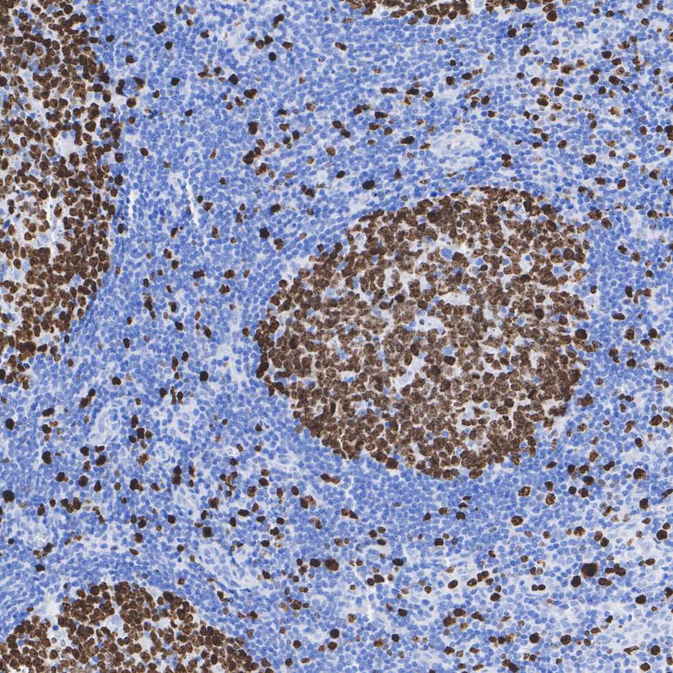
-
mIHC analysis of human spleen tissue (Formalin/PFA-fixed paraffin-embedded sections) with Rabbit anti-Ki67 antibody (IRS036) at 1/100 dilution. The immunostaining was performed with the IRISKit® HyperView mTSA Kit (MH900206). Heat mediated antigen retrieval with Tris-EDTA buffer (pH 9.0) for 30 mins at 95℃. DAPI (blue) was used as a nuclear counter stain. Image acquisition was performed with Olympus VS200 Slide Scanner.
-
mIHC analysis of human kidney tissue (Formalin/PFA-fixed paraffin-embedded sections) with Rabbit anti-Ki67 antibody (IRS036) at 1/100 dilution. The immunostaining was performed with the IRISKit® HyperView mTSA Kit (MH900206). Heat mediated antigen retrieval with Tris-EDTA buffer (pH 9.0) for 30 mins at 95℃. DAPI (blue) was used as a nuclear counter stain. Image acquisition was performed with Olympus VS200 Slide Scanner.
-
mIHC analysis of human tonsil tissue (Formalin/PFA-fixed paraffin-embedded sections) with Ki67 (IRS036), CD23 (IRS021), CD31 (IRS023) and PD-1 antibody at 1/100 dilution. The immunostaining was performed with the IRISKit® HyperView mTSA Kit (MH900206). Heat mediated antigen retrieval with Tris-EDTA buffer (pH 9.0) for 30 mins at 95℃. DAPI (blue) was used as a nuclear counter stain. Image acquisition was performed with Olympus VS200 Slide Scanner.
-
mIHC analysis of human tonsil tissue (Formalin/PFA-fixed paraffin-embedded sections) with Ki67 (IRS036), CD11c (IRS001), PD-1 and CD8 (IRS007) antibody at 1/100 dilution. The immunostaining was performed with the IRISKit® HyperView mTSA Kit (MH900206). Heat mediated antigen retrieval with Tris-EDTA buffer (pH 9.0) for 30 mins at 95℃. DAPI (blue) was used as a nuclear counter stain. Image acquisition was performed with Olympus VS200 Slide Scanner.
-
mIHC analysis of human lung cancer tissue (Formalin/PFA-fixed paraffin-embedded sections) with panCK (IRS010), CD3 (IRS022) and Ki67 (IRS036) antibody at 1/100 dilution. The immunostaining was performed with the IRISKit® HyperView mTSA Kit (MH900206). Heat mediated antigen retrieval with Tris-EDTA buffer (pH 9.0) for 30 mins at 95℃. DAPI (blue) was used as a nuclear counter stain. Image acquisition was performed with Olympus VS200 Slide Scanner.
-
mIHC analysis of human ovarian cancer tissue (Formalin/PFA-fixed paraffin-embedded sections) with Ki67 (IRS036), panCK (IRS010) and HER2 (IRS034) antibody at 1/100 dilution. The immunostaining was performed with the IRISKit® HyperView mTSA Kit (MH900206). Heat mediated antigen retrieval with Tris-EDTA buffer (pH 9.0) for 30 mins at 95℃. DAPI (blue) was used as a nuclear counter stain. Image acquisition was performed with Olympus VS200 Slide Scanner.
请注意: All products are "FOR RESEARCH USE ONLY AND ARE NOT INTENDED FOR DIAGNOSTIC OR THERAPEUTIC USE"
同靶点 & 同通路的产品

Ki67 Recombinant Rabbit Monoclonal Antibody [PSH0-02]
Application: IHC-P,FC
Reactivity: Human
Conjugate: unconjugated

Ki67 Rabbit Polyclonal Antibody
Application: WB,IHC-P,IF-Cell
Reactivity: Human
Conjugate: unconjugated

iFluor™ 647 Conjugated Ki67 Recombinant Rabbit Monoclonal Antibody [SR00-02]
Application: IF-Cell,IF-Tissue,FC,IHC-Fr
Reactivity: Human,Mouse
Conjugate: iFluor™ 647

Ki67 Rabbit Polyclonal Antibody
Application: WB,IF-Cell,IHC-P,FC
Reactivity: Human
Conjugate: unconjugated

Ki67 Recombinant Mouse Monoclonal Antibody [PD00-10]
Application: IHC-P,IF-Tissue
Reactivity: Human,Mouse
Conjugate: unconjugated

Ki67 Recombinant Rabbit Monoclonal Antibody [ST50-01]
Application: WB,IF-Cell,IHC-P,FC
Reactivity: Human
Conjugate: unconjugated

Ki67 Rabbit Polyclonal Antibody
Application: WB,IP,IF,IHC-P
Reactivity: Human
Conjugate: unconjugated
Ki67 Recombinant Rabbit Monoclonal Antibody [SR00-02]
Application: IHC-P,IF-Tissue,mIHC,IF-Cell,FC,IHC-Fr
Reactivity: Human,Mouse,Rat
Conjugate: unconjugated
