SIRT3 Recombinant Rabbit Monoclonal Antibody [PSH05-22]


Catalog# HA722251
SIRT3 Recombinant Rabbit Monoclonal Antibody [PSH05-22]
-
WB
-
IHC-P
-
IF-Cell
-
FC
-
IF-Tissue
-
IP
-
ChIP
-
Human
-
Mouse
-
Rat
概述
产品名称
SIRT3 Recombinant Rabbit Monoclonal Antibody [PSH05-22]
抗体类型
Recombinant Rabbit monoclonal Antibody
免疫原
Recombinant protein within human SIRT3 aa 51-399 / 399.
种属反应性
Human, Mouse, Rat
验证应用
WB, IHC-P, IF-Cell, FC, IF-Tissue, IP, ChIP
分子量
Predicted band size: 44 kDa
阳性对照
PANC-1 cell lysate, HepG2 cell lysate, A549 cell lysate, SiHa cell lysate, mouse liver tissue lysate, mouse kidney tissue lysate, rat liver tissue lysate, human liver tissue, human colon tissue, HeLa cell lysate, HEK-293 cell lysate, SW480 cell lysate, HT-29 cell lysate, rat kidney tissue lysate, human kidney tissue, mouse liver tissue, rat liver tissue, HepG2, NIH/3T3.
偶联
unconjugated
克隆号
PSH05-22
产品特性
形态
Liquid
存放说明
Shipped at 4℃. Store at +4℃ short term (1-2 weeks). It is recommended to aliquot into single-use upon delivery. Store at -20℃ long term.
存储缓冲液
PBS (pH7.4), 0.1% BSA, 40% Glycerol. Preservative: 0.05% Sodium Azide.
亚型
IgG
纯化方式
Protein A affinity purified.
应用稀释度
-
WB
-
1:2,000
-
IHC-P
-
1:1,000
-
IF-Cell
-
1:100
-
FC
-
1:1,000
-
IF-Tissue
-
1:200
-
IP
-
1:2000
-
ChIP
-
Use 0.5~2 μg for 25 μg of chromatin.
发表文章中的应用
| WB | 查看 3 篇文献如下 |
| IHC-P | 查看 1 篇文献如下 |
| IF | 查看 1 篇文献如下 |
发表文章中的种属
| Mouse | 查看 3 篇文献如下 |
靶点
功能
NAD-dependent deacetylase sirtuin-3, mitochondrial also known as SIRT3 is a protein that in humans is encoded by the SIRT3 gene. SIRT3 is member of the mammalian sirtuin family of proteins, which are homologs to the yeast Sir2 protein. SIRT3 exhibits NAD+-dependent deacetylase activity. Members of the sirtuin family are characterized by a sirtuin core domain and grouped into four classes, and the protein encoded by this gene is included in class I of the sirtuin family. The human sirtuins have a range of molecular functions and have emerged as important proteins in aging, stress resistance, and metabolic regulation. Yeast sirtuin proteins are known to regulate epigenetic gene silencing and suppress recombination of rDNA. In addition to protein deacetylation, studies have shown that the human sirtuins may also function as intracellular regulatory proteins with mono ADP ribosyltransferase activity.
背景文献
1. Zhou L et al. The Role of SIRT3 in Exercise and Aging. Cells. 2022 Aug
2. Diao Z et al. SIRT3 consolidates heterochromatin and counteracts senescence. Nucleic Acids Res. 2021 May
亚细胞定位
Mitochondrion matrix.
别名
hSIRT 3 antibody
hSIRT3 antibody
Mitochondrial nicotinamide adenine dinucleotide dependent deacetylase antibody
NAD dependent deacetylase sirtuin 3 mitochondrial antibody
NAD-dependent protein deacetylase sirtuin-3, mitochondrial antibody
Regulatory protein SIR2 homolog 3 antibody
Silent mating type information regulation 2 S.cerevisiae homolog 3 antibody
Sir 2 like 3 antibody
SIR 2 like protein 3 antibody
SIR 3 antibody
展开图片
-
Western blot analysis of SIRT3 on different lysates with Rabbit anti-SIRT3 antibody (HA722251) at 1/2,000 dilution and competitor's antibody at 1/2,000 dilution.
Lane 1: PANC-1 cell lysate
Lane 2: HepG2 cell lysate
Lane 3: A549 cell lysate
Lane 4: SiHa cell lysate
Lane 5: Mouse liver tissue lysate
Lane 6: Mouse kidney tissue lysate
Lane 7: Rat liver tissue lysate
Lysates/proteins at 20 µg/Lane.
Predicted band size: 44 kDa
Observed band size: 28 kDa
Exposure time: 30 seconds; ECL: K1801;
4-20% SDS-PAGE gel.
Proteins were transferred to a PVDF membrane and blocked with 5% NFDM/TBST for 1 hour at room temperature. The primary antibody (HA722251) at 1/2,000 dilution and competitor's antibody at 1/2,000 dilution were used in 5% NFDM/TBST at 4℃ overnight. Goat Anti-Rabbit IgG - HRP Secondary Antibody (HA1001) at 1/50,000 dilution was used for 1 hour at room temperature. -
Immunohistochemical analysis of paraffin-embedded human liver tissue with Rabbit anti-SIRT3 antibody (HA722251) at 1/1,000 dilution and competitor's antibody at 1/1,000 dilution.
The section was pre-treated using heat mediated antigen retrieval with Tris-EDTA buffer (pH 9.0) for 20 minutes. The tissues were blocked in 1% BSA for 20 minutes at room temperature, washed with ddH2O and PBS, and then probed with the primary antibody (HA722251) at 1/1,000 dilution and competitor's antibody at 1/1,000 dilution for 1 hour at room temperature. The detection was performed using an HRP conjugated compact polymer system. DAB was used as the chromogen. Tissues were counterstained with hematoxylin and mounted with DPX. -
Immunohistochemical analysis of paraffin-embedded human colon tissue with Rabbit anti-SIRT3 antibody (HA722251) at 1/1,000 dilution and competitor's antibody at 1/1,000 dilution.
The section was pre-treated using heat mediated antigen retrieval with Tris-EDTA buffer (pH 9.0) for 20 minutes. The tissues were blocked in 1% BSA for 20 minutes at room temperature, washed with ddH2O and PBS, and then probed with the primary antibody (HA722251) at 1/1,000 dilution and competitor's antibody at 1/1,000 dilution for 1 hour at room temperature. The detection was performed using an HRP conjugated compact polymer system. DAB was used as the chromogen. Tissues were counterstained with hematoxylin and mounted with DPX. -
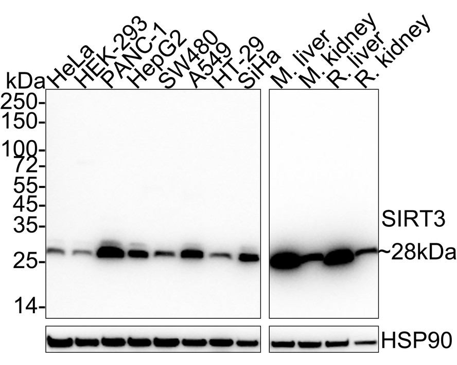
Western blot analysis of SIRT3 on different lysates with Rabbit anti-SIRT3 antibody (HA722251) at 1/2,000 dilution.
Lane 1: HeLa cell lysate (20 µg/Lane)
Lane 2: HEK-293 cell lysate (20 µg/Lane)
Lane 3: PANC-1 cell lysate (20 µg/Lane)
Lane 4: HepG2 cell lysate (20 µg/Lane)
Lane 5: SW480 cell lysate (20 µg/Lane)
Lane 6: A549 cell lysate (20 µg/Lane)
Lane 7: HT-29 cell lysate (20 µg/Lane)
Lane 8: SiHa cell lysate (20 µg/Lane)
Lane 9: Mouse liver tissue lysate (40 µg/Lane)
Lane 10: Mouse kidney tissue lysate (40 µg/Lane)
Lane 11: Rat liver tissue lysate (40 µg/Lane)
Lane 12: Rat kidney tissue lysate (40 µg/Lane)
Predicted band size: 44 kDa
Observed band size: 28 kDa
Exposure time: 25 seconds; ECL: K1801;
4-20% SDS-PAGE gel.
Proteins were transferred to a PVDF membrane and blocked with 5% NFDM/TBST for 1 hour at room temperature. The primary antibody (HA722251) at 1/2,000 dilution was used in 5% NFDM/TBST at 4℃ overnight. Goat Anti-Rabbit IgG - HRP Secondary Antibody (HA1001) at 1/50,000 dilution was used for 1 hour at room temperature. -
☑ Knockdown (KD)
Western blot analysis of SIRT3 on different lysates with Rabbit anti-SIRT3 antibody (HA722251) at 1/5,000 dilution.
Lane 1: HAP1-parental cell lysate
Lane 2: HAP1-SIRT3 KD cell lysate
Lysates/proteins at 10 µg/Lane.
Predicted band size: 44 kDa
Observed band size: 28 kDa
Exposure time: 2 minutes; ECL: K1801;
4-20% SDS-PAGE gel.
Proteins were transferred to a PVDF membrane and blocked with 5% NFDM/TBST for 1 hour at room temperature. The primary antibody (HA722251) at 1/5,000 dilution was used in primary antibody dilution (K1803) at 4℃ overnight. Goat Anti-Rabbit IgG - HRP Secondary Antibody (HA1001) at 1/50,000 dilution was used for 1 hour at room temperature. -
Immunohistochemical analysis of paraffin-embedded human kidney tissue with Rabbit anti-SIRT3 antibody (HA722251) at 1/1,000 dilution.
The section was pre-treated using heat mediated antigen retrieval with Tris-EDTA buffer (pH 9.0) for 20 minutes. The tissues were blocked in 1% BSA for 20 minutes at room temperature, washed with ddH2O and PBS, and then probed with the primary antibody (HA722251) at 1/1,000 dilution for 1 hour at room temperature. The detection was performed using an HRP conjugated compact polymer system. DAB was used as the chromogen. Tissues were counterstained with hematoxylin and mounted with DPX. -
Immunohistochemical analysis of paraffin-embedded mouse liver tissue with Rabbit anti-SIRT3 antibody (HA722251) at 1/1,000 dilution.
The section was pre-treated using heat mediated antigen retrieval with Tris-EDTA buffer (pH 9.0) for 20 minutes. The tissues were blocked in 1% BSA for 20 minutes at room temperature, washed with ddH2O and PBS, and then probed with the primary antibody (HA722251) at 1/1,000 dilution for 1 hour at room temperature. The detection was performed using an HRP conjugated compact polymer system. DAB was used as the chromogen. Tissues were counterstained with hematoxylin and mounted with DPX. -
Immunohistochemical analysis of paraffin-embedded rat liver tissue with Rabbit anti-SIRT3 antibody (HA722251) at 1/1,000 dilution.
The section was pre-treated using heat mediated antigen retrieval with Tris-EDTA buffer (pH 9.0) for 20 minutes. The tissues were blocked in 1% BSA for 20 minutes at room temperature, washed with ddH2O and PBS, and then probed with the primary antibody (HA722251) at 1/1,000 dilution for 1 hour at room temperature. The detection was performed using an HRP conjugated compact polymer system. DAB was used as the chromogen. Tissues were counterstained with hematoxylin and mounted with DPX. -
Immunocytochemistry analysis of HepG2 cells labeling SIRT3 with Rabbit anti-SIRT3 antibody (HA722251) at 1/100 dilution.
Cells were fixed in 4% paraformaldehyde for 20 minutes at room temperature, permeabilized with 0.1% Triton X-100 in PBS for 5 minutes at room temperature, then blocked with 1% BSA in 10% negative goat serum for 1 hour at room temperature. Cells were then incubated with Rabbit anti-SIRT3 antibody (HA722251) at 1/100 dilution in 1% BSA in PBST overnight at 4 ℃. Goat Anti-Rabbit IgG H&L (iFluor™ 488, HA1121) was used as the secondary antibody at 1/1,000 dilution. PBS instead of the primary antibody was used as the secondary antibody only control. Counterstained with Mitotracker (Red). Nuclear DNA was labelled in blue with DAPI. -
Flow cytometric analysis of HepG2 cells labeling SIRT3.
Cells were fixed and permeabilized. Then stained with the primary antibody (HA722251, 1μg/mL) (red) compared with Rabbit IgG Isotype Control (green). After incubation of the primary antibody at +4℃ for an hour, the cells were stained with a iFluor™ 488 conjugate-Goat anti-Rabbit IgG Secondary antibody (HA1121) at 1/1,000 dilution for 30 minutes at +4℃. Unlabelled sample was used as a control (cells without incubation with primary antibody; black). -
Flow cytometric analysis of NIH/3T3 cells labeling SIRT3.
Cells were fixed and permeabilized. Then stained with the primary antibody (HA722251, 1μg/mL) (red) compared with Rabbit IgG Isotype Control (green). After incubation of the primary antibody at +4℃ for an hour, the cells were stained with a iFluor™ 488 conjugate-Goat anti-Rabbit IgG Secondary antibody (HA1121) at 1/1,000 dilution for 30 minutes at +4℃. Unlabelled sample was used as a control (cells without incubation with primary antibody; black). -
SIRT3 was immunoprecipitated from 0.2 mg HepG2 cell lysate with HA722251 at 2 µg/10 µl beads. Western blot was performed from the immunoprecipitate using HA722251 at 1/2,000 dilution. Anti-Rabbit IgG for IP Nano-secondary antibody (NBI01H) at 1/5,000 dilution was used for 1 hour at room temperature.
Lane 1: HepG2 cell lysate (input)
Lane 2: HA722251 IP in HepG2 cell lysate
Lane 3: Rabbit IgG instead of HA722251 in HepG2 cell lysate
Blocking/Dilution buffer: 5% NFDM/TBST
Exposure time: 1 minute 17 seconds; ECL: K1801 -
Chromatin immunoprecipitations were performed with cross-linked chromatin from HeLa cells with SIRT3 (HA722251) or Normal Rabbit IgG according to the ChIP protocol. The enriched DNA was quantified by real-time PCR using indicated primers. The amount of immunoprecipitated DNA in each sample is represented as signal relative to the total amount of input chromatin, which is equivalent to one.
请注意: All products are "FOR RESEARCH USE ONLY AND ARE NOT INTENDED FOR DIAGNOSTIC OR THERAPEUTIC USE"
引文
-
Enhanced SIRT3 expression restores mitochondrial quality control mechanism to reverse osteogenic impairment in type 2 diabetes mellitus
Author: Xian Yansi, Liu Bin, Shen Tao, Yang Lin, Peng Rui, Shen Hongdou, An Xueying, Wang Yutian, Ben Yu, Jiang Qing, Guo Baosheng
PMID: 40025004

期刊: Bone Research
应用: IHC-P,IF,WB
反应种属: Mouse
发表时间: 2025 Mar
-
 Citation
-
PKM2 Drives Lactate-mediated Fibrosis in Liver Transplantation
Author: Liu Jun-yan, Liao Wei, Chen Zi-yan, Liu Tao, Wang Rui, Gong Jian-ping, Xu Xue-song, Wu Hao
PMID: 40571987
期刊: Transplantation
应用: WB
反应种属: Mouse
发表时间: 2025 Jun
-
Citation
-
Dual-Functional Nanoparticles to Reverse Osteoblast Senescence and Enhance Calcium Supplementation for Alleviating Senile Osteoporosis
Author: Caini Yu, Yanling Peng, Tong Yu, Jia Ke, Qi Jiang, Peirong Li, Renxiang Yuan, Tingting Meng, Fuqiang Hu, Jianwei Wang, Hong Yuan
PMID: 40662226
期刊: Advanced Healthcare Materials
应用: WB
反应种属: Mouse
发表时间: 2025 Jul
-
Citation