Glucosidase 2 subunit beta Rabbit Polyclonal Antibody
Catalog# HA500152
Glucosidase 2 subunit beta Rabbit Polyclonal Antibody
-
WB
-
IF-Cell
-
IHC-P
-
FC
-
Human
-
Rat
概述
产品名称
Glucosidase 2 subunit beta Rabbit Polyclonal Antibody
抗体类型
Rabbit Polyclonal Antibody
免疫原
Recombinant protein within human Glucosidase 2 subunit beta aa 30-230.
种属反应性
Human, Rat
验证应用
WB, IF-Cell, IHC-P, FC
分子量
Predicted band size: 59 kDa
阳性对照
Hela cell lysate, Jurkat cell lysate, A431 cell lysate, rat kidney tissue lysate, rat liver tissue lysate, human liver tissue, human kidney tissue, Hela, SiHa.
偶联
unconjugated
RRID
产品特性
形态
Liquid
浓度
1ug/ul
存放说明
Store at +4℃ after thawing. Aliquot store at -20℃. Avoid repeated freeze / thaw cycles.
存储缓冲液
1*TBS (pH7.4), 0.2% BSA, 50% Glycerol. Preservative: 0.05% Sodium Azide.
亚型
IgG
纯化方式
Immunogen affinity purified.
应用稀释度
-
WB
-
1:1,000-1:2,000
-
IF-Cell
-
1:200
-
IHC-P
-
1:100-1:500
-
FC
-
1:500-1:1,000
靶点
功能
This gene encodes the beta-subunit of glucosidase II, an N-linked glycan-processing enzyme in the endoplasmic reticulum. The encoded protein is an acidic phosphoprotein known to be a substrate for protein kinase C. Mutations in this gene have been associated with the autosomal dominant polycystic liver disease. Alternative splicing results in multiple transcript variants. Regulatory subunit of glucosidase II that cleaves sequentially the 2 innermost alpha-1,3-linked glucose residues from the Glc(2)Man(9)GlcNAc(2) oligosaccharide precursor of immature glycoproteins. Required for efficient PKD1/Polycystin-1 biogenesis and trafficking to the plasma membrane of the primary cilia (By similarity).
背景文献
1. Huang R. et. al. PRKCSH Alternative Splicing Involves in Silica-Induced Expression of Epithelial-Mesenchymal Transition Markers and Cell Proliferation. Dose Response. 2020 May
2. Shin GC. et. al. PRKCSH contributes to tumorigenesis by selective boosting of IRE1 signaling pathway. Nat Commun. 2019 Jul
亚细胞定位
Endoplasmic reticulum.
UNIPROT #
别名
80K-H protein antibody
AGE-binding receptor 2 antibody
AGE-R2 antibody
G19P1 antibody
GLU2B_HUMAN antibody
Glucosidase 2 subunit beta antibody
Glucosidase II beta subunit antibody
Glucosidase II subunit beta antibody
Hepatocystin antibody
PCLD antibody
展开图片
-
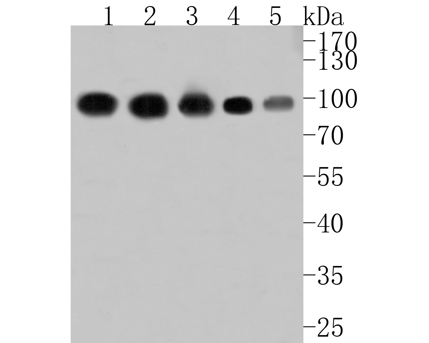
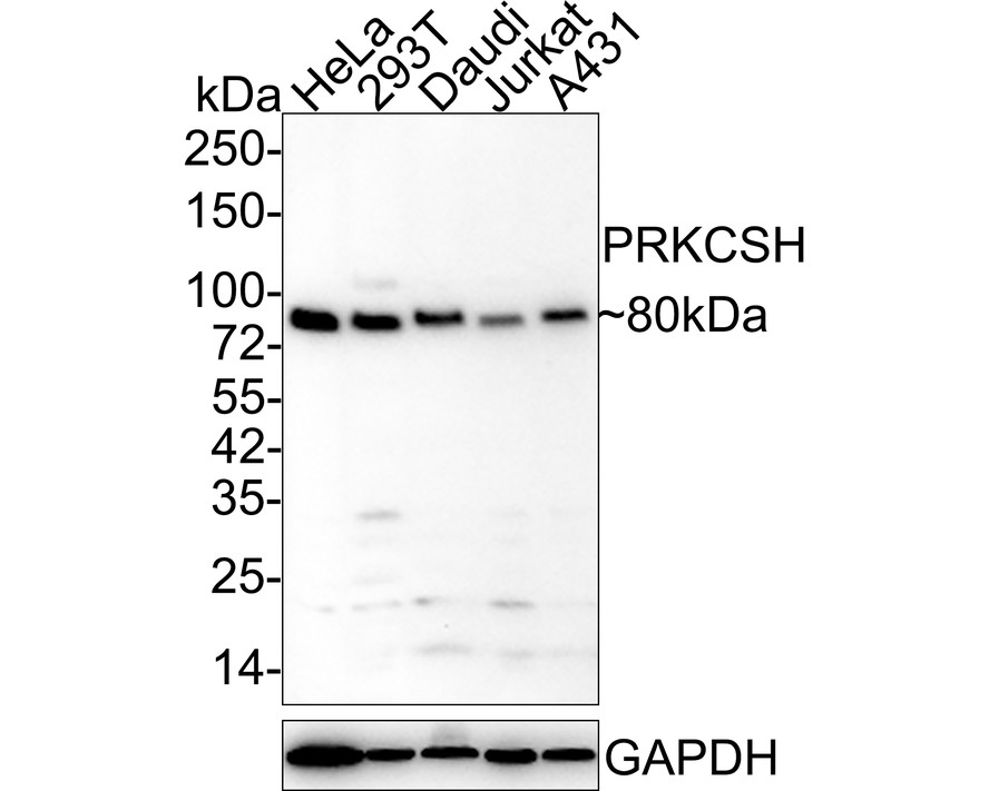
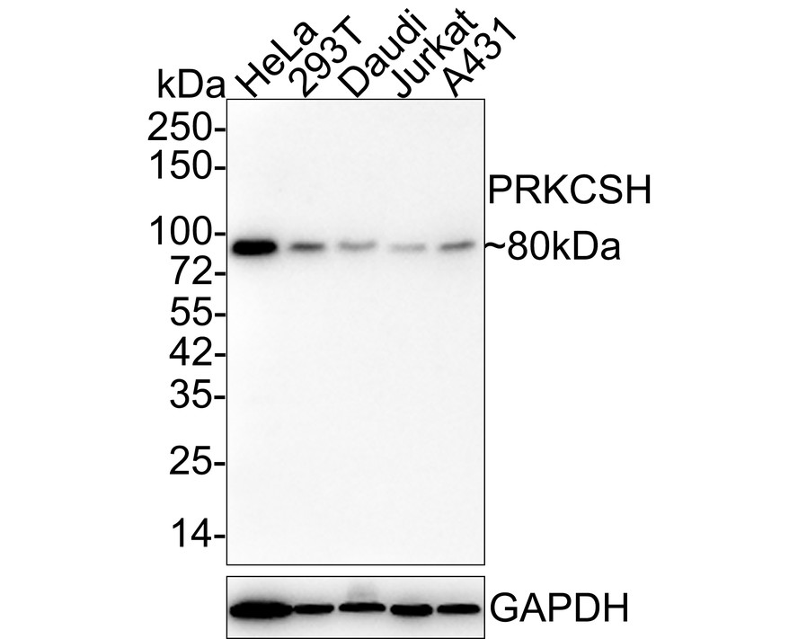
Western blot analysis of Glucosidase 2 subunit beta on different lysates. Proteins were transferred to a PVDF membrane and blocked with 5% BSA in PBS for 1 hour at room temperature. The primary antibody (HA500152, 1/1,000) was used in 5% BSA at room temperature for 2 hours. Goat Anti-Rabbit IgG - HRP Secondary Antibody (HA1001) at 1:200,000 dilution was used for 1 hour at room temperature.
Positive control:
Lane 1: Hela cell lysate
Lane 2: Jurkat cell lysate
Lane 3: A431 cell lysate
Lane 4: Rat kidney tissue lysate
Lane 5: Rat liver tissue lysate -
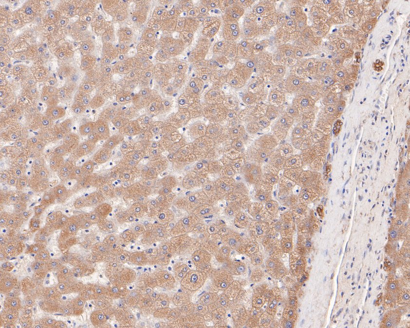
Immunohistochemical analysis of paraffin-embedded human liver tissue using anti-Glucosidase 2 subunit beta antibody. The section was pre-treated using heat mediated antigen retrieval with Tris-EDTA buffer (pH 8.0-8.4) for 20 minutes.The tissues were blocked in 5% BSA for 30 minutes at room temperature, washed with ddH2O and PBS, and then probed with the primary antibody (HA500152, 1/400) for 30 minutes at room temperature. The detection was performed using an HRP conjugated compact polymer system. DAB was used as the chromogen. Tissues were counterstained with hematoxylin and mounted with DPX.
-
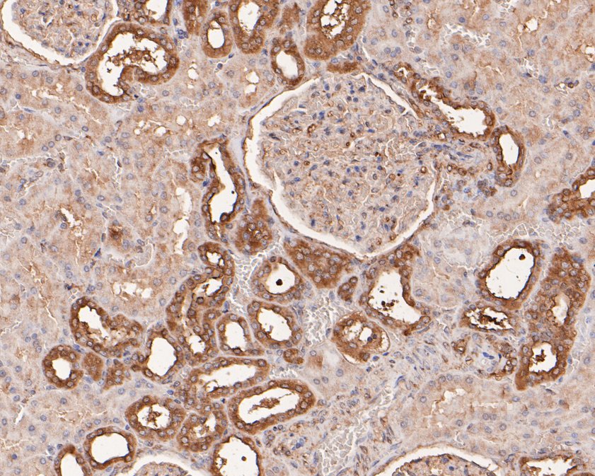
Immunohistochemical analysis of paraffin-embedded human kidney tissue using anti-Glucosidase 2 subunit beta antibody. The section was pre-treated using heat mediated antigen retrieval with Tris-EDTA buffer (pH 8.0-8.4) for 20 minutes.The tissues were blocked in 5% BSA for 30 minutes at room temperature, washed with ddH2O and PBS, and then probed with the primary antibody (HA500152, 1/400) for 30 minutes at room temperature. The detection was performed using an HRP conjugated compact polymer system. DAB was used as the chromogen. Tissues were counterstained with hematoxylin and mounted with DPX.
-
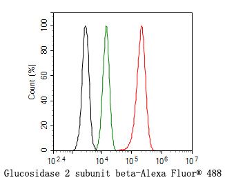
Flow cytometric analysis of Hela cells labeling Glucosidase 2 subunit beta.
Cells were fixed and permeabilized. Then stained with the primary antibody (HA500152, 1ug/ml) (red) compared with Rabbit IgG Isotype Control (green). After incubation of the primary antibody at +4℃ for an hour, the cells were stained with a Alexa Fluor® 488 conjugate-Goat anti-Rabbit IgG Secondary antibody at 1/1,000 dilution for 30 minutes at +4℃. Unlabelled sample was used as a control (cells without incubation with primary antibody; black). -
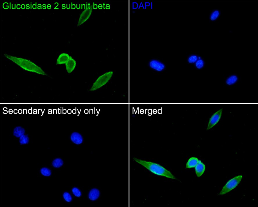
Immunocytochemistry analysis of SiHa cells labeling Glucosidase 2 subunit beta with Rabbit anti-Glucosidase 2 subunit beta antibody (HA500152) at 1/200 dilution.
Cells were fixed in 4% paraformaldehyde for 10 minutes at 37 ℃, permeabilized with 0.05% Triton X-100 in PBS for 20 minutes, and then blocked with 2% negative goat serum for 30 minutes at room temperature. Cells were then incubated with Rabbit anti-Glucosidase 2 subunit beta antibody (HA500152) at 1/200 dilution in 2% negative goat serum overnight at 4 ℃. Goat Anti-Rabbit IgG H&L (iFluor™ 488, HA1121) was used as the secondary antibody at 1/1,000 dilution. PBS instead of the primary antibody was used as the secondary antibody only control. Nuclear DNA was labelled in blue with DAPI.
Please note: All products are "FOR RESEARCH USE ONLY AND ARE NOT INTENDED FOR DIAGNOSTIC OR THERAPEUTIC USE"
Alternative Products
Glucosidase 2 subunit beta Mouse Monoclonal Antibody [A6H9]
Application: WB,IF-Cell,IHC-P,FC
Reactivity: Human
Conjugate: unconjugated
Glucosidase 2 subunit beta Recombinant Mouse Monoclonal Antibody [A6H9-R]
Application: WB
Reactivity: Human
Conjugate: unconjugated
