CD163 Recombinant Rabbit Monoclonal Antibody [JA51-30]


Catalog# ET1704-43
CD163 Recombinant Rabbit Monoclonal Antibody [JA51-30]
-
WB
-
IHC-P
-
mIHC
-
IF-Tissue
-
IP
-
Human
概述
产品名称
CD163 Recombinant Rabbit Monoclonal Antibody [JA51-30]
抗体类型
Recombinant Rabbit monoclonal Antibody
免疫原
Recombinant protein within Human CD163 aa 1012-1149 / 1156.
种属反应性
Human
验证应用
WB, IHC-P, mIHC, IF-Tissue, IP
分子量
Predicted band size: 125 kDa
阳性对照
Human pancreatic carcinoma, human cervical cancer, human lung tissue lysates, human liver tissue lysates, human liver tissue, human spleen tissue, human placenta tissue, human tonsil tissue.
偶联
unconjugated
克隆号
JA51-30
RRID
产品特性
形态
Liquid
浓度
1ug/ul
存放说明
Store at +4℃ after thawing. Aliquot store at -20℃ or -80℃. Avoid repeated freeze / thaw cycles.
存储缓冲液
1*TBS (pH7.4), 0.05% BSA, 40% Glycerol. Preservative: 0.05% Sodium Azide.
亚型
IgG
纯化方式
Protein A affinity purified.
应用稀释度
-
WB
-
1:500-1:2,000
-
IHC-P
-
1:1,000
-
mIHC
-
1:2,000-1:3,000
-
IF-Tissue
-
1:200
-
IP
-
Use at an assay dependent concentration.
发表文章中的应用
发表文章中的种属
| Mouse | See 1 publications below |
| Human | See 1 publications below |
靶点
功能
CD163, also designated M130, is a macrophage-associated antigen that is a member of the scavenger receptor cysteine-rich (SRCR) superfamily. It is highly expressed on macrogphages and to a lesser extent on monocytes. The acute phase-regulated and signal-inducing macrophage protein, CD163, is a receptor that scavenges hemoglobin by mediating endocytosis of haptoglobin-hemoglobin complexes. CD163 binds only haptoglobin and hemoglobin in complex, which indicates the exposure of a receptor-binding neoepitope. The receptor-ligand interaction is calcium-dependent and of high affinity. The existence of several CD163 isoforms, which differ in the structure of their cytoplasmic domains and putative phosphorylation sites, suggests that these isoforms also differ in their signaling mechanism. The gene which encodes CD163 maps to human chromosome 12p13.31.
背景文献
1. Sato Y et al. The PD-1/PD-L1 axis may be aberrantly activated in occupational cholangiocarcinoma.Pathol Int 67(3):163-170 (2017).
2. Chen H et al. An Agonist of the Protective Factor SIRT1 Improves Functional Recovery and Promotes Neuronal Survival by Attenuating Inflammation after Spinal Cord Injury. J Neurosci 37:2916-2930 (2017).
组织特异性
Expressed in monocytes and mature macrophages such as Kupffer cells in the liver, red pulp macrophages in the spleen, cortical macrophages in the thymus, resident bone marrow macrophages and meningeal macrophages of the central nervous system. Expressed also in blood. Isoform 1 is the lowest abundant in the blood. Isoform 2 is the lowest abundant in the liver and the spleen. Isoform 3 is the predominant isoform detected in the blood.
翻译后修饰
A soluble form (sCD163) is produced by proteolytic shedding which can be induced by lipopolysaccharide, phorbol ester and Fc region of immunoglobulin gamma. This cleavage is dependent on protein kinase C and tyrosine kinases and can be blocked by protease inhibitors. The shedding is inhibited by the tissue inhibitor of metalloproteinase TIMP3, and thus probably induced by membrane-bound metalloproteinases ADAMs.; Phosphorylated.
亚细胞定位
Secreted, Cell membrane.
UNIPROT #
别名
C163A_HUMAN antibody
CD 163 antibody
CD163 antibody
CD163 antigen antibody
CD163 molecule antibody
Hemoglobin scavenger receptor antibody
M130 antibody
M130 antigen precursor antibody
Macrophage associated antigen antibody
MM130 antibody
展开图片
-
Fluorescence multiplex immunohistochemical analysis of the human cervical cancer (Formalin/PFA-fixed paraffin-embedded sections). Panel A: the merged image of anti-CD14 (ET1610-85, red), anti-S100A9 (ET1702-73, green), anti-CD68 (HA601115, cyan), anti-panCK (HA601138, magenta) and anti-CD163 (ET1704-43, yellow) on human cervical cancer. Panel B: anti- CD14 stained on monocyte and MDSCs. Panel C: anti-S100A9 stained on MDSCs. Panel D: anti-CD68 stained on macrophage M1 and macrophage M2. Panel E: anti-panCK stained on tumor cells. Panel F: anti-CD163 stained on macrophage M2. HRP Conjugated UltraPolymer Goat Polyclonal Antibody HA1119/HA1120 was used as a secondary antibody. The immunostaining was performed with the Sequential Immuno-staining Kit (IRISKit™MH010101, www.luminiris.cn). The section was incubated in five rounds of staining: in the order of ET1610-85 (1/1,000 dilution), ET1702-73 (1/1,000 dilution), HA601115 (1/2,000 dilution), HA601138 (1/3,000 dilution), and ET1704-43 (1/2,000 dilution) for 20 mins at room temperature. Each round was followed by a separate fluorescent tyramide signal amplification system. Heat mediated antigen retrieval with Tris-EDTA buffer (pH 9.0) for 30 mins at 95℃. DAPI (blue) was used as a nuclear counter stain. Image acquisition was performed with Olympus VS200 Slide Scanner.
-
Fluorescence multiplex immunohistochemical analysis of the human pancreatic carcinoma (Formalin/PFA-fixed paraffin-embedded sections). Panel A: the merged image of anti-CD68 (EM1901-95, green), anti-CD163 (ET1704-43, red) and anti-PanCK (HA601094, violet) on human pancreatic carcinoma. Panel B: anti- CD68 stained on M1 macrophages. Panel C: anti-CD163 stained on M2 macrophages cells. Panel D: anti-panCK stained on cancer cells. HRP Conjugated UltraPolymer Goat Polyclonal Antibody HA1119/HA1120 was used as a secondary antibody. The immunostaining was performed with the Sequential Immuno-staining Kit (IRISKit™MH010101, www.luminiris.cn). The section was incubated in three rounds of staining: in the order of EM1901-95 (1/3,000 dilution), ET1704-43 (1/3,000 dilution), and HA601094 (1/3,000 dilution) for 20 mins at room temperature. Each round was followed by a separate fluorescent tyramide signal amplification system. Heat mediated antigen retrieval with Tris-EDTA buffer (pH 9.0) for 30 mins at 95℃. DAPI (blue) was used as a nuclear counter stain. Image acquisition was performed with Nikon ECLIPSE Ni-E microscope.
-
Fluorescence multiplex immunohistochemical analysis of human cervical carcinoma (Formalin/PFA-fixed paraffin-embedded sections). Panel A: the merged image of anti-S100A9 (ET1702-73, White), anti-CD117 (HA721154, Red) and anti-CD163(ET1704-43, Yellow) on human cervical carcinoma. HRP Conjugated UltraPolymer Goat Polyclonal Antibody HA1119/HA1120 was used as a secondary antibody. The immunostaining was performed with the Sequential Immuno-staining Kit (IRISKit™MH010101, www.luminiris.cn). The section was incubated in three rounds of staining: in the order of ET1702-73 (1/1,000 dilution), HA721154 (1/1,000 dilution) and ET1704-43 (1/2,000 dilution) for 20 mins at room temperature. Each round was followed by a separate fluorescent tyramide signal amplification system. Heat mediated antigen retrieval with Tris-EDTA buffer (pH 9.0) for 30 mins at 95℃. DAPI (blue) was used as a nuclear counter stain. Image acquisition was performed with Zeiss Observer 7 Inverted Fluorescence Microscope.
-
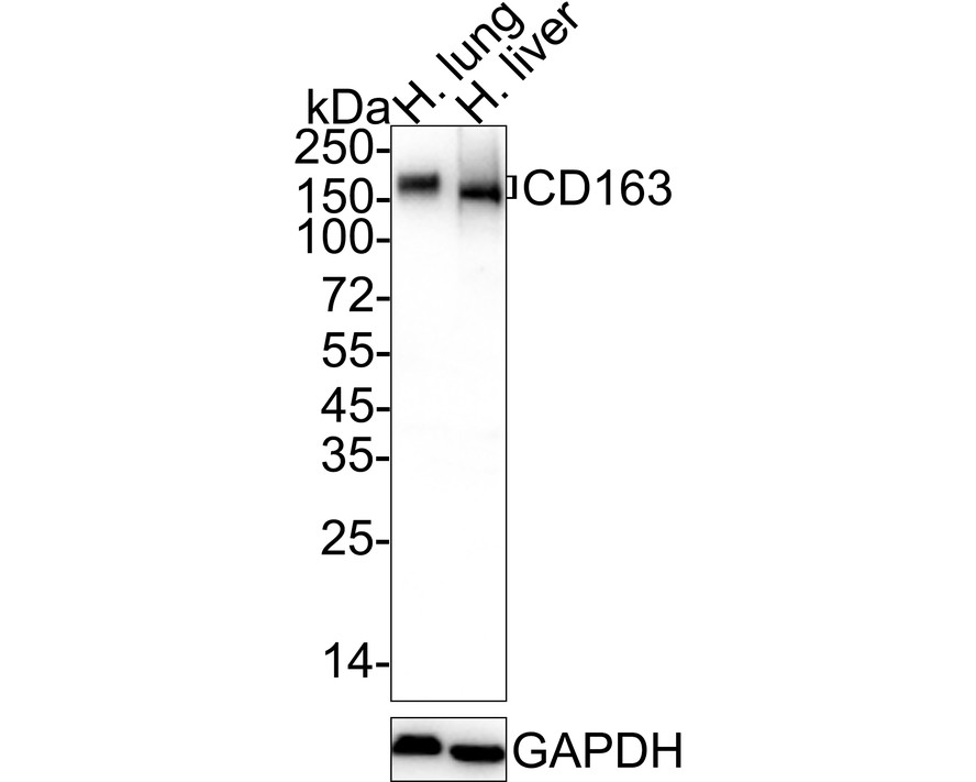
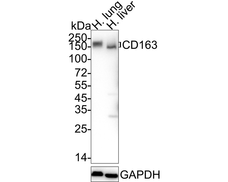
Western blot analysis of CD163 on different lysates with Rabbit anti-CD163 antibody (ET1704-43) at 1/1,000 dilution.
Lane 1: Human lung tissue lysate
Lane 2: Human liver tissue lysate
Lysates/proteins at 40 µg/Lane.
Predicted band size: 125 kDa
Observed band size: 150-170 kDa
Exposure time: 1 minute; ECL: K1801;
4-20% SDS-PAGE gel.
Proteins were transferred to a PVDF membrane and blocked with 5% NFDM/TBST for 1 hour at room temperature. The primary antibody (ET1704-43) at 1/1,000 dilution was used in 5% NFDM/TBST at 4℃ overnight. Goat Anti-Rabbit IgG - HRP Secondary Antibody (HA1001) at 1/50,000 dilution was used for 1 hour at room temperature. -
Immunohistochemical analysis of paraffin-embedded human liver tissue with Rabbit anti-CD163 antibody (ET1704-43) at 1/1,000 dilution.
The section was pre-treated using heat mediated antigen retrieval with Tris-EDTA buffer (pH 9.0) for 20 minutes. The tissues were blocked in 1% BSA for 20 minutes at room temperature, washed with ddH2O and PBS, and then probed with the primary antibody (ET1704-43) at 1/1,000 dilution for 1 hour at room temperature. The detection was performed using an HRP conjugated compact polymer system. DAB was used as the chromogen. Tissues were counterstained with hematoxylin and mounted with DPX. -
Immunohistochemical analysis of paraffin-embedded human spleen tissue with Rabbit anti-CD163 antibody (ET1704-43) at 1/1,000 dilution.
The section was pre-treated using heat mediated antigen retrieval with Tris-EDTA buffer (pH 9.0) for 20 minutes. The tissues were blocked in 1% BSA for 20 minutes at room temperature, washed with ddH2O and PBS, and then probed with the primary antibody (ET1704-43) at 1/1,000 dilution for 1 hour at room temperature. The detection was performed using an HRP conjugated compact polymer system. DAB was used as the chromogen. Tissues were counterstained with hematoxylin and mounted with DPX. -
Immunohistochemical analysis of paraffin-embedded human placenta tissue with Rabbit anti-CD163 antibody (ET1704-43) at 1/1,000 dilution.
The section was pre-treated using heat mediated antigen retrieval with Tris-EDTA buffer (pH 9.0) for 20 minutes. The tissues were blocked in 1% BSA for 20 minutes at room temperature, washed with ddH2O and PBS, and then probed with the primary antibody (ET1704-43) at 1/1,000 dilution for 1 hour at room temperature. The detection was performed using an HRP conjugated compact polymer system. DAB was used as the chromogen. Tissues were counterstained with hematoxylin and mounted with DPX. -
Immunohistochemical analysis of paraffin-embedded human tonsil tissue with Rabbit anti-CD163 antibody (ET1704-43) at 1/1,000 dilution.
The section was pre-treated using heat mediated antigen retrieval with Tris-EDTA buffer (pH 9.0) for 20 minutes. The tissues were blocked in 1% BSA for 20 minutes at room temperature, washed with ddH2O and PBS, and then probed with the primary antibody (ET1704-43) at 1/1,000 dilution for 1 hour at room temperature. The detection was performed using an HRP conjugated compact polymer system. DAB was used as the chromogen. Tissues were counterstained with hematoxylin and mounted with DPX.
Please note: All products are "FOR RESEARCH USE ONLY AND ARE NOT INTENDED FOR DIAGNOSTIC OR THERAPEUTIC USE"
引文
-
Transcription factor EHF drives cholangiocarcinoma development through transcriptional activation of glioma-associated oncogene homolog 1 and chemokine CCL2
Author: Luo Yiming,et al
PMID: 38741887
应用: WB
反应种属: Mouse
发表时间: 2024 May
-
 Citation
-
BRAFV600E mutant colorectal cancer cells mediate local immunosuppressive microenvironment through exosomal long noncoding RNAs
Author: Zhi, J., Jia, X. J., Yan, J., Wang, H. C., Feng, B., Xing, H. Y., & Jia, Y. T.
PMID: 35070047

应用: IHC
反应种属: Human
发表时间: 2021 Dec
-
Citation
同靶点&同通路的产品

CD163 Mouse Monoclonal Antibody [A3B5]
Application: IHC-P,FC
Reactivity: Human
Conjugate: unconjugated
CD163 Rabbit Polyclonal Antibody
Application: WB,IHC-P,IF-Cell
Reactivity: Human,Mouse,Rat
Conjugate: unconjugated