Dnmt1 Recombinant Rabbit Monoclonal Antibody [JF09-89]

Catalog# ET1702-77
Dnmt1 Recombinant Rabbit Monoclonal Antibody [JF09-89]
-
WB
-
IF-Cell
-
IF-Tissue
-
IHC-P
-
Human
-
Mouse
-
Rat
概述
产品名称
Dnmt1 Recombinant Rabbit Monoclonal Antibody [JF09-89]
抗体类型
Recombinant Rabbit monoclonal Antibody
免疫原
Synthetic peptide within Human Dnmt1 aa 1,506-1,549 / 1,616.
种属反应性
Human, Mouse, Rat
验证应用
WB, IF-Cell, IF-Tissue, IHC-P
分子量
Predicted band size: 183 kDa
阳性对照
HepG2 cell lysates, Hela, HepG2, 293T, human lymph nodes tissue, F9.
偶联
unconjugated
克隆号
JF09-89
RRID
产品特性
形态
Liquid
浓度
1ug/ul
存放说明
Store at +4℃ after thawing. Aliquot store at -20℃ or -80℃. Avoid repeated freeze / thaw cycles.
存储缓冲液
1*TBS (pH7.4), 0.05% BSA, 40% Glycerol. Preservative: 0.05% Sodium Azide.
亚型
IgG
纯化方式
Protein A affinity purified.
应用稀释度
-
WB
-
1:500-1:2,000
-
IF-Cell
-
1:50-1:200
-
IF-Tissue
-
1:50-1:200
-
IHC-P
-
1:50-1:1.000
发表文章中的应用
靶点
功能
Methylation at the 5'-position of cytosine is the only known naturally occurring covalent modification of the mammalian genome. DNA methylation requires the enzymatic activity of DNA 5-cytosine methyltransferase (Dnmt) proteins, which catalyze the transfer of a methyl group from S-adenosyl methionine to the 5'-position of cytosines residing in the dinucleotide CpG motif, and this methylation results in transcriptional repression of the target gene. The Dnmt enzymes are encoded by independent genes. Dnmt1 is the most abundant, and it preferentially methylates hemimethylated DNA and coordinates gene expression during development. Additional mammalian Dnmt proteins include Dnmt2 and Dnmt3. Dnmt2 lacks the large N-terminal regulator domain of Dnmt1, is expressed at substantially lower levels in adult tissues, and is likely involved in methylating newly integrated retroviral DNA. Dnmt3a and Dnmt3b are encoded by two distinct genes, but both are abundantly expressed in embryonic stem cells, where they also methylate CpG motifs on DNA.
背景文献
1. Liu R et al. Dnmt1 regulates the myogenic lineage specification of muscle stem cells. Sci Rep 6:35355 (2016).
2. Chalertpet K et al. Human papillomavirus type 16 E7 oncoprotein mediates CCNA1 promoter methylation. Cancer Sci 106:1333-40 (2015).
序列相似性
Belongs to the class I-like SAM-binding methyltransferase superfamily. C5-methyltransferase family.
组织特异性
Ubiquitous; highly expressed in fetal tissues, heart, kidney, placenta, peripheral blood mononuclear cells, and expressed at lower levels in spleen, lung, brain, small intestine, colon, liver, and skeletal muscle. Isoform 2 is less expressed than isoform 1.
翻译后修饰
Sumoylated; sumoylation increases activity.; Acetylation on multiple lysines, mainly by KAT2B/PCAF, regulates cell cycle G(2)/M transition. Deacetylation of Lys-1349 and Lys-1415 by SIRT1 increases methyltransferase activity.; Phosphorylation of Ser-154 by CDKs is important for enzymatic activity and protein stability. Phosphorylation of Ser-143 by AKT1 prevents methylation by SETD7 therebye increasing DNMT1 stability.; Methylation at Lys-142 by SETD7 promotes DNMT1 proteasomal degradation.; Ubiquitinated by UHRF1; interaction with USP7 counteracts ubiquitination by UHRF1 by promoting deubiquitination and preventing degradation by the proteasome.
亚细胞定位
Nucleus.
UNIPROT #
别名
ADCADN antibody
AIM antibody
CXXC finger protein 9 antibody
CXXC-type zinc finger protein 9 antibody
CXXC9 antibody
DNA (cytosine 5 ) methyltransferase 1 antibody
DNA (cytosine-5)-methyltransferase 1 antibody
DNA methyltransferase 1 antibody
DNA methyltransferase HsaI antibody
DNA methyltransferase M.HsaI. antibody
展开图片
-
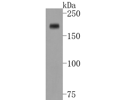
Western blot analysis of Dnmt1 on HepG2 cell lysates. Proteins were transferred to a PVDF membrane and blocked with 5% BSA in PBS for 1 hour at room temperature. The primary antibody (ET1702-77, 1/500) was used in 5% BSA at room temperature for 2 hours. Goat Anti-Rabbit IgG - HRP Secondary Antibody (HA1001) at 1:5,000 dilution was used for 1 hour at room temperature.
-
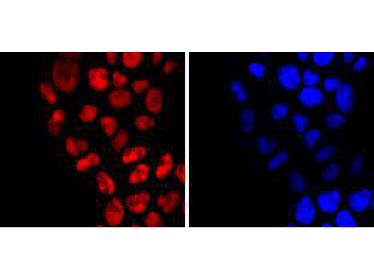
ICC staining of Dnmt1 in Hela cells (red). Formalin fixed cells were permeabilized with 0.1% Triton X-100 in TBS for 10 minutes at room temperature and blocked with 1% Blocker BSA for 15 minutes at room temperature. Cells were probed with the primary antibody (ET1702-77, 1/50) for 1 hour at room temperature, washed with PBS. Alexa Fluor®594 Goat anti-Rabbit IgG was used as the secondary antibody at 1/1,000 dilution. The nuclear counter stain is DAPI (blue).
-
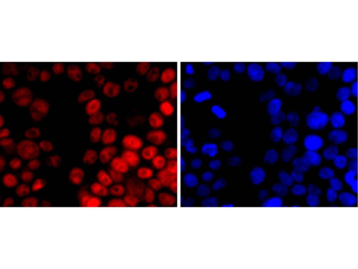
ICC staining of Dnmt1 in 293T cells (red). Formalin fixed cells were permeabilized with 0.1% Triton X-100 in TBS for 10 minutes at room temperature and blocked with 1% Blocker BSA for 15 minutes at room temperature. Cells were probed with the primary antibody (ET1702-77, 1/50) for 1 hour at room temperature, washed with PBS. Alexa Fluor®594 Goat anti-Rabbit IgG was used as the secondary antibody at 1/1,000 dilution. The nuclear counter stain is DAPI (blue).
-
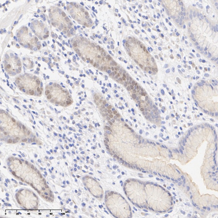
Immunohistochemical analysis of paraffin-embedded human lymph nodes tissue with Rabbit anti-Dnmt1 antibody (ET1702-77) at 1/200 dilution.
The section was pre-treated using heat mediated antigen retrieval with sodium citrate buffer (pH 6.0) for 2 minutes. The tissues were blocked in 1% BSA for 20 minutes at room temperature, washed with ddH2O and PBS, and then probed with the primary antibody (ET1702-77) at 1/200 dilution for 1 hour at room temperature. The detection was performed using an HRP conjugated compact polymer system. DAB was used as the chromogen. Tissues were counterstained with hematoxylin and mounted with DPX. -
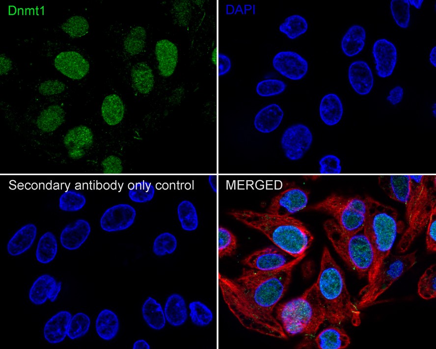
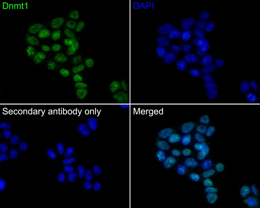
Immunocytochemistry analysis of HepG2 cells labeling Dnmt1 with Rabbit anti-Dnmt1 antibody (ET1702-77) at 1/100 dilution.
Cells were fixed in 4% paraformaldehyde for 20 minutes at room temperature, permeabilized with 0.1% Triton X-100 in PBS for 5 minutes at room temperature, then blocked with 1% BSA in 10% negative goat serum for 1 hour at room temperature. Cells were then incubated with Rabbit anti-Dnmt1 antibody (ET1702-77) at 1/100 dilution in 1% BSA in PBST overnight at 4 ℃. Goat Anti-Rabbit IgG H&L (iFluor™ 488, HA1121) was used as the secondary antibody at 1/1,000 dilution. PBS instead of the primary antibody was used as the secondary antibody only control. Nuclear DNA was labelled in blue with DAPI.
Beta tubulin (M1305-2, red) was stained at 1/100 dilution overnight at +4℃. Goat Anti-Mouse IgG H&L (iFluor™ 594, HA1126) was used as the secondary antibody at 1/1,000 dilution. -
Immunocytochemistry analysis of F9 cells labeling Dnmt1 with Rabbit anti-Dnmt1 antibody (ET1702-77) at 1/50 dilution.
Cells were fixed in 4% paraformaldehyde for 10 minutes at 37 ℃, permeabilized with 0.05% Triton X-100 in PBS for 20 minutes, and then blocked with 2% negative goat serum for 30 minutes at room temperature. Cells were then incubated with Rabbit anti-Dnmt1 antibody (ET1702-77) at 1/50 dilution in 2% negative goat serum overnight at 4 ℃. Goat Anti-Rabbit IgG H&L (iFluor™ 488, HA1121) was used as the secondary antibody at 1/1,000 dilution. PBS instead of the primary antibody was used as the secondary antibody only control. Nuclear DNA was labelled in blue with DAPI. -
Immunohistochemical analysis of paraffin-embedded human stomach tissue with Rabbit anti-Dnmt1 antibody (ET1702-77) at 1/200 dilution.
The section was pre-treated using heat mediated antigen retrieval with sodium citrate buffer (pH 6.0) for 2 minutes. The tissues were blocked in 1% BSA for 20 minutes at room temperature, washed with ddH2O and PBS, and then probed with the primary antibody (ET1702-77) at 1/200 dilution for 1 hour at room temperature. The detection was performed using an HRP conjugated compact polymer system. DAB was used as the chromogen. Tissues were counterstained with hematoxylin and mounted with DPX. -
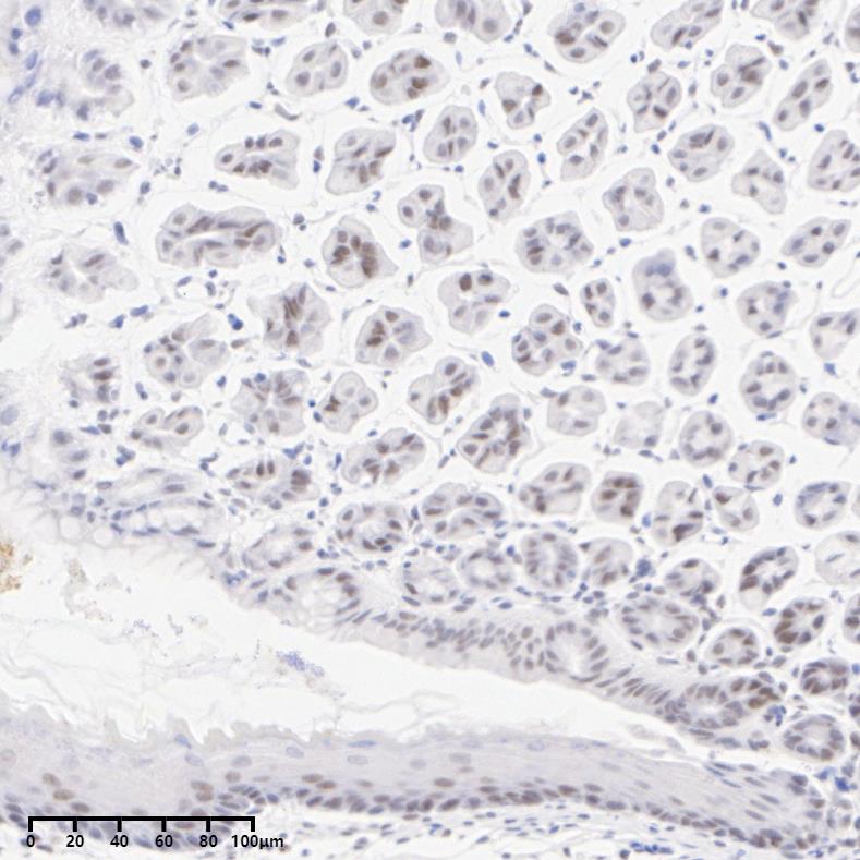
Immunohistochemical analysis of paraffin-embedded mouse stomach tissue with Rabbit anti-Dnmt1 antibody (ET1702-77) at 1/1,000 dilution.
The section was pre-treated using heat mediated antigen retrieval with sodium citrate buffer (pH 6.0) for 2 minutes. The tissues were blocked in 1% BSA for 20 minutes at room temperature, washed with ddH2O and PBS, and then probed with the primary antibody (ET1702-77) at 1/1,000 dilution for 1 hour at room temperature. The detection was performed using an HRP conjugated compact polymer system. DAB was used as the chromogen. Tissues were counterstained with hematoxylin and mounted with DPX. -
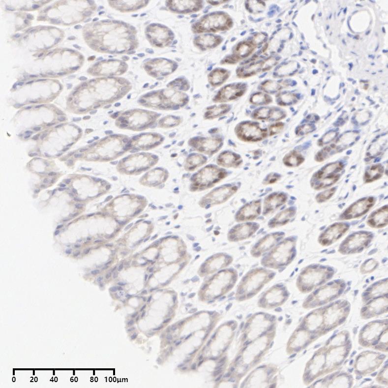
Immunohistochemical analysis of paraffin-embedded rat stomach tissue with Rabbit anti-Dnmt1 antibody (ET1702-77) at 1/200 dilution.
The section was pre-treated using heat mediated antigen retrieval with sodium citrate buffer (pH 6.0) for 2 minutes. The tissues were blocked in 1% BSA for 20 minutes at room temperature, washed with ddH2O and PBS, and then probed with the primary antibody (ET1702-77) at 1/200 dilution for 1 hour at room temperature. The detection was performed using an HRP conjugated compact polymer system. DAB was used as the chromogen. Tissues were counterstained with hematoxylin and mounted with DPX.
Please note: All products are "FOR RESEARCH USE ONLY AND ARE NOT INTENDED FOR DIAGNOSTIC OR THERAPEUTIC USE"
引文
-
Punicalagin protects against impaired skeletal muscle function in high-fat-diet-induced obese mice by regulating TET2
Author:
PMID: 36929898

应用: WB
反应种属: Mouse
发表时间: 2023 Apr
-
 Citation
-
Moderate DNA hypomethylation suppresses intestinal tumorigenesis by promoting caspase-3 expression and apoptosis. Oncogenesis, 10(5), 38.
Author: Duan, X., Huang, Y., Chen, X., Wang, W., Chen, J., Li, J., Yang, W., Li, J., Wu, Q., & Wong, J.
PMID: 33947834

应用: WB
反应种属: Mouse
发表时间: 2021 May
-
Citation
-
TXNIP, a novel key factor to cause Schwann cell dysfunction in diabetic peripheral neuropathy, under the regulation of PI3K/Akt pathway inhibition-induced DNMT1 and DNMT3a overexpression. Cell death & disease, 12(7), 642.
Author: Zhang, X., Zhao, S., Yuan, Q., Zhu, L., Li, F., Wang, H., Kong, D., & Hao, J.
PMID: 34162834

应用: WB,IHC,IF
反应种属:
发表时间: 2021 Jun
-
Citation
-
DNMT1, a Novel Regulator Mediating mTORC1/mTORC2 Pathway-Induced NGF Expression in Schwann Cells.
Author:
PMID: 30229399

应用: WB、ICC
反应种属: Rat
发表时间: 2018 Nov
-
Citation