PDHA1 Recombinant Rabbit Monoclonal Antibody [JF996-0]


Catalog# ET1702-75
PDHA1 Recombinant Rabbit Monoclonal Antibody [JF996-0]
-
WB
-
IF-Cell
-
IF-Tissue
-
IHC-P
-
IP
-
FC
-
Human
-
Mouse
-
Rat
概述
产品名称
PDHA1 Recombinant Rabbit Monoclonal Antibody [JF996-0]
抗体类型
Recombinant Rabbit monoclonal Antibody
免疫原
Synthetic peptide within Human PDHA1 aa 100-145 / 390.
种属反应性
Human, Mouse, Rat
验证应用
WB, IF-Cell, IF-Tissue, IHC-P, IP, FC
分子量
43 kDa
阳性对照
Rat kidney tissue lysate, mouse stomach tissue lysate, 293T cell lysate, A431 cell lysate, mouse heart tissue lysate, Hela, HepG2, SH-SY5Y, human breast carcinoma tissue, human kidney tissue, mouse skeletal muscle tissue, human lung carcinoma tissue, mouse colon tissue, mouse stomach tissue.
偶联
unconjugated
克隆号
JF996-0
RRID
产品特性
形态
Liquid
浓度
1ug/ul
存放说明
Store at +4℃ after thawing. Aliquot store at -20℃ or -80℃. Avoid repeated freeze / thaw cycles.
存储缓冲液
1*TBS (pH7.4), 0.05% BSA, 40% Glycerol. Preservative: 0.05% Sodium Azide.
亚型
IgG
纯化方式
Protein A affinity purified.
应用稀释度
-
WB
-
1:500-1:2,000
-
IF-Cell
-
1:100-1:500
-
IF-Tissue
-
1:100-1:500
-
IHC-P
-
1:50-1:1,000
-
FC
-
1:50-1:100
-
IP
-
Use at an assay dependent concentration.
靶点
功能
The pyruvate dehydrogenase (PDH) complex is a nuclear-encoded mitochondrial matrix enzyme complex that functions as the primary link between glycolysis and the tricarboxylic acid (TCA) cycle by catalyzing the irreversible conversion of pyruvate into acetyl-CoA. The E1 enzyme of the PDH complex is made up of a heterotetramer of two α and two β subunits. The E1-α subunit (PDH-E1α) contains the E1 active site and plays a key role in the function of the PDH complex. The PDH complex is regulated by phosphorylation and dephosphorylation of PDH-E1α. The gene encoding for PDH-E1α maps to chromosome Xp22.12, and a 20bp deletion in the last exon of this gene is sufficient to cause PDH deficiency, which causes a broad range of symptoms including the development of seizures, mental retardation and spasticity, as well as intermittent episodes of lactic acidosis associated with cerebellar ataxia.
背景文献
1. Zhang Y et al. Induction of Posttranslational Modifications of Mitochondrial Proteins by ATP Contributes to Negative Regulation of Mitochondrial Function. PLoS One 11:e0150454 (2016).
2. Vavrova E et al. Muscle expression of a malonyl-CoA-insensitive carnitine palmitoyltransferase-1 protects mice against high-fat/high-sucrose diet-induced insulin resistance. Am J Physiol Endocrinol Metab 311:E649-60 (2016).
组织特异性
Ubiquitous.
翻译后修饰
Phosphorylation at Ser-232, Ser-293 and Ser-300 by PDK family kinases inactivates the enzyme; for this phosphorylation at a single site is sufficient. Dephosphorylation at all three sites, i.e. at Ser-232, Ser-293 and Ser-300, is required for reactivation.; Acetylation alters the phosphorylation pattern. Deacetylated by SIRT3 (By similarity).
亚细胞定位
Mitochondrion matrix.
UNIPROT #
别名
ODPA_HUMAN antibody
PDH antibody
PDHA antibody
PDHA1 antibody
PDHCE1A antibody
PDHE1 A type I antibody
PDHE1-A type I antibody
PHE1A antibody
Pyruvate Dehydrogenase (lipoamide) alpha 1 antibody
Pyruvate dehydrogenase complex, E1 alpha polypeptide 1 antibody
展开图片
-
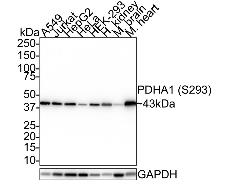
Western blot analysis of PDHA1 on different lysates. Proteins were transferred to a PVDF membrane and blocked with 5% BSA in PBS for 1 hour at room temperature. The primary antibody (ET1702-75, 1/500) was used in 5% BSA at room temperature for 2 hours. Goat Anti-Rabbit IgG - HRP Secondary Antibody (HA1001) at 1:5,000 dilution was used for 1 hour at room temperature.
Positive control:
Lane 1: rat kidney tissue lysate
Lane 2: mouse stomach tissue lysate -
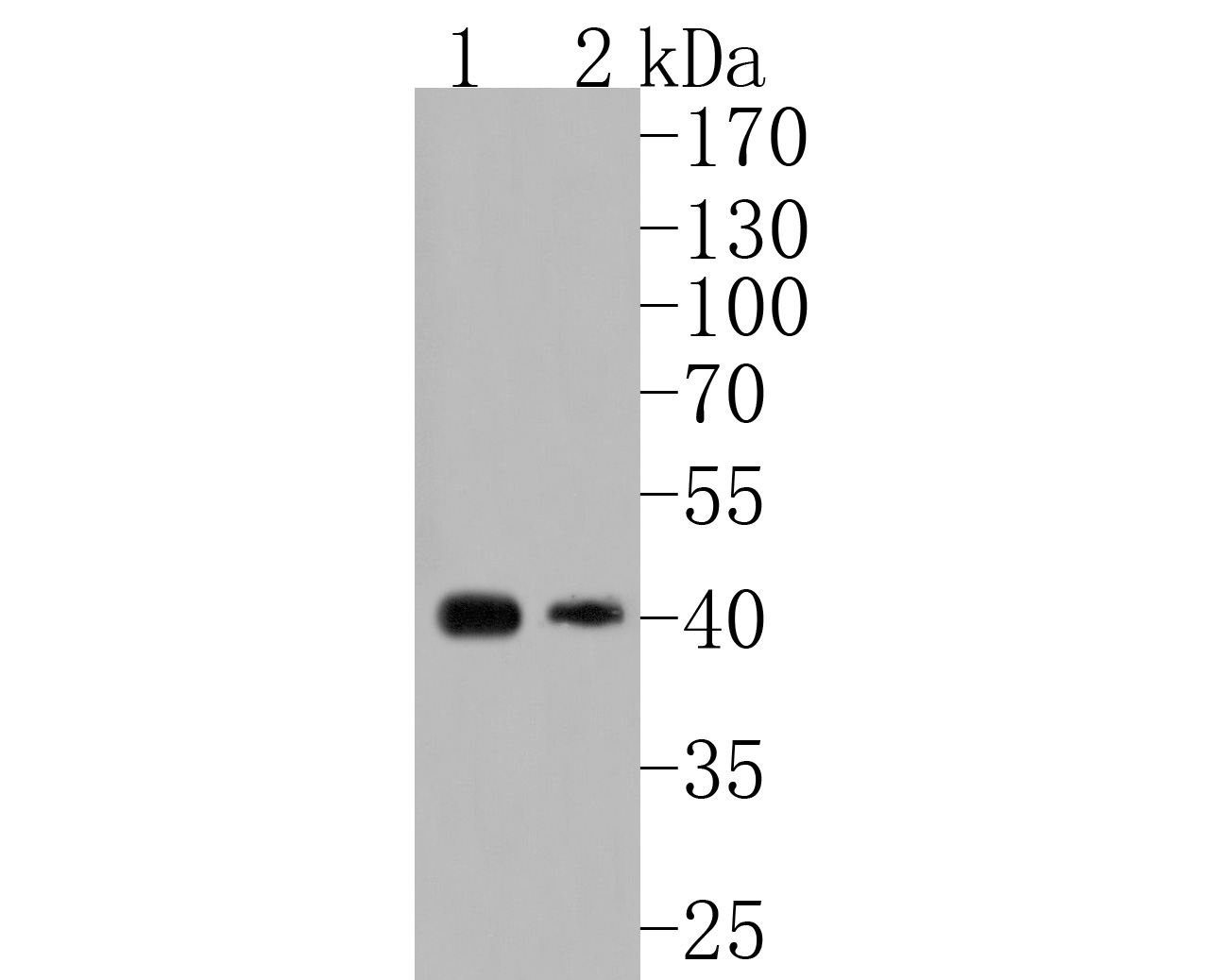
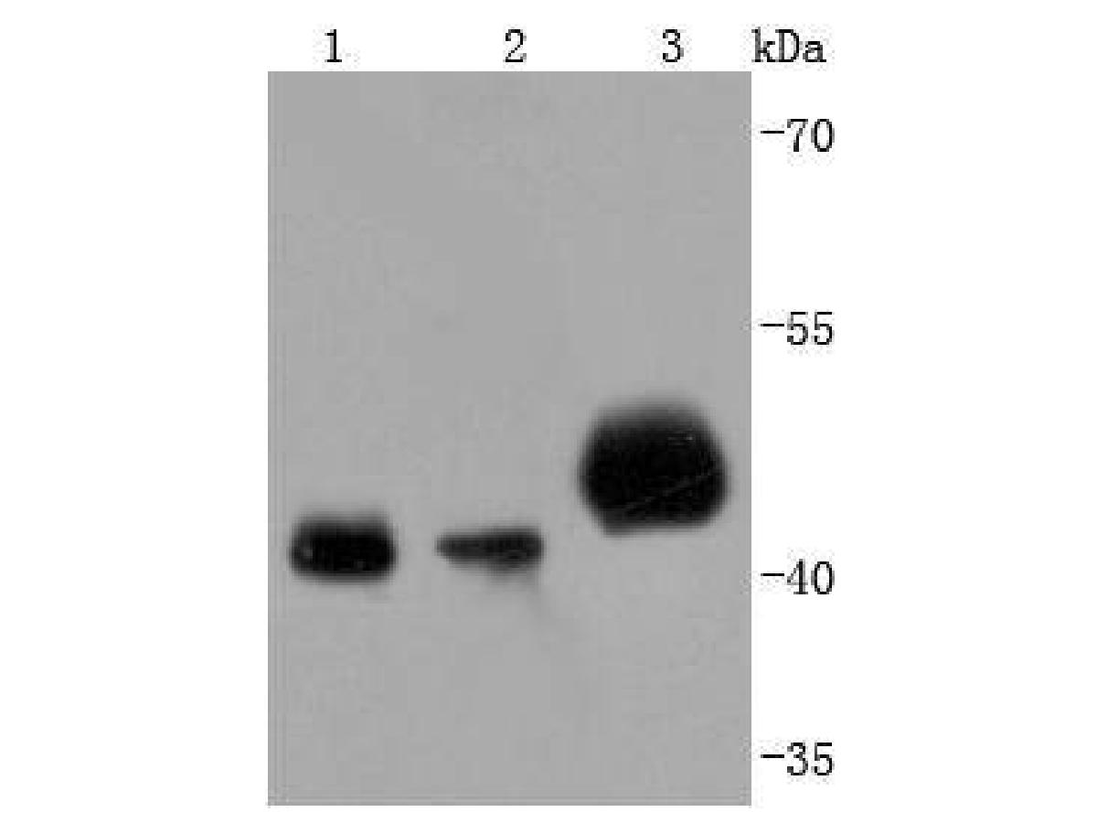
Western blot analysis of PDHA1 on different lysates. Proteins were transferred to a PVDF membrane and blocked with 5% BSA in PBS for 1 hour at room temperature. The primary antibody (ET1702-75, 1/500) was used in 5% BSA at room temperature for 2 hours. Goat Anti-Rabbit IgG - HRP Secondary Antibody (HA1001) at 1:5,000 dilution was used for 1 hour at room temperature.
Positive control:
Lane 1: 293T cell lysate
Lane 2: A431 cell lysate
Lane 3: mouse heart tissue lysate -
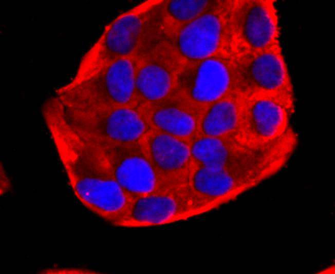
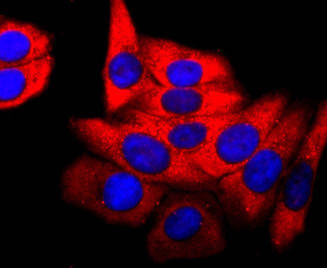
ICC staining of PDHA1 in Hela cells (red). Formalin fixed cells were permeabilized with 0.1% Triton X-100 in TBS for 10 minutes at room temperature and blocked with 1% Blocker BSA for 15 minutes at room temperature. Cells were probed with the primary antibody (ET1702-75, 1/50) for 1 hour at room temperature, washed with PBS. Alexa Fluor®594 Goat anti-Rabbit IgG was used as the secondary antibody at 1/1,000 dilution. The nuclear counter stain is DAPI (blue).
-
ICC staining of PDHA1 in HepG2 cells (red). Formalin fixed cells were permeabilized with 0.1% Triton X-100 in TBS for 10 minutes at room temperature and blocked with 1% Blocker BSA for 15 minutes at room temperature. Cells were probed with the primary antibody (ET1702-75, 1/50) for 1 hour at room temperature, washed with PBS. Alexa Fluor®594 Goat anti-Rabbit IgG was used as the secondary antibody at 1/1,000 dilution. The nuclear counter stain is DAPI (blue).
-
ICC staining of PDHA1 in SH-SY5Y cells (red). Formalin fixed cells were permeabilized with 0.1% Triton X-100 in TBS for 10 minutes at room temperature and blocked with 1% Blocker BSA for 15 minutes at room temperature. Cells were probed with the primary antibody (ET1702-75, 1/50) for 1 hour at room temperature, washed with PBS. Alexa Fluor®594 Goat anti-Rabbit IgG was used as the secondary antibody at 1/1,000 dilution. The nuclear counter stain is DAPI (blue).
-
Immunohistochemical analysis of paraffin-embedded human breast carcinoma tissue using anti-PDHA1 antibody. The section was pre-treated using heat mediated antigen retrieval with Tris-EDTA buffer (pH 8.0-8.4) for 20 minutes.The tissues were blocked in 5% BSA for 30 minutes at room temperature, washed with ddH2O and PBS, and then probed with the primary antibody (ET1702-75, 1/50) for 30 minutes at room temperature. The detection was performed using an HRP conjugated compact polymer system. DAB was used as the chromogen. Tissues were counterstained with hematoxylin and mounted with DPX.
-
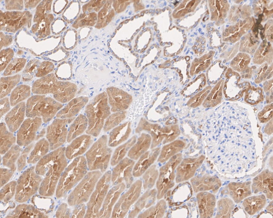
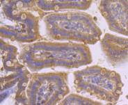
Immunohistochemical analysis of paraffin-embedded human kidney tissue with Rabbit anti-PDHA1 antibody (ET1702-75) at 1/1,000 dilution.
The section was pre-treated using heat mediated antigen retrieval with Tris-EDTA buffer (pH 9.0) for 20 minutes. The tissues were blocked in 1% BSA for 20 minutes at room temperature, washed with ddH2O and PBS, and then probed with the primary antibody (ET1702-75) at 1/1,000 dilution for 1 hour at room temperature. The detection was performed using an HRP conjugated compact polymer system. DAB was used as the chromogen. Tissues were counterstained with hematoxylin and mounted with DPX. -
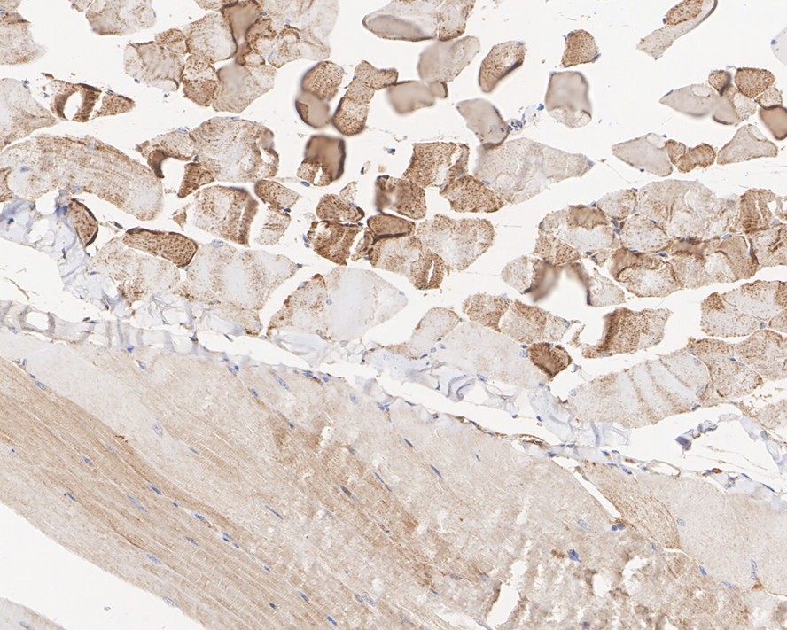
Immunohistochemical analysis of paraffin-embedded mouse skeletal muscle tissue with Rabbit anti-PDHA1 antibody (ET1702-75) at 1/1,000 dilution.
The section was pre-treated using heat mediated antigen retrieval with Tris-EDTA buffer (pH 9.0) for 20 minutes. The tissues were blocked in 1% BSA for 20 minutes at room temperature, washed with ddH2O and PBS, and then probed with the primary antibody (ET1702-75) at 1/1,000 dilution for 1 hour at room temperature. The detection was performed using an HRP conjugated compact polymer system. DAB was used as the chromogen. Tissues were counterstained with hematoxylin and mounted with DPX. -
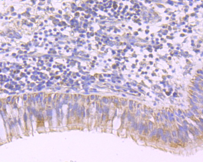
Immunohistochemical analysis of paraffin-embedded human lung carcinoma tissue using anti-PDHA1 antibody. The section was pre-treated using heat mediated antigen retrieval with Tris-EDTA buffer (pH 8.0-8.4) for 20 minutes.The tissues were blocked in 5% BSA for 30 minutes at room temperature, washed with ddH2O and PBS, and then probed with the primary antibody (ET1702-75, 1/50) for 30 minutes at room temperature. The detection was performed using an HRP conjugated compact polymer system. DAB was used as the chromogen. Tissues were counterstained with hematoxylin and mounted with DPX.
-
Immunohistochemical analysis of paraffin-embedded mouse colon tissue using anti-PDHA1 antibody. The section was pre-treated using heat mediated antigen retrieval with Tris-EDTA buffer (pH 8.0-8.4) for 20 minutes.The tissues were blocked in 5% BSA for 30 minutes at room temperature, washed with ddH2O and PBS, and then probed with the primary antibody (ET1702-75, 1/50) for 30 minutes at room temperature. The detection was performed using an HRP conjugated compact polymer system. DAB was used as the chromogen. Tissues were counterstained with hematoxylin and mounted with DPX.
-
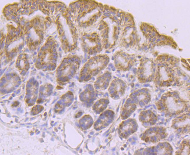
Immunohistochemical analysis of paraffin-embedded mouse stomach tissue using anti-PDHA1 antibody. The section was pre-treated using heat mediated antigen retrieval with Tris-EDTA buffer (pH 8.0-8.4) for 20 minutes.The tissues were blocked in 5% BSA for 30 minutes at room temperature, washed with ddH2O and PBS, and then probed with the primary antibody (ET1702-75, 1/50) for 30 minutes at room temperature. The detection was performed using an HRP conjugated compact polymer system. DAB was used as the chromogen. Tissues were counterstained with hematoxylin and mounted with DPX.
-
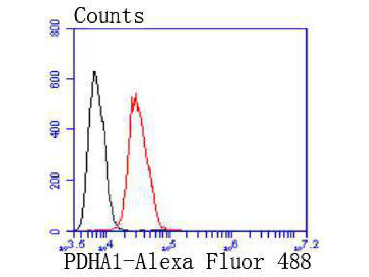
Flow cytometric analysis of PDHA1 was done on Hela cells. The cells were fixed, permeabilized and stained with the primary antibody (ET1702-75, 1/50) (red). After incubation of the primary antibody at room temperature for an hour, the cells were stained with a Alexa Fluor 488-conjugated Goat anti-Rabbit IgG Secondary antibody at 1/1000 dilution for 30 minutes.Unlabelled sample was used as a control (cells without incubation with primary antibody; black).
Please note: All products are "FOR RESEARCH USE ONLY AND ARE NOT INTENDED FOR DIAGNOSTIC OR THERAPEUTIC USE"
同靶点&同通路的产品