概述
产品名称
Histone H3 Recombinant Rabbit Monoclonal Antibody [JJ090-07]
抗体类型
Recombinant Rabbit monoclonal Antibody
免疫原
Recombinant protein within human Histone H3 aa 85-136/136.
种属反应性
Human, Mouse, Rat
验证应用
WB, IF-Cell, IF-Tissue, IHC-P, ChIP, IP
分子量
Predicted band size: 15 kDa
阳性对照
HeLa cell lysate, A549 cell lysate, HT-29 cell lysate, HEK-293 cell lysate, C2C12 cell lysate, L-929 cell lysate, C6 cell lysate, HeLa, human kidney tissue, human skin tissue, mouse liver tissue, mouse kidney tissue, rat liver tissue, rat kidney tissue, rat skin tissue, human liver tissue, mouse testis tissue, rat testis tissue.
偶联
unconjugated
克隆号
JJ090-07
RRID
产品特性
形态
Liquid
浓度
1ug/ul
存放说明
Store at +4℃ after thawing. Aliquot store at -20℃ or -80℃. Avoid repeated freeze / thaw cycles.
存储缓冲液
1*TBS (pH7.4), 0.05% BSA, 40% Glycerol. Preservative: 0.05% Sodium Azide.
亚型
IgG
纯化方式
Protein A affinity purified.
应用稀释度
-
WB
-
1:20,000
-
IF-Cell
-
1:100
-
IF-Tissue
-
1:100
-
IHC-P
-
1:5,000-1:10,000
-
ChIP
-
Use 0.5~2 μg for 25 μg of chromatin.
-
IP
-
1-2μg/sample
发表文章中的应用
| WB | 查看 6 篇文献如下 |
发表文章中的种属
| Mouse | 查看 3 篇文献如下 |
| Human | 查看 2 篇文献如下 |
| Rice | 查看 1 篇文献如下 |
靶点
功能
In eukaryotes, DNA is wrapped around histone octamers to form the basic unit of chromatin structure. The octamer is composed of histones H2A, H2B, H3 and H4, and it associates with approximately 200 base pairs of DNA to form the nucleosome. The association of DNA with histones results in dense packing of chromatin, which restricts proteins involved in gene transcription from binding to DNA. p300 preferentially acetylates Histone H3 at lysines 14 and 18 and Histone H4 at lysines 5 and 8. PCAF in its native form, primarily acetylates Histone H3 at lysine 14 to a monoacetylated form, and less efficiently acetylates Histone H4 at lysine 8. Histone H4 may also be acetylated at lysines 12 and 16, and the involvement of acetylated H4 with Histones H2A, H2B and H3 suggests that acetylated histones may be involved in dynamic chromatin remodeling.
背景文献
1. Wani S et al. Human SCP4 is a chromatin-associated CTD phosphatase and exhibits the dynamic translocation during erythroid differentiation. J Biochem 160:111-20 (2016).
2. Ni JZ et al. A transgenerational role of the germline nuclear RNAi pathway in repressing heat stress-induced transcriptional activation in C. elegans. Epigenetics Chromatin 9:3 (2016).
序列相似性
Belongs to the histone H3 family.
翻译后修饰
Acetylation is generally linked to gene activation. Acetylation on Lys-10 (H3K9ac) impairs methylation at Arg-9 (H3R8me2s). Acetylation on Lys-19 (H3K18ac) and Lys-24 (H3K24ac) favors methylation at Arg-18 (H3R17me). Acetylation at Lys-123 (H3K122ac) by EP300/p300 plays a central role in chromatin structure: localizes at the surface of the histone octamer and stimulates transcription, possibly by promoting nucleosome instability.; Citrullination at Arg-9 (H3R8ci) and/or Arg-18 (H3R17ci) by PADI4 impairs methylation and represses transcription.; Asymmetric dimethylation at Arg-18 (H3R17me2a) by CARM1 is linked to gene activation. Symmetric dimethylation at Arg-9 (H3R8me2s) by PRMT5 is linked to gene repression. Asymmetric dimethylation at Arg-3 (H3R2me2a) by PRMT6 is linked to gene repression and is mutually exclusive with H3 Lys-5 methylation (H3K4me2 and H3K4me3). H3R2me2a is present at the 3' of genes regardless of their transcription state and is enriched on inactive promoters, while it is absent on active promoters.; Methylation at Lys-5 (H3K4me), Lys-37 (H3K36me) and Lys-80 (H3K79me) are linked to gene activation. Methylation at Lys-5 (H3K4me) facilitates subsequent acetylation of H3 and H4. Methylation at Lys-80 (H3K79me) is associated with DNA double-strand break (DSB) responses and is a specific target for TP53BP1. Methylation at Lys-10 (H3K9me) and Lys-28 (H3K27me) are linked to gene repression. Methylation at Lys-10 (H3K9me) is a specific target for HP1 proteins (CBX1, CBX3 and CBX5) and prevents subsequent phosphorylation at Ser-11 (H3S10ph) and acetylation of H3 and H4. Methylation at Lys-5 (H3K4me) and Lys-80 (H3K79me) require preliminary monoubiquitination of H2B at 'Lys-120'. Methylation at Lys-10 (H3K9me) and Lys-28 (H3K27me) are enriched in inactive X chromosome chromatin. Monomethylation at Lys-57 (H3K56me1) by EHMT2/G9A in G1 phase promotes interaction with PCNA and is required for DNA replication.; Phosphorylated at Thr-4 (H3T3ph) by HASPIN during prophase and dephosphorylated during anaphase. Phosphorylation at Ser-11 (H3S10ph) by AURKB is crucial for chromosome condensation and cell-cycle progression during mitosis and meiosis. In addition phosphorylation at Ser-11 (H3S10ph) by RPS6KA4 and RPS6KA5 is important during interphase because it enables the transcription of genes following external stimulation, like mitogens, stress, growth factors or UV irradiation and result in the activation of genes, such as c-fos and c-jun. Phosphorylation at Ser-11 (H3S10ph), which is linked to gene activation, prevents methylation at Lys-10 (H3K9me) but facilitates acetylation of H3 and H4. Phosphorylation at Ser-11 (H3S10ph) by AURKB mediates the dissociation of HP1 proteins (CBX1, CBX3 and CBX5) from heterochromatin. Phosphorylation at Ser-11 (H3S10ph) is also an essential regulatory mechanism for neoplastic cell transformation. Phosphorylated at Ser-29 (H3S28ph) by MAP3K20 isoform 1, RPS6KA5 or AURKB during mitosis or upon ultraviolet B irradiation. Phosphorylation at Thr-7 (H3T6ph) by PRKCB is a specific tag for epigenetic transcriptional activation that prevents demethylation of Lys-5 (H3K4me) by LSD1/KDM1A. At centromeres, specifically phosphorylated at Thr-12 (H3T11ph) from prophase to early anaphase, by DAPK3 and PKN1. Phosphorylation at Thr-12 (H3T11ph) by PKN1 is a specific tag for epigenetic transcriptional activation that promotes demethylation of Lys-10 (H3K9me) by KDM4C/JMJD2C. Phosphorylation at Thr-12 (H3T11ph) by chromatin-associated CHEK1 regulates the transcription of cell cycle regulatory genes by modulating acetylation of Lys-10 (H3K9ac). Phosphorylation at Tyr-42 (H3Y41ph) by JAK2 promotes exclusion of CBX5 (HP1 alpha) from chromatin.; Monoubiquitinated by RAG1 in lymphoid cells, monoubiquitination is required for V(D)J recombination (By similarity). Ubiquitinated by the CUL4-DDB-RBX1 complex in response to ultraviolet irradiation. This may weaken the interaction between histones and DNA and facilitate DNA accessibility to repair proteins.; Lysine deamination at Lys-5 (H3K4all) to form allysine is mediated by LOXL2. Allysine formation by LOXL2 only takes place on H3K4me3 and results in gene repression.; Crotonylation (Kcr) is specifically present in male germ cells and marks testis-specific genes in post-meiotic cells, including X-linked genes that escape sex chromosome inactivation in haploid cells. Crotonylation marks active promoters and enhancers and confers resistance to transcriptional repressors. It is also associated with post-meiotically activated genes on autosomes.; Butyrylation of histones marks active promoters and competes with histone acetylation. It is present during late spermatogenesis.; Succinylation at Lys-80 (H3K79succ) by KAT2A takes place with a maximum frequency around the transcription start sites of genes. It gives a specific tag for epigenetic transcription activation. Desuccinylation at Lys-123 (H3K122succ) by SIRT7 in response to DNA damage promotes chromatin condensation and double-strand breaks (DSBs) repair.; Serine ADP-ribosylation constitutes the primary form of ADP-ribosylation of proteins in response to DNA damage. Serine ADP-ribosylation at Ser-11 (H3S10ADPr) is mutually exclusive with phosphorylation at Ser-11 (H3S10ph) and impairs acetylation at Lys-10 (H3K9ac).
亚细胞定位
Nucleus, Chromosome.
UNIPROT
别名
H3 histone family, member A antibody
H3/A antibody
H31_HUMAN antibody
H3FA antibody
Hist1h3a antibody
HIST1H3B antibody
HIST1H3C antibody
HIST1H3D antibody
HIST1H3E antibody
HIST1H3F antibody
展开H3 histone family, member A antibody
H3/A antibody
H31_HUMAN antibody
H3FA antibody
Hist1h3a antibody
HIST1H3B antibody
HIST1H3C antibody
HIST1H3D antibody
HIST1H3E antibody
HIST1H3F antibody
HIST1H3G antibody
HIST1H3H antibody
HIST1H3I antibody
HIST1H3J antibody
histone 1, H3a antibody
Histone cluster 1, H3a antibody
Histone H3.1 antibody
Histone H3/a antibody
Histone H3/b antibody
Histone H3/c antibody
Histone H3/d antibody
Histone H3/f antibody
Histone H3/h antibody
Histone H3/i antibody
Histone H3/j antibody
Histone H3/k antibody
Histone H3/l antibody
折叠图片
-

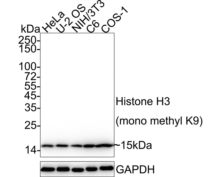
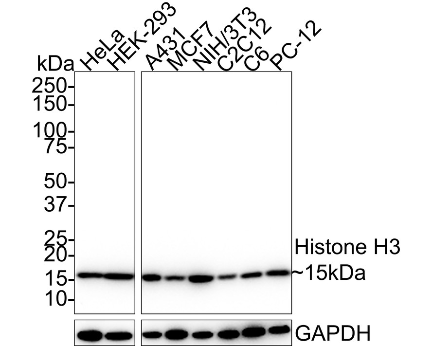
Western blot analysis of Histone H3 on different lysates with Rabbit anti-Histone H3 antibody (ET1701-64) at 1/20,000 dilution and competitor's antibody at 1/5,000 dilution.
Lane 1: HeLa cell lysate
Lane 2: A549 cell lysate
Lane 3: HT-29 cell lysate
Lane 4: HEK-293 cell lysate
Lane 5: C2C12 cell lysate
Lane 6: L-929 cell lysate
Lane 7: C6 cell lysate
Lysates/proteins at 10 µg/Lane.
Predicted band size: 15 kDa
Observed band size: 15 kDa
Exposure time: 18 seconds; ECL: K1801;
4-20% SDS-PAGE gel.
Proteins were transferred to a PVDF membrane and blocked with 5% NFDM/TBST for 1 hour at room temperature. The primary antibody (ET1701-64) at 1/20,000 dilution and competitor's antibody at 1/5,000 dilution were used in 5% NFDM/TBST at 4℃ overnight. Goat Anti-Rabbit IgG - HRP Secondary Antibody (HA1001) at 1/50,000 dilution was used for 1 hour at room temperature. -

Immunocytochemistry analysis of HeLa cells labeling Histone H3 with Rabbit anti-Histone H3 antibody (ET1701-64) at 1/100 dilution.
Cells were fixed in 4% paraformaldehyde for 10 minutes at 37 ℃, permeabilized with 0.05% Triton X-100 in PBS for 20 minutes, and then blocked with 2% negative goat serum for 30 minutes at room temperature. Cells were then incubated with Rabbit anti-Histone H3 antibody (ET1701-64) at 1/100 dilution in 2% negative goat serum overnight at 4 ℃. Goat Anti-Rabbit IgG H&L (iFluor™ 488, HA1121) was used as the secondary antibody at 1/1,000 dilution. PBS instead of the primary antibody was used as the secondary antibody only control. Nuclear DNA was labelled in blue with DAPI. -

Immunohistochemical analysis of paraffin-embedded human kidney tissue with Rabbit anti-Histone H3 antibody (ET1701-64) at 1/10,000 dilution.
The section was pre-treated using heat mediated antigen retrieval with sodium citrate buffer (pH 6.0) for 2 minutes. The tissues were blocked in 1% BSA for 20 minutes at room temperature, washed with ddH2O and PBS, and then probed with the primary antibody (ET1701-64) at 1/10,000 dilution for 1 hour at room temperature. The detection was performed using an HRP conjugated compact polymer system. DAB was used as the chromogen. Tissues were counterstained with hematoxylin and mounted with DPX. -

Immunohistochemical analysis of paraffin-embedded human skin tissue with Rabbit anti-Histone H3 antibody (ET1701-64) at 1/5,000 dilution.
The section was pre-treated using heat mediated antigen retrieval with sodium citrate buffer (pH 6.0) for 2 minutes. The tissues were blocked in 1% BSA for 20 minutes at room temperature, washed with ddH2O and PBS, and then probed with the primary antibody (ET1701-64) at 1/5,000 dilution for 1 hour at room temperature. The detection was performed using an HRP conjugated compact polymer system. DAB was used as the chromogen. Tissues were counterstained with hematoxylin and mounted with DPX. -

Immunohistochemical analysis of paraffin-embedded mouse liver tissue with Rabbit anti-Histone H3 antibody (ET1701-64) at 1/10,000 dilution.
The section was pre-treated using heat mediated antigen retrieval with sodium citrate buffer (pH 6.0) for 2 minutes. The tissues were blocked in 1% BSA for 20 minutes at room temperature, washed with ddH2O and PBS, and then probed with the primary antibody (ET1701-64) at 1/10,000 dilution for 1 hour at room temperature. The detection was performed using an HRP conjugated compact polymer system. DAB was used as the chromogen. Tissues were counterstained with hematoxylin and mounted with DPX. -

Immunohistochemical analysis of paraffin-embedded mouse kidney tissue with Rabbit anti-Histone H3 antibody (ET1701-64) at 1/10,000 dilution.
The section was pre-treated using heat mediated antigen retrieval with sodium citrate buffer (pH 6.0) for 2 minutes. The tissues were blocked in 1% BSA for 20 minutes at room temperature, washed with ddH2O and PBS, and then probed with the primary antibody (ET1701-64) at 1/10,000 dilution for 1 hour at room temperature. The detection was performed using an HRP conjugated compact polymer system. DAB was used as the chromogen. Tissues were counterstained with hematoxylin and mounted with DPX. -

Immunohistochemical analysis of paraffin-embedded rat liver tissue with Rabbit anti-Histone H3 antibody (ET1701-64) at 1/10,000 dilution.
The section was pre-treated using heat mediated antigen retrieval with sodium citrate buffer (pH 6.0) for 2 minutes. The tissues were blocked in 1% BSA for 20 minutes at room temperature, washed with ddH2O and PBS, and then probed with the primary antibody (ET1701-64) at 1/10,000 dilution for 1 hour at room temperature. The detection was performed using an HRP conjugated compact polymer system. DAB was used as the chromogen. Tissues were counterstained with hematoxylin and mounted with DPX. -

Immunohistochemical analysis of paraffin-embedded rat kidney tissue with Rabbit anti-Histone H3 antibody (ET1701-64) at 1/10,000 dilution.
The section was pre-treated using heat mediated antigen retrieval with sodium citrate buffer (pH 6.0) for 2 minutes. The tissues were blocked in 1% BSA for 20 minutes at room temperature, washed with ddH2O and PBS, and then probed with the primary antibody (ET1701-64) at 1/10,000 dilution for 1 hour at room temperature. The detection was performed using an HRP conjugated compact polymer system. DAB was used as the chromogen. Tissues were counterstained with hematoxylin and mounted with DPX. -

Immunohistochemical analysis of paraffin-embedded rat skin tissue with Rabbit anti-Histone H3 antibody (ET1701-64) at 1/5,000 dilution.
The section was pre-treated using heat mediated antigen retrieval with sodium citrate buffer (pH 6.0) for 2 minutes. The tissues were blocked in 1% BSA for 20 minutes at room temperature, washed with ddH2O and PBS, and then probed with the primary antibody (ET1701-64) at 1/5,000 dilution for 1 hour at room temperature. The detection was performed using an HRP conjugated compact polymer system. DAB was used as the chromogen. Tissues were counterstained with hematoxylin and mounted with DPX. -

Chromatin immunoprecipitations were performed with cross-linked chromatin from HeLa cells with Histone H3 (ET1701-64) / Competitor's antibody / Normal Rabbit IgG according to the ChIP protocol. The enriched DNA was quantified by real-time PCR using indicated primers. The amount of immunoprecipitated DNA in each sample is represented as signal relative to the total amount of input chromatin, which is equivalent to one.
-

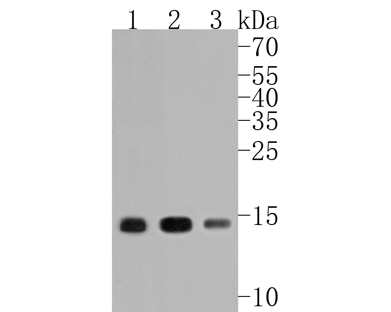
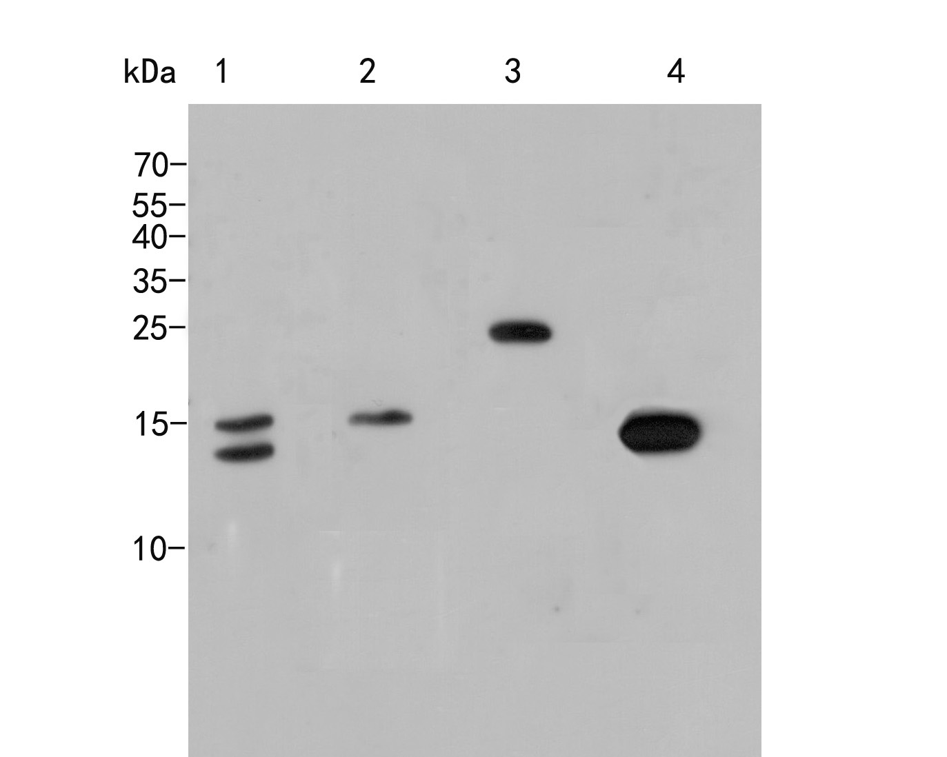
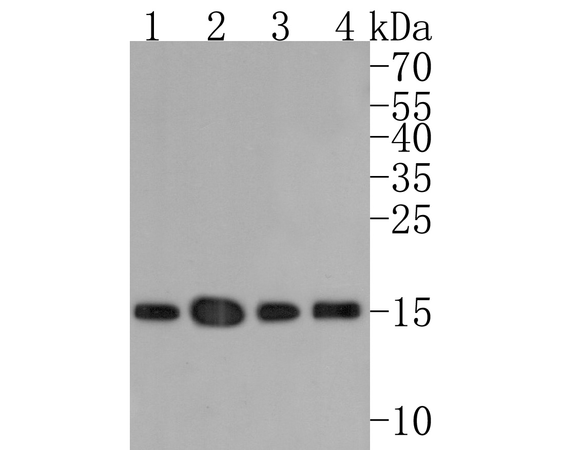
Histone H3 was immunoprecipitated in 0.2mg HeLa cell lysate with ET1701-64 at 2 µg/25 µl agarose. Western blot was performed from the immunoprecipitate using ET1701-64 at 1/20,000 dilution. Anti-Rabbit IgG for IP Nano-secondary antibody (NBI01H) at 1/5,000 dilution was used for 1 hour at room temperature.
Lane 1: HeLa cell lysate (input)
Lane 2: ET1701-64 IP in HeLa cell lysate
Lane 3: Rabbit IgG instead of ET1701-64 in HeLa cell lysate
Blocking/Dilution buffer: 5% NFDM/TBST
Exposure time: 1 minute 59 seconds; ECL: K1801 -

Immunofluorescence analysis of paraffin-embedded human liver tissue labeling Histone H3 with Rabbit anti-Histone H3 antibody (ET1701-64) at 1/100 dilution.
The section was pre-treated using heat mediated antigen retrieval with sodium citrate buffer (pH 6.0) for 2 minutes. The tissues were blocked in 10% negative goat serum for 1 hour at room temperature, washed with PBS, and then probed with the primary antibody (ET1701-64, green) at 1/100 dilution overnight at 4 ℃, washed with PBS. Goat Anti-Rabbit IgG H&L (iFluor™ 488, HA1121) was used as the secondary antibody at 1/1,000 dilution. Nuclei were counterstained with DAPI (blue). -

Immunofluorescence analysis of paraffin-embedded mouse testis tissue labeling Histone H3 with Rabbit anti-Histone H3 antibody (ET1701-64) at 1/100 dilution.
The section was pre-treated using heat mediated antigen retrieval with sodium citrate buffer (pH 6.0) for 2 minutes. The tissues were blocked in 10% negative goat serum for 1 hour at room temperature, washed with PBS, and then probed with the primary antibody (ET1701-64, green) at 1/100 dilution overnight at 4 ℃, washed with PBS. Goat Anti-Rabbit IgG H&L (iFluor™ 488, HA1121) was used as the secondary antibody at 1/1,000 dilution. Nuclei were counterstained with DAPI (blue). -

Immunofluorescence analysis of paraffin-embedded rat testis tissue labeling Histone H3 with Rabbit anti-Histone H3 antibody (ET1701-64) at 1/100 dilution.
The section was pre-treated using heat mediated antigen retrieval with sodium citrate buffer (pH 6.0) for 2 minutes. The tissues were blocked in 10% negative goat serum for 1 hour at room temperature, washed with PBS, and then probed with the primary antibody (ET1701-64, green) at 1/100 dilution overnight at 4 ℃, washed with PBS. Goat Anti-Rabbit IgG H&L (iFluor™ 488, HA1121) was used as the secondary antibody at 1/1,000 dilution. Nuclei were counterstained with DAPI (blue).
请注意: All products are "FOR RESEARCH USE ONLY AND ARE NOT INTENDED FOR DIAGNOSTIC OR THERAPEUTIC USE"
引文
-
The role of apoptosis, immunogenic cell death, and macrophage polarization in carbon ion radiotherapy for keloids: Targeting the TGF-β1/SMADs signaling pathway
Author: Heng Zhou,et al
PMID: 39245184
应用: WB
反应种属: Mouse
发表时间: 2024 Sep
-
Citation
-
QBT improved cognitive dysfunction in rats with vascular dementia by regulating the Nrf2/xCT/GPX4 and NLRP3/Caspase-1/GSDMD pathways to inhibit ferroptosis and pyroptosis of neurons
Author: Lu Feng,et al
PMID: 39265351
应用:
反应种属:
发表时间: 2024 Sep
-
Citation
-
Nuclear KRT19 is a transcriptional corepressor promoting histone deacetylation and liver tumorigenesis
Author: Shixun Han, Haonan Fan, Guoxuan Zhong,et al
PMID: 38557414
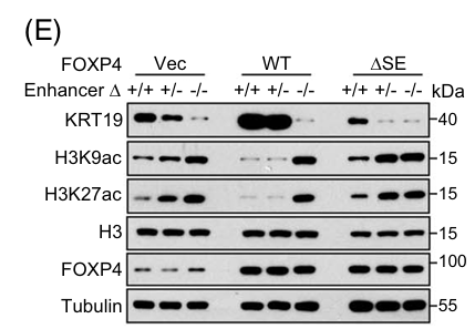
应用: WB
反应种属: Human
发表时间: 2024 Apr
-
Citation
-
Different viral effectors suppress hormone-mediated antiviral immunity of rice coordinated by OsNPR1
Author:
PMID: 37230965
应用: WB
反应种属: Rice
发表时间: 2023 May
-
Citation
-
LIF and bFGF enhanced chicken primordial follicle activation by Wnt/β-catenin pathway
Author:
PMID: 34555602

应用: WB
反应种属: Mouse
发表时间: 2021 Dec
-
Citation
-
Tumor-derived HMGB1 induces CD62Ldim neutrophil polarization and promotes lung metastasis in triple-negative breast cancer
Author: Jian Huang
PMID: 32943604
应用: WB
反应种属: Mouse
发表时间: 2020 Sep
-
Citation
-
The role of Ca2+/NFAT in Dysfunction and Infammation of Human Coronary Endothelial Cells induced by Sera from patients with Kawasaki disease
Author:
PMID: 32170198

应用: WB
反应种属: Human
发表时间: 2020 Mar
-
Citation
同靶点 & 同通路的产品
Histone H3 (mono methyl K18) Recombinant Rabbit Monoclonal Antibody [SA42-07]
Application: WB,IF-Cell,IF-Tissue,IHC-P
Reactivity: Human,Mouse,Rat
Conjugate: unconjugated
Histone H3 (acetyl K18) Rabbit Polyclonal Antibody
Application: WB,IHC-P,IF-Cell
Reactivity: Human,Mouse
Conjugate: unconjugated
Histone H3 (acetyl K14) Recombinant Rabbit Monoclonal Antibody [JU43-26]
Application: WB,IF-Cell,IF-Tissue,IHC-P,IP,SNAP-ChIP,CUT&Tag-seq
Reactivity: Human,Mouse,Rat
Conjugate: unconjugated
Histone H3 Mouse Monoclonal Antibody [3-C4]
Application: WB,IHC-P,mIHC,IF-Tissue,ChIP
Reactivity: Human,Mouse,Rat
Conjugate: unconjugated
Histone H3 (mono methyl K9) Recombinant Rabbit Monoclonal Antibody [JE43-27]
Application: WB,IF-Cell,IHC-P,FC,ChIP,IF-Tissue,Dot Blot
Reactivity: Human,Mouse,Rat,Monkey
Conjugate: unconjugated
Histone H3 (mono+di+tri methyl K36) Recombinant Rabbit Monoclonal Antibody [PSH07-14]
Application: WB,IHC-P,IF-Cell,FC,ChIP
Reactivity: Human,Mouse,Rat
Conjugate: unconjugated
Histone H3 (mono methyl K4) Recombinant Rabbit Monoclonal Antibody [PSH07-15]
Application: WB,IF-Cell,ChIP,Dot Blot
Reactivity: Human,Mouse,Rat
Conjugate: unconjugated
Histone H3 (acetyl K27) Mouse Monoclonal Antibody [A6D6]
Application: WB,IF-Cell,IHC-P,FC,ChIP
Reactivity: Human,Mouse,Rat
Conjugate: unconjugated
Histone H3 Recombinant Mouse Monoclonal Antibody [6-A7-R] - Loading control
Application: WB,IHC-P,IF-Tissue,ChIP
Reactivity: Human,Mouse,Rat
Conjugate: unconjugated
Histone H3 (acetyl K23) Recombinant Rabbit Monoclonal Antibody [JE46-39]
Application: WB,IF-Cell
Reactivity: Human,Mouse
Conjugate: unconjugated
Histone H3 Rabbit Polyclonal Antibody
Application: WB
Reactivity: Human,Mouse,Rat
Conjugate: unconjugated
Histone H3 (di methyl K4) Rabbit Polyclonal Antibody
Application: WB,IF-Cell,IHC-P,FC,Dot Blot
Reactivity: Human,Mouse,Rat
Conjugate: unconjugated
Histone H3 (acetyl K36) Recombinant Rabbit Monoclonal Antibody [JE77-32]
Application: WB,IHC-P,IF-Cell,ChIP
Reactivity: Human,Mouse,Rat
Conjugate: unconjugated
Histone H3 (acetyl K9) Recombinant Rabbit Monoclonal Antibody [PSH04-47] - ChIP Grade
Application: WB,IF-Cell,IHC-P,IF-Tissue,FC,ChIP,Dot Blot,IP
Reactivity: Human,Mouse,Rat
Conjugate: unconjugated
Histone H3 (mono methyl K36) Recombinant Rabbit Monoclonal Antibody [SR04-20]
Application: WB,IF-Cell,IF-Tissue,ChIP
Reactivity: Human,Mouse
Conjugate: unconjugated
Histone H3 (acetyl K56) Recombinant Rabbit Monoclonal Antibody [SU30-10]
Application: WB,IF-Cell,IF-Tissue,IHC-P,ChIP,CUT&Tag-seq
Reactivity: Human,Mouse,Rat
Conjugate: unconjugated
Histone H3 (mono methyl R2) Recombinant Rabbit Monoclonal Antibody [ST0427]
Application: WB,IF-Cell,IF-Tissue,IHC-P,ChIP
Reactivity: Human,Mouse,Rat
Conjugate: unconjugated
Histone H3 Rabbit Polyclonal Antibody
Application: WB
Reactivity: Zebrafish,Human,Mouse
Conjugate: unconjugated
Histone H3 (acetyl K18) Mouse Monoclonal Antibody [A7B6]
Application: WB,IF-Cell
Reactivity: Human
Conjugate: unconjugated
Phospho-Histone H3 (T3) Recombinant Rabbit Monoclonal Antibody [JE42-48]
Application: WB,IHC-P
Reactivity: Human,Mouse,Rat
Conjugate: unconjugated
Histone H3 (mono methyl K14) Recombinant Rabbit Monoclonal Antibody [JE43-29]
Application: WB,IF-Cell,ChIP,Dot Blot
Reactivity: Human,Mouse,Rat
Conjugate: unconjugated
Histone H3 (mono methyl K27) Recombinant Rabbit Monoclonal Antibody [PSH05-67]
Application: WB,IF-Cell,FC,ChIP
Reactivity: Human,Mouse,Rat
Conjugate: unconjugated
Histone H3 (mono+di+tri methyl K4) Recombinant Rabbit Monoclonal Antibody [PSH06-30]
Application: WB,IHC-P,IF-Cell,FC,ChIP
Reactivity: Human,Mouse,Rat
Conjugate: unconjugated
Histone H3 Mouse Monoclonal Antibody [A11-D7]
Application: WB,IF-Cell,IHC-P,IF-Tissue
Reactivity: Human,Mouse,Rat,Zebrafish
Conjugate: unconjugated
Histone H3 (acetyl K4) Recombinant Rabbit Monoclonal Antibody [PSH03-71]
Application: WB,IF-Cell,ChIP,Dot Blot
Reactivity: Human,Mouse,Rat
Conjugate: unconjugated
Histone H3 (mono methyl K79) Recombinant Rabbit Monoclonal Antibody [PSH07-66]
Application: WB,IF-Cell,IHC-P,FC,ChIP
Reactivity: Human,Mouse,Rat,Monkey
Conjugate: unconjugated
Histone H3 (di methyl K9) Recombinant Rabbit Monoclonal Antibody [SN07-30]
Application: WB,IF-Cell,IF-Tissue,IHC-P
Reactivity: Human,Mouse,Rat
Conjugate: unconjugated
Histone H3 (acetyl K27) Rabbit Polyclonal Antibody
Application: WB,IHC-P,ChIP
Reactivity: Human,Mouse,Rat
Conjugate: unconjugated
Histone H3 (mono+di+tri methyl K79) Recombinant Rabbit Monoclonal Antibody [SR42-06]
Application: WB,IHC-P,ChIP
Reactivity: Human,Mouse,Rat
Conjugate: unconjugated
Histone H3 (acetyl K18) Mouse Monoclonal Antibody [A7B5]
Application: WB,IHC-P,ChIP
Reactivity: Human,Rat
Conjugate: unconjugated
Histone H3 (tri methyl K9) Mouse Monoclonal Antibody [2G1]
Application: WB,IF
Reactivity: Human
Conjugate: unconjugated
Histone H3 Recombinant Mouse Monoclonal Antibody [A11-D7-R]
Application: WB,IF-Cell,IHC-P
Reactivity: Human,Mouse,Rat
Conjugate: unconjugated
Histone H3 Mouse Monoclonal Antibody [6-A7]
Application: WB,IHC-P,IF-Tissue,ChIP
Reactivity: Human,Mouse,Rat
Conjugate: unconjugated
Histone H3 (tri methyl K9) Mouse Monoclonal Antibody [1-6]
Application: WB,IF-Cell,IHC-P,IF-Tissue
Reactivity: Human,Mouse,Rat
Conjugate: unconjugated
Histone H3 (tri methyl K27) Recombinant Rabbit Monoclonal Antibody [PSH05-15]
Application: WB,IF-Cell,IHC-P,IF-Tissue,ChIP
Reactivity: Human,Mouse,Rat
Conjugate: unconjugated
Histone H3 (di methyl K4) Recombinant Rabbit Monoclonal Antibody [JE00-98]
Application: WB,IF-Cell,ChIP,Dot Blot,IHC-P
Reactivity: Human,Mouse,Rat,Monkey
Conjugate: unconjugated
Phospho-Histone H3 (S28) Recombinant Rabbit Monoclonal Antibody [JE56-06]
Application: WB,IF-Cell,IHC-P,FC,ChIP
Reactivity: Human,Rat,Mouse
Conjugate: unconjugated
Histone H3 (di methyl K27) Recombinant Rabbit Monoclonal Antibody [PSH05-88]
Application: WB,IF-Cell,ChIP
Reactivity: Human,Mouse,Rat
Conjugate: unconjugated
Histone H3 (acetyl K27) Mouse Monoclonal Antibody [A6D7]
Application: WB,IF-Cell,IHC-P,FC,ChIP
Reactivity: Human,Mouse,Rat
Conjugate: unconjugated
Histone H3 Recombinant Rabbit Monoclonal Antibody [JJ092-08]
Application: WB,IF-Tissue,IHC-P,ChIP
Reactivity: Human,Mouse,Rat
Conjugate: unconjugated
Histone H3 (mono+di+tri methyl K79) Recombinant Rabbit Monoclonal Antibody [PSH09-10]
Application: WB,IHC-P,IF-Cell,ChIP
Reactivity: Human,Mouse,Rat,Monkey
Conjugate: unconjugated
Histone H3 (mono methyl K4) Rabbit Polyclonal Antibody
Application: WB,IF-Cell,IHC-P,FC,Dot Blot
Reactivity: Human,Mouse,Rat
Conjugate: unconjugated
Phospho-Histone H3 (S10) Recombinant Rabbit Monoclonal Antibody [SA31-01]
Application: WB,IF-Cell,IHC-P,IP,FC,ChIP
Reactivity: Human,Mouse,Rat
Conjugate: unconjugated


