PAX8 Mouse Monoclonal Antibody [6G5]

Catalog# EM1701-81
PAX8 Mouse Monoclonal Antibody [6G5]
-
WB
-
IHC-P
-
IF-Cell
-
Human
概述
产品名称
PAX8 Mouse Monoclonal Antibody [6G5]
抗体类型
Mouse Monoclonal Antibody
免疫原
Synthetic peptide corresponding to human PAX8 aa 400-450.
种属反应性
Human
验证应用
WB, IHC-P, IF-Cell
分子量
Predicted band size: 48 kDa
阳性对照
Skov-3 cells lysates, NIH:OVCAR-3, human kidney tissue, human thyroid tissue, human thyroid cancer tissue, mouse thyroid tissue.
偶联
unconjugated
克隆号
6G5
RRID
产品特性
形态
Liquid
浓度
2ug/ul
存放说明
Store at +4℃ after thawing. Aliquot store at -20℃. Avoid repeated freeze / thaw cycles.
存储缓冲液
1*PBS (pH7.4), 0.2% BSA, 50% Glycerol. Preservative: 0.05% Sodium Azide.
亚型
IgG1
纯化方式
Immunogen affinity purified.
应用稀释度
-
WB
-
1:500-1:2,000
-
IHC-P
-
1:2,000
-
IF-Cell
-
1:100
靶点
功能
The PAX gene family has an important role in the formation of tissues and organs during embryonic development and maintaining the normal function of some cells after birth. The PAX genes give instructions for making proteins that attach themselves to certain areas of DNA. This nuclear protein is involved in thyroid follicular cell development and expression of thyroid-specific genes. PAX8 releases the hormones important for regulating growth, brain development, and metabolism. Also functions in very early stages of kidney organogenesis, the müllerian system, and the thymus. Additionally, PAX8 is expressed in the renal excretory system, epithelial cells of the endocervix, endometrium, ovary, Fallopian tube, seminal vesicle, epididymis, pancreatic islet cells and lymphoid cells. PAX8 and other transcription factors play a role in binding to DNA and regulating the genes that drive thyroid hormone synthesis (Tg, TPO, Slc5a5 and Tshr). This PAX-8 antibody is intended for qualified laboratories to qualitatively identify by light microscopy the presence of associated antigens in sections of formalinfixed, paraffin-embedded tissue sections using IHC test methods.
背景文献
1. Laury AR et al. A comprehensive analysis of PAX8 expression in human epithelial tumors. The American Journal of Surgical Pathology 35 (6): 816–26 (2011).
2. Fernández LP et al. Thyroid transcription factors in development, differentiation and disease. Nature Reviews Endocrinology 11 (1): 29-42 (2015).
组织特异性
Expressed in the excretory system, thyroid gland and Wilms tumors.
亚细胞定位
Nucleus.
UNIPROT #
别名
OTTHUMP00000158659 antibody
OTTHUMP00000158660 antibody
OTTHUMP00000203723 antibody
OTTHUMP00000203724 antibody
Paired box 8 antibody
Paired box gene 8 antibody
paired box homeotic gene 8 antibody
Paired box protein Pax 8 antibody
Paired box protein Pax-8 antibody
Paired domain gene 8 antibody
展开图片
-
Western blot analysis of Pax8 on skov-3 cells lysates using anti-Pax8 antibody at 1/500 dilution.
-
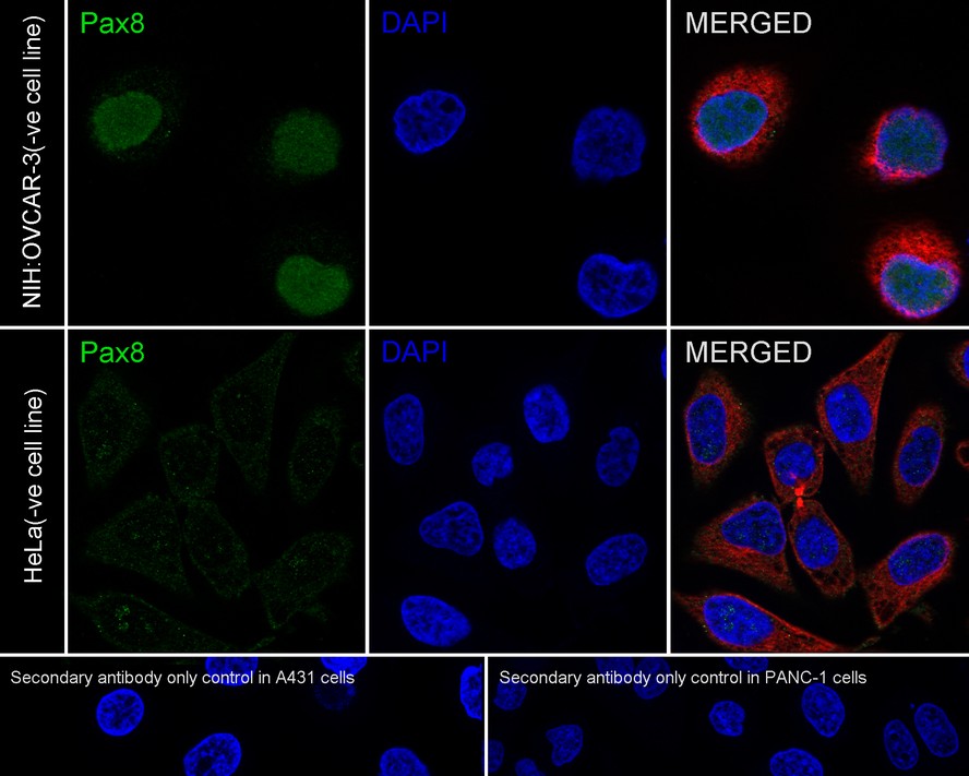
Immunocytochemistry analysis of NIH:OVCAR-3 (positive) and HeLa (negative) labeling PAX8 with Mouse anti-PAX8 antibody (EM1701-81) at 1/100 dilution.
Cells were fixed in 4% paraformaldehyde for 20 minutes at room temperature, permeabilized with 0.1% Triton X-100 in PBS for 5 minutes at room temperature, then blocked with 1% BSA in 10% negative goat serum for 1 hour at room temperature. Cells were then incubated with Mouse anti-PAX8 antibody (EM1701-81) at 1/100 dilution in 1% BSA in PBST overnight at 4 ℃. Goat Anti-Mouse IgG H&L (iFluor™ 488, HA1125) was used as the secondary antibody at 1/1,000 dilution. PBS instead of the primary antibody was used as the secondary antibody only control. Nuclear DNA was labelled in blue with DAPI.
beta Tubulin (ET1602-4, red) was stained at 1/100 dilution overnight at +4℃. Goat Anti-Rabbit IgG H&L (iFluor™ 594, HA1122) were used as the secondary antibody at 1/1,000 dilution. -
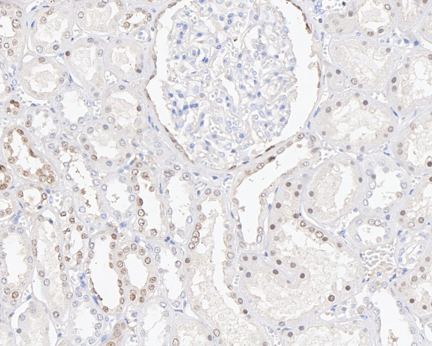
Immunohistochemical analysis of paraffin-embedded human kidney tissue with Mouse anti-PAX8 antibody (EM1701-81) at 1/2,000 dilution.
The section was pre-treated using heat mediated antigen retrieval with sodium citrate buffer (pH 6.0) for 2 minutes. The tissues were blocked in 1% BSA for 20 minutes at room temperature, washed with ddH2O and PBS, and then probed with the primary antibody (EM1701-81) at 1/2,000 dilution for 1 hour at room temperature. The detection was performed using an HRP conjugated compact polymer system. DAB was used as the chromogen. Tissues were counterstained with hematoxylin and mounted with DPX. -
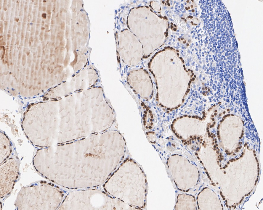
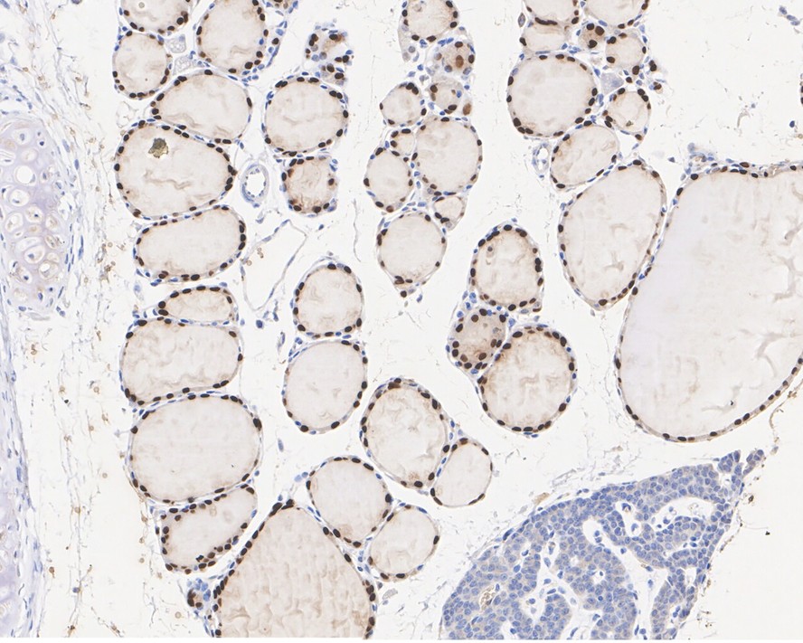
Immunohistochemical analysis of paraffin-embedded human thyroid tissue with Mouse anti-PAX8 antibody (EM1701-81) at 1/2,000 dilution.
The section was pre-treated using heat mediated antigen retrieval with sodium citrate buffer (pH 6.0) for 2 minutes. The tissues were blocked in 1% BSA for 20 minutes at room temperature, washed with ddH2O and PBS, and then probed with the primary antibody (EM1701-81) at 1/2,000 dilution for 1 hour at room temperature. The detection was performed using an HRP conjugated compact polymer system. DAB was used as the chromogen. Tissues were counterstained with hematoxylin and mounted with DPX. -
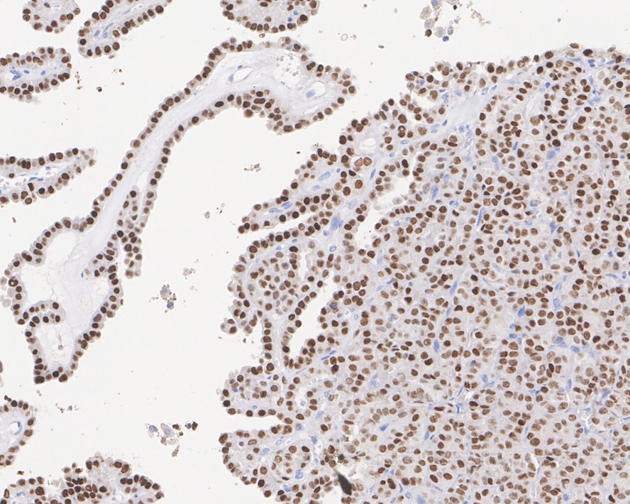
Immunohistochemical analysis of paraffin-embedded human thyroid cancer tissue with Mouse anti-PAX8 antibody (EM1701-81) at 1/2,000 dilution.
The section was pre-treated using heat mediated antigen retrieval with sodium citrate buffer (pH 6.0) for 2 minutes. The tissues were blocked in 1% BSA for 20 minutes at room temperature, washed with ddH2O and PBS, and then probed with the primary antibody (EM1701-81) at 1/2,000 dilution for 1 hour at room temperature. The detection was performed using an HRP conjugated compact polymer system. DAB was used as the chromogen. Tissues were counterstained with hematoxylin and mounted with DPX. -
Immunohistochemical analysis of paraffin-embedded mouse thyroid tissue with Mouse anti-PAX8 antibody (EM1701-81) at 1/2,000 dilution.
The section was pre-treated using heat mediated antigen retrieval with sodium citrate buffer (pH 6.0) for 2 minutes. The tissues were blocked in 1% BSA for 20 minutes at room temperature, washed with ddH2O and PBS, and then probed with the primary antibody (EM1701-81) at 1/2,000 dilution for 1 hour at room temperature. The detection was performed using an HRP conjugated compact polymer system. DAB was used as the chromogen. Tissues were counterstained with hematoxylin and mounted with DPX.
Please note: All products are "FOR RESEARCH USE ONLY AND ARE NOT INTENDED FOR DIAGNOSTIC OR THERAPEUTIC USE"