iFluor™ 647 Conjugated Cytokeratin 19 Recombinant Rabbit Monoclonal Antibody [SA30-06]


Catalog# HA720152F
iFluor™ 647 Conjugated Cytokeratin 19 Recombinant Rabbit Monoclonal Antibody [SA30-06]
-
IF-Cell
-
IF-Tissue
-
FC
-
Human
-
Mouse
概述
产品名称
iFluor™ 647 Conjugated Cytokeratin 19 Recombinant Rabbit Monoclonal Antibody [SA30-06]
抗体类型
Recombinant Rabbit monoclonal Antibody
免疫原
Synthetic peptide within Human Cytokeratin 19 aa 348-400.
种属反应性
Human, Mouse
验证应用
IF-Cell, IF-Tissue, FC
分子量
Predicted band size: 44 kDa
阳性对照
SK-Br-3, human liver tissue, human breast tissue, human kidney tissue, MCF-7.
偶联
iFluor™ 647
克隆号
SA30-06
RRID
产品特性
形态
Liquid
浓度
1ug/ul
存放说明
Store at +4℃ after thawing. Aliquot store at -20℃. Avoid repeated freeze / thaw cycles.
存储缓冲液
Preservative: 0.02% Sodium azide Constituents: 30% Glycerol, 1% BSA, 68.98% PBS
亚型
IgG
纯化方式
Protein A affinity purified.
应用稀释度
-
IF-Cell
-
1:100
-
IF-Tissue
-
1:50-1:100
-
FC
-
1 ug/mL
靶点
功能
Cytokeratins comprise a diverse group of intermediate filament proteins (IFPs) that are expressed as pairs in both keratinized and non-keratinized epithelial tissue. Cytokeratins play a critical role in differentiation and tissue specialization and function to maintain the overall structural integrity of epithelial cells and have been found to be useful markers of tissue differentiation, which is directly applicable to the characterization of malignant tumors. For example, many types of cancer cells express Cytokeratin 19 (CK19), an epithelial cytoskeletal protein within the suprabasal squamous epithelium. Cytokeratin 19 is a specific marker of moderate to severe dysplasia and carcinoma in situ in oral cavity squamous epithelium, and measurement of Cytokeratin 19 may be a useful marker in diagnosing hepatoma. Cytokeratin 19 fragment levels in serum have been documented as a marker for lung cancer. Clinical investigations have suggested that serum CYFRA 21-1, a fragment of Cytokeratin 19, may be among the most useful tumor markers.
背景文献
1. Guye P. et. al. Genetically engineering self-organization of human pluripotent stem cells into a liver bud-like tissue using Gata6. Nat Commun 7:10243 (2016).
2. Cui M. et. al. PTEN is a potent suppressor of small cell lung cancer. Mol Cancer Res 12:654-9 (2014).
亚细胞定位
Cytoskeleton.
UNIPROT #
别名
40 kDa keratin intermediate filament antibody
CK 19 antibody
CK-19 antibody
CK19 antibody
Cytokeratin 19 antibody
Cytokeratin-19 antibody
K19 antibody
K1C19_HUMAN antibody
K1CS antibody
Keratin 19 antibody
展开图片
-
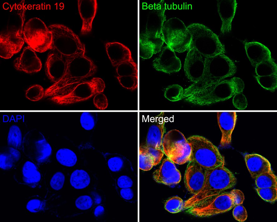
Immunocytochemistry analysis of SK-Br-3 cells labeling Cytokeratin 19 with Rabbit anti-Cytokeratin 19 antibody (HA720152F) at 1/100 dilution.
Cells were fixed in 100% methanol for 10 minutes, permeabilized with 0.1% Triton X-100 in PBS for 15 minutes, and then blocked with 2% normal goat serum for 1 hour at 37℃. Cells were then incubated with Rabbit anti-Cytokeratin 19 antibody (HA720152F, red) at 1/100 dilution in 2% normal goat serum overnight at 4 ℃. Nuclear DNA was labelled in blue with DAPI.
Beta tubulin (M1305-2, green) was stained at 1/200 dilution overnight at +4℃. Goat Anti-Mouse IgG H&L (iFluor™ 488, HA1125) was used as the secondary antibody at 1/800 dilution. -
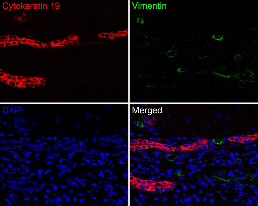
Immunofluorescence analysis of paraffin-embedded human liver tissue labeling Cytokeratin 19 (HA720152F) and Vimentin (EM0401).
The section was pre-treated using heat mediated antigen retrieval with Tris-EDTA buffer (pH 9.0) for 20 minutes. The tissues were blocked in 10% negative goat serum for 1 hour at room temperature, washed with PBS. And then probed with the primary antibodies Cytokeratin 19 (HA720152F, red) at 1/50 dilution and Vimentin (EM0401, green) at 1/1,000 dilution overnight at 4 ℃, washed with PBS.
iFluor™ 488 conjugate-Goat anti-Mouse IgG (HA1125) was used as the secondary antibody at 1/1,000 dilution. DAPI was used as nuclear counterstain. -
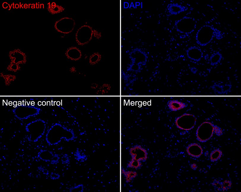
Immunofluorescence analysis of paraffin-embedded human breast tissue labeling Cytokeratin 19 (HA720152F).
The section was pre-treated using heat mediated antigen retrieval with Tris-EDTA buffer (pH 9.0) for 20 minutes. The tissues were blocked in 10% negative goat serum for 1 hour at room temperature, washed with PBS. And then probed with the primary antibody Cytokeratin 19 (HA720152F, iFluor™ 647) at 1/100 dilution overnight at 4 ℃, washed with PBS. DAPI was used as nuclear counterstain. -
Immunofluorescence analysis of paraffin-embedded human kidney tissue labeling Cytokeratin 19 (HA720152F).
The section was pre-treated using heat mediated antigen retrieval with Tris-EDTA buffer (pH 9.0) for 20 minutes. The tissues were blocked in 10% negative goat serum for 1 hour at room temperature, washed with PBS. And then probed with the primary antibody Cytokeratin 19 (HA720152F, iFluor™ 647) at 1/100 dilution overnight at 4 ℃, washed with PBS. DAPI was used as nuclear counterstain. -
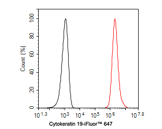
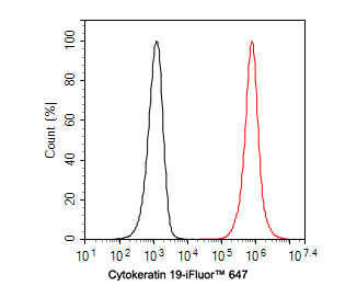
Flow cytometric analysis of MCF-7 cells labeling Cytokeratin 19.
Cells were fixed and permeabilized. Then incubated for 1 hour at +4℃ with Cytokeratin 19 (HA720152F, red, 1ug/ml). Unlabelled sample was used as a control (cells without incubation with primary antibody; black). -
Flow cytometric analysis of SK-Br-3 cells labeling Cytokeratin 19.
Cells were fixed and permeabilized. Then incubated for 1 hour at +4℃ with Cytokeratin 19 (HA720152F, red, 1ug/ml). Unlabelled sample was used as a control (cells without incubation with primary antibody; black).
Please note: All products are "FOR RESEARCH USE ONLY AND ARE NOT INTENDED FOR DIAGNOSTIC OR THERAPEUTIC USE"