Cytokeratin 19 Rabbit Polyclonal Antibody


Catalog# ER1803-79
Cytokeratin 19 Rabbit Polyclonal Antibody
-
WB
-
IF-Cell
-
IHC-P
-
FC
-
Human
-
Mouse
-
Rat
概述
产品名称
Cytokeratin 19 Rabbit Polyclonal Antibody
抗体类型
Rabbit Polyclonal Antibody
免疫原
Recombinant protein within Human Cytokeratin 19 aa 100-350.
种属反应性
Human, Mouse, Rat
验证应用
WB, IF-Cell, IHC-P, FC
分子量
44 kDa
阳性对照
MCF-7, mouse lung tissue, HepG2, SK-Br-3, rat kidney tissue, human lung cancer tissue, human appendix tissue, human breast tissue, mouse colon tissue.
偶联
unconjugated
RRID
产品特性
形态
Liquid
浓度
1ug/ul
存放说明
Store at +4℃ after thawing. Aliquot store at -20℃. Avoid repeated freeze / thaw cycles.
存储缓冲液
1*PBS (pH7.4), 0.2% BSA, 50% Glycerol. Preservative: 0.05% Sodium Azide.
亚型
IgG
纯化方式
Immunogen affinity purified.
应用稀释度
-
WB
-
1:2,000-1:20,000
-
IF-Cell
-
1:50-1:200
-
IHC-P
-
1:50-1:200
-
FC
-
1:50-1:100
靶点
功能
Cytokeratins comprise a diverse group of intermediate filament proteins (IFPs) that are expressed as pairs in both keratinized and non-keratinized epithelial tissue. Cytokeratins play a critical role in differentiation and tissue specialization and function to maintain the overall structural integrity of epithelial cells and have been found to be useful markers of tissue differentiation, which is directly applicable to the characterization of malignant tumors. For example, many types of cancer cells express Cytokeratin 19 (CK19), an epithelial cytoskeletal protein within the suprabasal squamous epithelium. Cytokeratin 19 is a specific marker of moderate to severe dysplasia and carcinoma in situ in oral cavity squamous epithelium, and measurement of Cytokeratin 19 may be a useful marker in diagnosing hepatoma. Cytokeratin 19 fragment levels in serum have been documented as a marker for lung cancer. Clinical investigations have suggested that serum CYFRA 21-1, a fragment of Cytokeratin 19, may be among the most useful tumor markers.
背景文献
1. Guye P et al. Genetically engineering self-organization of human pluripotent stem cells into a liver bud-like tissue using Gata6. Nat Commun 7:10243 (2016).
2. Cui M et al. PTEN is a potent suppressor of small cell lung cancer. Mol Cancer Res 12:654-9 (2014).
序列相似性
Belongs to the intermediate filament family.
组织特异性
Expressed in a defined zone of basal keratinocytes in the deep outer root sheath of hair follicles. Also observed in sweat gland and mammary gland ductal and secretory cells, bile ducts, gastrointestinal tract, bladder urothelium, oral epithelia, esophagus, ectocervical epithelium (at protein level). Expressed in epidermal basal cells, in nipple epidermis and a defined region of the hair follicle. Also seen in a subset of vascular wall cells in both the veins and artery of human umbilical cord, and in umbilical cord vascular smooth muscle. Observed in muscle fibers accumulating in the costameres of myoplasm at the sarcolemma in structures that contain dystrophin and spectrin.
亚细胞定位
Cytoplasm.
UNIPROT #
别名
40 kDa keratin intermediate filament antibody
CK 19 antibody
CK-19 antibody
CK19 antibody
Cytokeratin 19 antibody
Cytokeratin-19 antibody
K19 antibody
K1C19_HUMAN antibody
K1CS antibody
Keratin 19 antibody
展开图片
-
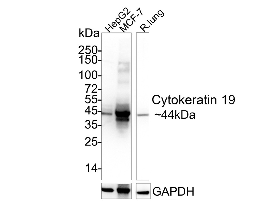
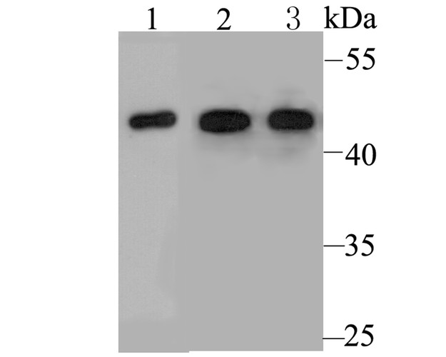
Western blot analysis of Cytokeratin 19 on rat lung tissue lysate. Proteins were transferred to a PVDF membrane and blocked with 5% BSA in PBS for 1 hour at room temperature. The primary antibody was used at a 1:20,000 dilution in 5% BSA at room temperature for 2 hours. Goat Anti-Rabbit IgG - HRP Secondary Antibody (HA1001) at 1:5,000 dilution was used for 1 hour at room temperature.
Positive control:
Lane 1: Rat lung tissue lysate
Lane 2: MCF-7 cell lysate
Lane 3: Mouse lung tissue lysate -
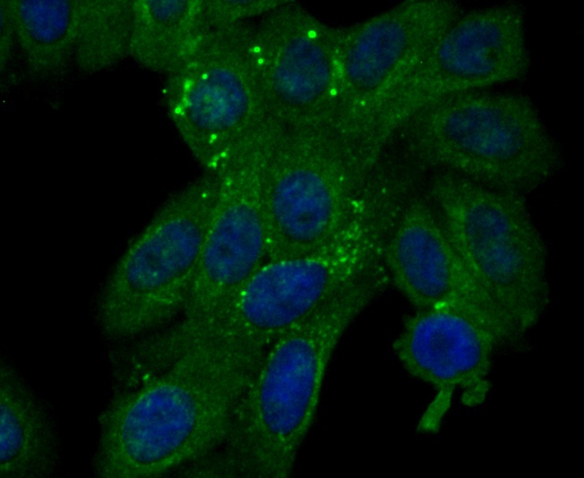
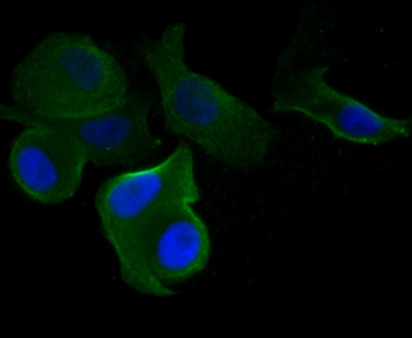
ICC staining Cytokeratin 19 in HepG2 cells (green). Formalin fixed cells were permeabilized with 0.1% Triton X-100 in TBS for 10 minutes at room temperature and blocked with 1% Blocker BSA for 15 minutes at room temperature. Cells were probed with Cytokeratin 19 polyclonal antibody at a dilution of 1:100 for 1 hour at room temperature, washed with PBS. Alexa Fluorc™ 488 Goat anti-Rabbit IgG was used as the secondary antibody at 1/100 dilution. The nuclear counter stain is DAPI (blue).
-
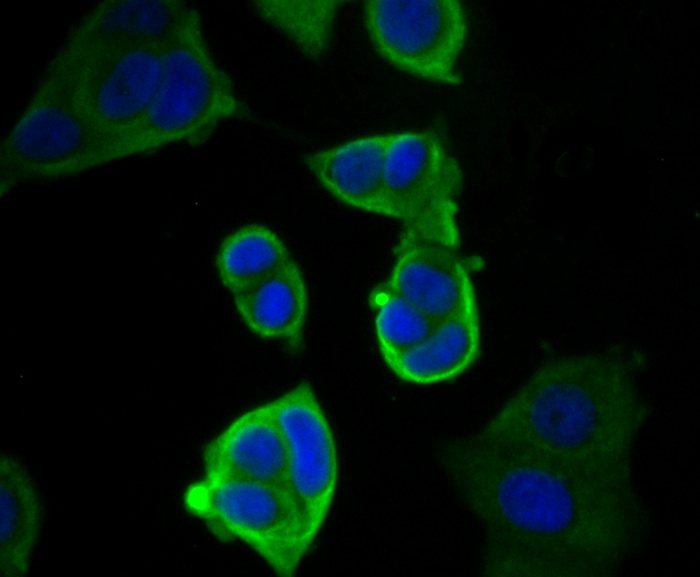
ICC staining Cytokeratin 19 in MCF-7 cells (green). Formalin fixed cells were permeabilized with 0.1% Triton X-100 in TBS for 10 minutes at room temperature and blocked with 1% Blocker BSA for 15 minutes at room temperature. Cells were probed with Cytokeratin 19 polyclonal antibody at a dilution of 1:200 for 1 hour at room temperature, washed with PBS. Alexa Fluorc™ 488 Goat anti-Rabbit IgG was used as the secondary antibody at 1/100 dilution. The nuclear counter stain is DAPI (blue).
-
ICC staining Cytokeratin 19 in SK-Br-3 cells (green). Formalin fixed cells were permeabilized with 0.1% Triton X-100 in TBS for 10 minutes at room temperature and blocked with 1% Blocker BSA for 15 minutes at room temperature. Cells were probed with Cytokeratin 19 polyclonal antibody at a dilution of 1:50 for 1 hour at room temperature, washed with PBS. Alexa Fluorc™ 488 Goat anti-Rabbit IgG was used as the secondary antibody at 1/100 dilution. The nuclear counter stain is DAPI (blue).
-
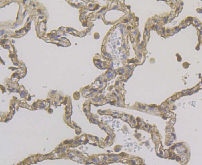
Immunohistochemical analysis of paraffin-embedded rat kidney tissue using anti-Cytokeratin 19 antibody. The section was pre-treated using heat mediated antigen retrieval with sodium citrate buffer (pH 6.0) for 20 minutes. The tissues were blocked in 5% BSA for 30 minutes at room temperature, washed with ddH2O and PBS, and then probed with the antibody (ER1803-79) at 1/200 dilution, for 30 minutes at room temperature and detected using an HRP conjugated compact polymer system. DAB was used as the chrogen. Counter stained with hematoxylin and mounted with DPX.
-
Immunohistochemical analysis of paraffin-embedded human lung cancer tissue using anti-Cytokeratin 19 antibody. The section was pre-treated using heat mediated antigen retrieval with sodium citrate buffer (pH 6.0) for 20 minutes. The tissues were blocked in 5% BSA for 30 minutes at room temperature, washed with ddH2O and PBS, and then probed with the antibody (ER1803-79) at 1/200 dilution, for 30 minutes at room temperature and detected using an HRP conjugated compact polymer system. DAB was used as the chrogen. Counter stained with hematoxylin and mounted with DPX.
-
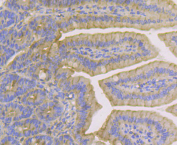
Immunohistochemical analysis of paraffin-embedded human appendix tissue using anti-Cytokeratin 19 antibody. The section was pre-treated using heat mediated antigen retrieval with sodium citrate buffer (pH 6.0) for 20 minutes. The tissues were blocked in 5% BSA for 30 minutes at room temperature, washed with ddH2O and PBS, and then probed with the antibody (ER1803-79) at 1/200 dilution, for 30 minutes at room temperature and detected using an HRP conjugated compact polymer system. DAB was used as the chrogen. Counter stained with hematoxylin and mounted with DPX.
-
Immunohistochemical analysis of paraffin-embedded human breast tissue using anti-Cytokeratin 19 antibody. The section was pre-treated using heat mediated antigen retrieval with sodium citrate buffer (pH 6.0) for 20 minutes. The tissues were blocked in 5% BSA for 30 minutes at room temperature, washed with ddH2O and PBS, and then probed with the antibody (ER1803-79) at 1/200 dilution, for 30 minutes at room temperature and detected using an HRP conjugated compact polymer system. DAB was used as the chrogen. Counter stained with hematoxylin and mounted with DPX.
-
Immunohistochemical analysis of paraffin-embedded mouse colon tissue using anti-Cytokeratin 19 antibody. The section was pre-treated using heat mediated antigen retrieval with sodium citrate buffer (pH 6.0) for 20 minutes. The tissues were blocked in 5% BSA for 30 minutes at room temperature, washed with ddH2O and PBS, and then probed with the antibody (ER1803-79) at 1/200 dilution, for 30 minutes at room temperature and detected using an HRP conjugated compact polymer system. DAB was used as the chrogen. Counter stained with hematoxylin and mounted with DPX.
-
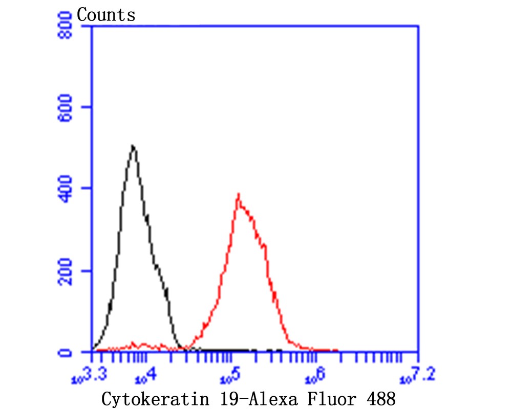
Flow cytometric analysis of Cytokeratin 19 was done on MCF-7 cells. The cells were fixed, permeabilized and stained with Cytokeratin 19 antibody at 1/100 dilution (red) compared with an unlabelled control (cells without incubation with primary antibody; black). After incubation of the primary antibody on room temperature for an hour, the cells was stained with a Alexa Fluor™ 488-conjugated goat anti-rabbit IgG Secondary antibody at 1/500 dilution for 30 minutes.
Please note: All products are "FOR RESEARCH USE ONLY AND ARE NOT INTENDED FOR DIAGNOSTIC OR THERAPEUTIC USE"
Alternative Products