MSH2 Mouse Monoclonal Antibody [10G2]


Catalog# EM1801-05
MSH2 Mouse Monoclonal Antibody [10G2]
-
WB
-
IHC-P
-
IF-Tissue
-
Human
-
Mouse
-
Rat
概述
产品名称
MSH2 Mouse Monoclonal Antibody [10G2]
抗体类型
Mouse Monoclonal Antibody
免疫原
Synthetic peptide within N-terminal human MSH2.
种属反应性
Human, Mouse, Rat
验证应用
WB, IHC-P, IF-Tissue
分子量
Predicted band size: 105 kDa
阳性对照
HeLa cell lysate, HEK-293 cell lysate, A549 cell lysate, A431 cell lysate, HCT 116 cell lysate, SW480 cell lysate, PC-3M cell lysate, NIH/3T3 cell lysate, RAW264.7 cell lysate, PC-12 cell lysate, mouse testis tissue lysate, rat testis tissue lysate, human breast cancer tissue, human colon cancer tissue, human stomach cancer tissue, human appendix tissue, human appendicular lymph nodes tissue, mouse colon tissue, rat colon tissue.
偶联
unconjugated
克隆号
10G2
RRID
产品特性
形态
Liquid
浓度
2ug/ul
存放说明
Store at +4℃ after thawing. Aliquot store at -20℃. Avoid repeated freeze / thaw cycles.
存储缓冲液
1*PBS (pH7.4), 0.2% BSA, 50% Glycerol. Preservative: 0.05% Sodium Azide.
亚型
IgG2b
纯化方式
Protein G affinity purified.
应用稀释度
-
WB
-
1:1,000-1:5,000
-
IHC-P
-
1:1,000
-
IF-Tissue
-
1:200
靶点
功能
Component of the post-replicative DNA mismatch repair system (MMR). Forms two different heterodimers: MutS alpha (MSH2-MSH6 heterodimer) and MutS beta (MSH2-MSH3 heterodimer) which binds to DNA mismatches thereby initiating DNA repair. When bound, heterodimers bend the DNA helix and shields approximately 20 base pairs. MutS alpha recognizes single base mismatches and dinucleotide insertion-deletion loops (IDL) in the DNA. MutS beta recognizes larger insertion-deletion loops up to 13 nucleotides long. After mismatch binding, MutS alpha or beta forms a ternary complex with the MutL alpha heterodimer, which is thought to be responsible for directing the downstream MMR events, including strand discrimination, excision, and resynthesis. Recruits DNA helicase MCM9 to chromatin which unwinds the mismatch containg DNA strand. ATP binding and hydrolysis play a pivotal role in mismatch repair functions. The ATPase activity associated with MutS alpha regulates binding similar to a molecular switch: mismatched DNA provokes ADP>ATP exchange, resulting in a discernible conformational transition that converts MutS alpha into a sliding clamp capable of hydrolysis-independent diffusion along the DNA backbone. This transition is crucial for mismatch repair. MutS alpha may also play a role in DNA homologous recombination repair. In melanocytes may modulate both UV-B-induced cell cycle regulation and apoptosis.
背景文献
1. Kansikas M. et al. Verification of the three-step model in assessing the pathogenicity of mismatch repair gene variants. Hum. Mutat. 32:107-115(2011).
2. Traver S. et al. MCM9 Is Required for Mammalian DNA Mismatch Repair. Mol. Cell 59:831-839(2015).
序列相似性
Belongs to the DNA mismatch repair MutS family.
组织特异性
Ubiquitously expressed.
翻译后修饰
Phosphorylated by PRKCZ, which may prevent MutS alpha degradation by the ubiquitin-proteasome pathway.
亚细胞定位
Nucleus, Chromosome.
UNIPROT #
别名
BAT26 antibody
COCA 1 antibody
COCA1 antibody
DNA mismatch repair protein Msh2 antibody
FCC 1 antibody
FCC1 antibody
hMSH2 antibody
HNPCC 1 antibody
HNPCC antibody
HNPCC1 antibody
展开图片
-
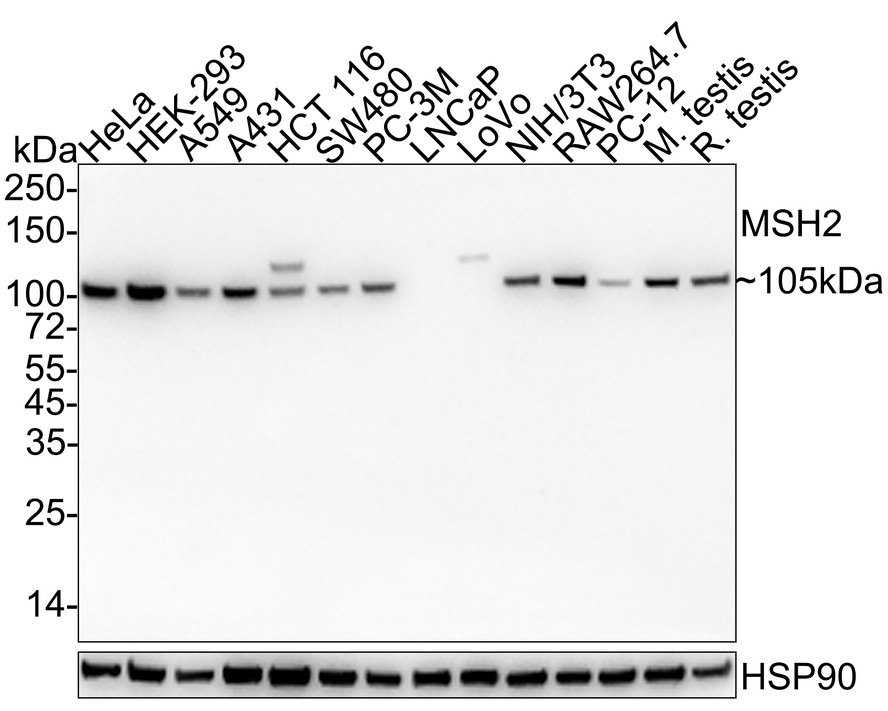
Western blot analysis of MSH2 on different lysates with Mouse anti-MSH2 antibody (EM1801-05) at 1/1,000 dilution.
Lane 1: HeLa cell lysate (15 µg/Lane)
Lane 2: HEK-293 cell lysate (15 µg/Lane)
Lane 3: A549 cell lysate (15 µg/Lane)
Lane 4: A431 cell lysate (15 µg/Lane)
Lane 5: HCT 116 cell lysate (15 µg/Lane)
Lane 6: SW480 cell lysate (15 µg/Lane)
Lane 7: PC-3M cell lysate (15 µg/Lane)
Lane 8: LNCaP cell lysate (negative) (15 µg/Lane)
Lane 9: LoVo cell lysate (negative) (15 µg/Lane)
Lane 10: NIH/3T3 cell lysate (15 µg/Lane)
Lane 11: RAW264.7 cell lysate (15 µg/Lane)
Lane 12: PC-12 cell lysate (15 µg/Lane)
Lane 13: Mouse testis tissue lysate (25 µg/Lane)
Lane 14: Rat testis tissue lysate (25 µg/Lane)
Predicted band size: 105 kDa
Observed band size: 105 kDa
Exposure time: 43 seconds;
4-20% SDS-PAGE gel.
Proteins were transferred to a PVDF membrane and blocked with 5% NFDM/TBST for 1 hour at room temperature. The primary antibody (EM1801-05) at 1/1,000 dilution was used in 5% NFDM/TBST at 4℃ overnight. Goat Anti-Mouse IgG - HRP Secondary Antibody (HA1006) at 1/50,000 dilution was used for 1 hour at room temperature. -
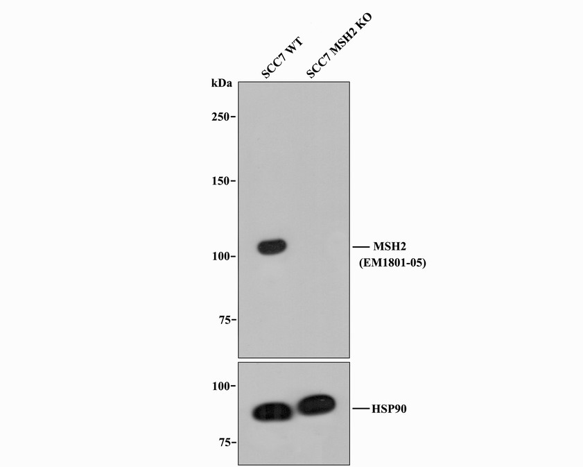
All lanes: Western blot analysis of MSH2 with anti-MSH2 antibody (EM1801-05) at 1/1,000 dilution.
Lane 1: Wild-type SCC7 whole cell lysate.
Lane 2: MSH2 knockout SCC7 whole cell lysate.
EM1801-05 was shown to specifically react with MSH2 in Wild-type SCC7 cells. No band was observed when MSH2 knockout sample was tested. Wild-type and MSH2 knockout samples were subjected to SDS-PAGE. Proteins were transferred to a PVDF membrane and blocked with 5% NFDM in TBST for 1 hour at room temperature. The primary Anti-MSH2 antibody (EM1801-05, 1/1,000) and Anti-HSP90 antibody (ET1605-56, 1/10,000) were used in 5% BSA at room temperature for 2 hours. Goat Anti-Mouse IgG HRP Secondary Antibody (HA1006) at 1:20,000 dilution was used for 1 hour at room temperature. -
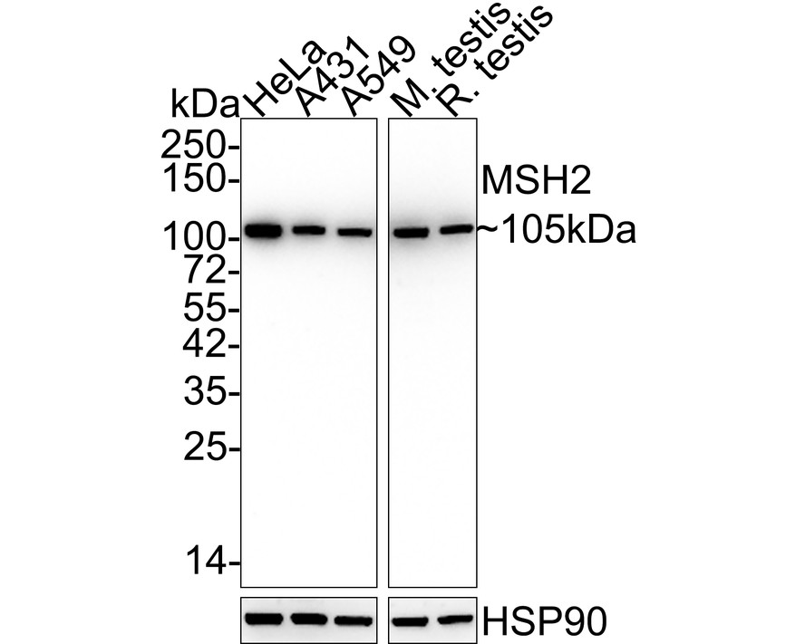
Western blot analysis of MSH2 on different lysates with Mouse anti-MSH2 antibody (EM1801-05) at 1/5,000 dilution.
Lane 1: HeLa cell lysate (15 µg/Lane)
Lane 2: A431 cell lysate (15 µg/Lane)
Lane 3: A549 cell lysate (15 µg/Lane)
Lane 4: Mouse testis tissue lysate (20 µg/Lane)
Lane 5: Rat testis tissue lysate (20 µg/Lane)
Predicted band size: 105 kDa
Observed band size: 105 kDa
Exposure time: 28 seconds;
4-20% SDS-PAGE gel.
Proteins were transferred to a PVDF membrane and blocked with 5% NFDM/TBST for 1 hour at room temperature. The primary antibody (EM1801-05) at 1/5,000 dilution was used in 5% NFDM/TBST at 4℃ overnight. Goat Anti-Mouse IgG - HRP Secondary Antibody (HA1006) at 1/50,000 dilution was used for 1 hour at room temperature. -
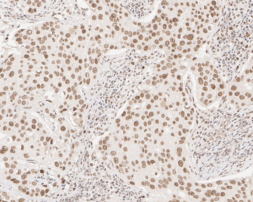
Immunohistochemical analysis of paraffin-embedded human breast cancer tissue with Mouse anti-MSH2 antibody (EM1801-05) at 1/1,000 dilution.
The section was pre-treated using heat mediated antigen retrieval with sodium citrate buffer (pH 6.0) for 2 minutes. The tissues were blocked in 1% BSA for 20 minutes at room temperature, washed with ddH2O and PBS, and then probed with the primary antibody (EM1801-05) at 1/1,000 dilution for 1 hour at room temperature. The detection was performed using an HRP conjugated compact polymer system. DAB was used as the chromogen. Tissues were counterstained with hematoxylin and mounted with DPX. -
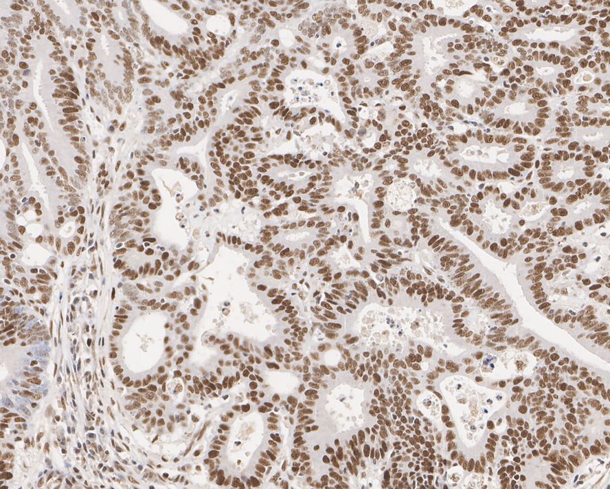
Immunohistochemical analysis of paraffin-embedded human colon cancer tissue with Mouse anti-MSH2 antibody (EM1801-05) at 1/1,000 dilution.
The section was pre-treated using heat mediated antigen retrieval with sodium citrate buffer (pH 6.0) for 2 minutes. The tissues were blocked in 1% BSA for 20 minutes at room temperature, washed with ddH2O and PBS, and then probed with the primary antibody (EM1801-05) at 1/1,000 dilution for 1 hour at room temperature. The detection was performed using an HRP conjugated compact polymer system. DAB was used as the chromogen. Tissues were counterstained with hematoxylin and mounted with DPX. -
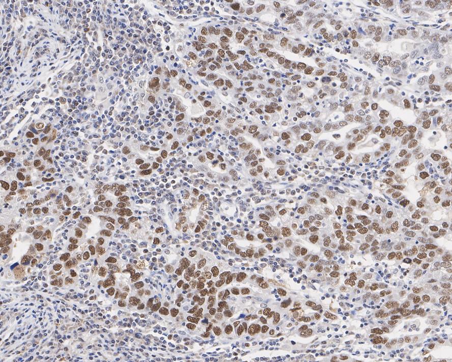
Immunohistochemical analysis of paraffin-embedded human stomach cancer tissue with Mouse anti-MSH2 antibody (EM1801-05) at 1/1,000 dilution.
The section was pre-treated using heat mediated antigen retrieval with sodium citrate buffer (pH 6.0) for 2 minutes. The tissues were blocked in 1% BSA for 20 minutes at room temperature, washed with ddH2O and PBS, and then probed with the primary antibody (EM1801-05) at 1/1,000 dilution for 1 hour at room temperature. The detection was performed using an HRP conjugated compact polymer system. DAB was used as the chromogen. Tissues were counterstained with hematoxylin and mounted with DPX. -
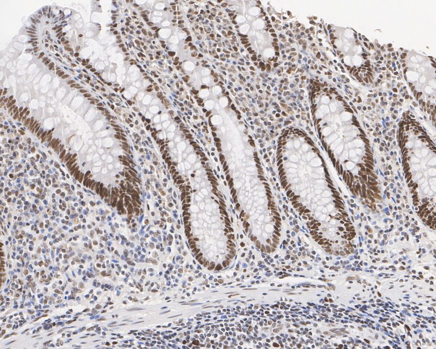
Immunohistochemical analysis of paraffin-embedded human appendix tissue with Mouse anti-MSH2 antibody (EM1801-05) at 1/1,000 dilution.
The section was pre-treated using heat mediated antigen retrieval with sodium citrate buffer (pH 6.0) for 2 minutes. The tissues were blocked in 1% BSA for 20 minutes at room temperature, washed with ddH2O and PBS, and then probed with the primary antibody (EM1801-05) at 1/1,000 dilution for 1 hour at room temperature. The detection was performed using an HRP conjugated compact polymer system. DAB was used as the chromogen. Tissues were counterstained with hematoxylin and mounted with DPX. -
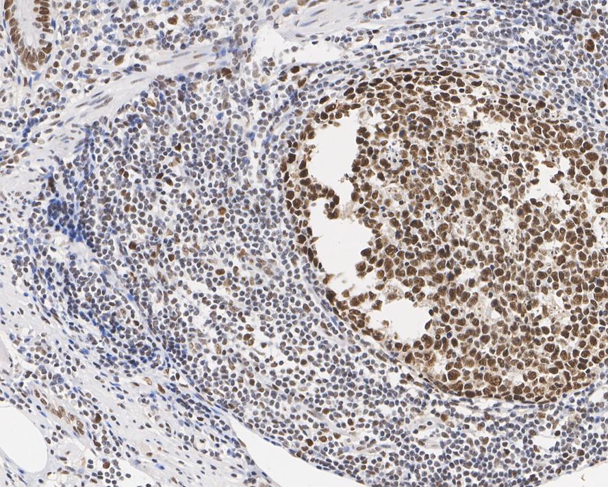
Immunohistochemical analysis of paraffin-embedded human appendicular lymph nodes tissue with Mouse anti-MSH2 antibody (EM1801-05) at 1/1,000 dilution.
The section was pre-treated using heat mediated antigen retrieval with sodium citrate buffer (pH 6.0) for 2 minutes. The tissues were blocked in 1% BSA for 20 minutes at room temperature, washed with ddH2O and PBS, and then probed with the primary antibody (EM1801-05) at 1/1,000 dilution for 1 hour at room temperature. The detection was performed using an HRP conjugated compact polymer system. DAB was used as the chromogen. Tissues were counterstained with hematoxylin and mounted with DPX. -
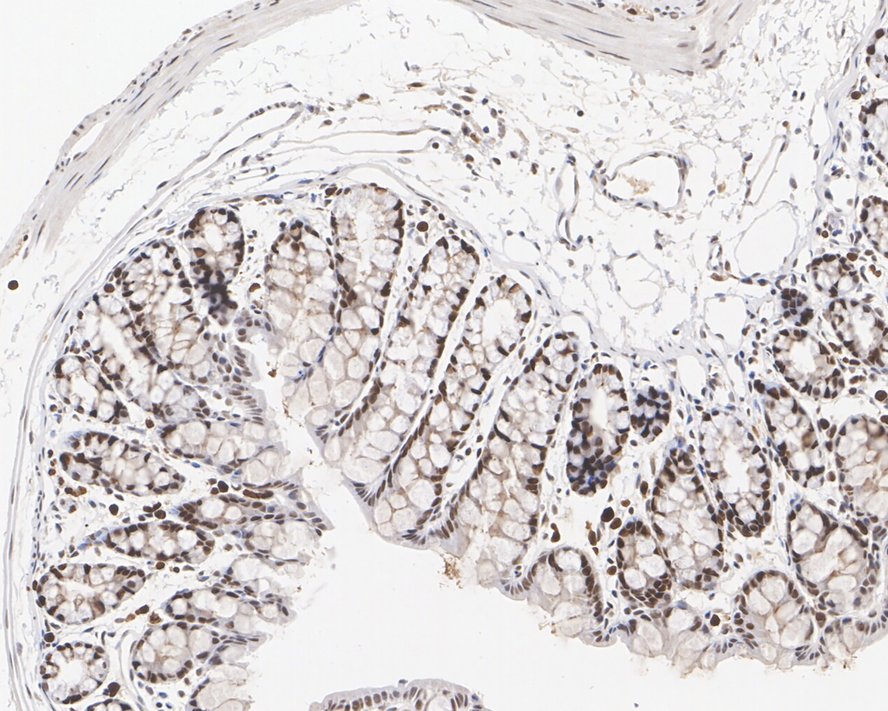
Immunohistochemical analysis of paraffin-embedded mouse colon tissue with Mouse anti-MSH2 antibody (EM1801-05) at 1/1,000 dilution.
The section was pre-treated using heat mediated antigen retrieval with sodium citrate buffer (pH 6.0) for 2 minutes. The tissues were blocked in 1% BSA for 20 minutes at room temperature, washed with ddH2O and PBS, and then probed with the primary antibody (EM1801-05) at 1/1,000 dilution for 1 hour at room temperature. The detection was performed using an HRP conjugated compact polymer system. DAB was used as the chromogen. Tissues were counterstained with hematoxylin and mounted with DPX. -
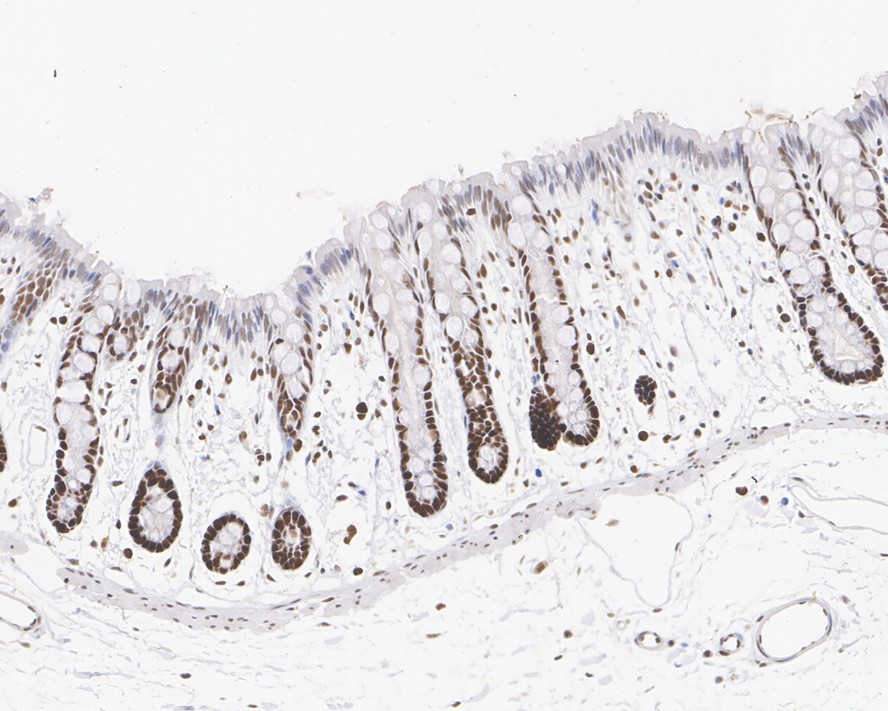
Immunohistochemical analysis of paraffin-embedded rat colon tissue with Mouse anti-MSH2 antibody (EM1801-05) at 1/1,000 dilution.
The section was pre-treated using heat mediated antigen retrieval with sodium citrate buffer (pH 6.0) for 2 minutes. The tissues were blocked in 1% BSA for 20 minutes at room temperature, washed with ddH2O and PBS, and then probed with the primary antibody (EM1801-05) at 1/1,000 dilution for 1 hour at room temperature. The detection was performed using an HRP conjugated compact polymer system. DAB was used as the chromogen. Tissues were counterstained with hematoxylin and mounted with DPX.
Please note: All products are "FOR RESEARCH USE ONLY AND ARE NOT INTENDED FOR DIAGNOSTIC OR THERAPEUTIC USE"