GLUT2 Recombinant Rabbit Monoclonal Antibody [JJ20-21]


Catalog# ET1701-34
GLUT2 Recombinant Rabbit Monoclonal Antibody [JJ20-21]
-
WB
-
IHC-P
-
FC
-
Human
概述
产品名称
GLUT2 Recombinant Rabbit Monoclonal Antibody [JJ20-21]
抗体类型
Recombinant Rabbit monoclonal Antibody
免疫原
Synthetic peptide within Human Glucose Transporter GLUT2 aa 35-84 / 524.
种属反应性
Human
验证应用
WB, IHC-P, FC
分子量
Predicted band size: 57 kDa
阳性对照
A549 cell lysates, human liver tissue, human kidney tissue, human pancreas tissue, HepG2.
偶联
unconjugated
克隆号
JJ20-21
RRID
产品特性
形态
Liquid
浓度
1ug/ul
存放说明
Store at +4℃ after thawing. Aliquot store at -20℃ or -80℃. Avoid repeated freeze / thaw cycles.
存储缓冲液
1*TBS (pH7.4), 0.05% BSA, 40% Glycerol. Preservative: 0.05% Sodium Azide.
亚型
IgG
纯化方式
Protein A affinity purified.
应用稀释度
-
WB
-
1:1,000
-
IHC-P
-
1:50-1:200
-
FC
-
1:50-1:100
靶点
功能
Glucose transporter 2 (GLUT2) also known as solute carrier family 2 (facilitated glucose transporter), member 2 (SLC2A2) is a transmembrane carrier protein that enables protein facilitated glucose movement across cell membranes. It is the principal transporter for transfer of glucose between liver and blood. Unlike GLUT4, it does not rely on insulin for facilitated diffusion. GLUT2 has high capacity for glucose but low affinity (high KM, ca. 15–20 mM) and thus functions as part of the "glucose sensor" in the pancreatic β-cells of rodents, though in human β-cells the role of GLUT2 seems to be a minor one. It is a very efficient carrier for glucose. GLUT2 also carries glucosamine. When the glucose concentration in the lumen of the small intestine goes above 30 mM, such as occurs in the fed-state, GLUT2 is up-regulated at the brush border membrane, enhancing the capacity of glucose transport. Basolateral GLUT2 in enterocytes also aids in the transport of fructose into the bloodstream through glucose-dependent cotransport.
背景文献
1. Pettinato G et al. ROCK inhibitor is not required for embryoid body formation from singularized human embryonic stem cells. PLoS One 9:e100742 (2014).
2. Pettinato G et al. Formation of well-defined embryoid bodies from dissociated human induced pluripotent stem cells using microfabricated cell-repellent microwell arrays. Sci Rep 4:7402 (2014).
序列相似性
Belongs to the major facilitator superfamily. Sugar transporter (TC 2.A.1.1) family. Glucose transporter subfamily.
组织特异性
Liver, insulin-producing beta cell, small intestine and kidney.
翻译后修饰
N-glycosylated; required for stability and retention at the cell surface of pancreatic beta cells.
亚细胞定位
Membrane.
UNIPROT #
别名
liver antibody
Glucose Transporter 2 antibody
Glucose Transporter GLUT2 antibody
Glucose transporter type 2 antibody
Glucose transporter type 2 liver antibody
Glucose transporter, liver/islet antibody
GLUT-2 antibody
GLUT2 antibody
GTR2_HUMAN antibody
GTT2 antibody
展开图片
-
Western blot analysis of GLUT2 on A549 cell lysates with Rabbit anti-GLUT2 antibody (ET1701-34) at 1/1,000 dilution.
Lysates/proteins at 30 µg/Lane.
Predicted band size: 57 kDa
Observed band size: 50 kDa
Exposure time: 28 seconds;
4-20% SDS-PAGE gel.
Proteins were transferred to a PVDF membrane and blocked with 5% NFDM/TBST for 1 hour at room temperature. The primary antibody (ET1701-34) at 1/1,000 dilution was used in 5% NFDM/TBST at room temperature for 2 hours. Goat Anti-Rabbit IgG - HRP Secondary Antibody (HA1001) at 1:100,000 dilution was used for 1 hour at room temperature. -
Immunohistochemical analysis of paraffin-embedded human liver tissue using anti-GLUT2 antibody. The section was pre-treated using heat mediated antigen retrieval with Tris-EDTA buffer (pH 9.0) for 20 minutes.The tissues were blocked in 5% BSA for 30 minutes at room temperature, washed with ddH2O and PBS, and then probed with the primary antibody (ET1701-34, 1/50) for 30 minutes at room temperature. The detection was performed using an HRP conjugated compact polymer system. DAB was used as the chromogen. Tissues were counterstained with hematoxylin and mounted with DPX.
-
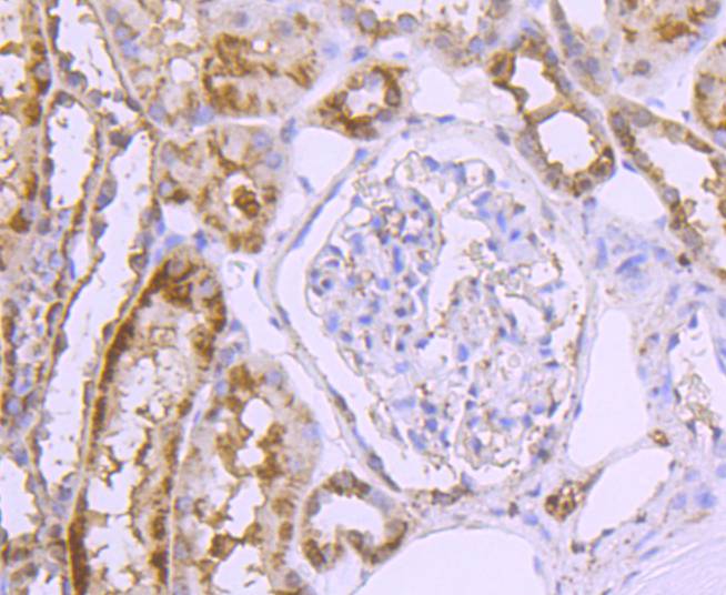
Immunohistochemical analysis of paraffin-embedded human kidney tissue using anti-GLUT2 antibody. The section was pre-treated using heat mediated antigen retrieval with Tris-EDTA buffer (pH 9.0) for 20 minutes.The tissues were blocked in 5% BSA for 30 minutes at room temperature, washed with ddH2O and PBS, and then probed with the primary antibody (ET1701-34, 1/50) for 30 minutes at room temperature. The detection was performed using an HRP conjugated compact polymer system. DAB was used as the chromogen. Tissues were counterstained with hematoxylin and mounted with DPX.
-
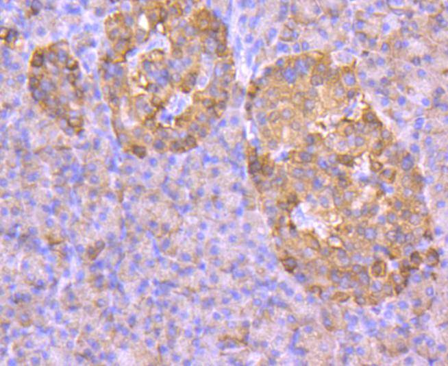
Immunohistochemical analysis of paraffin-embedded human pancreas tissue using anti-GLUT2 antibody. The section was pre-treated using heat mediated antigen retrieval with Tris-EDTA buffer (pH 9.0) for 20 minutes.The tissues were blocked in 5% BSA for 30 minutes at room temperature, washed with ddH2O and PBS, and then probed with the primary antibody (ET1701-34, 1/50) for 30 minutes at room temperature. The detection was performed using an HRP conjugated compact polymer system. DAB was used as the chromogen. Tissues were counterstained with hematoxylin and mounted with DPX.
-
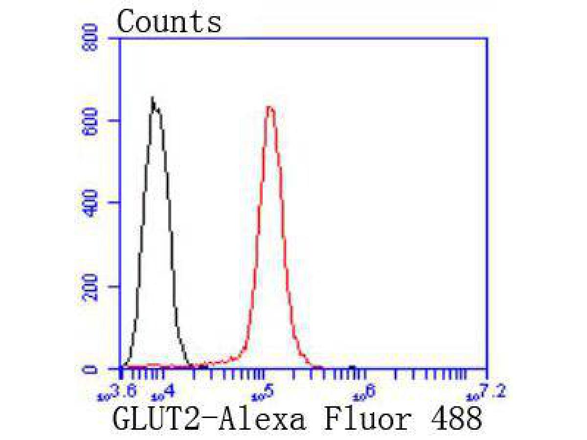
Flow cytometric analysis of GLUT2 was done on HepG2 cells. The cells were fixed, permeabilized and stained with the primary antibody (ET1701-34, 1/50) (red). After incubation of the primary antibody at room temperature for an hour, the cells were stained with a Alexa Fluor 488-conjugated Goat anti-Rabbit IgG Secondary antibody at 1/1,000 dilution for 30 minutes.Unlabelled sample was used as a control (cells without incubation with primary antibody; black).
Please note: All products are "FOR RESEARCH USE ONLY AND ARE NOT INTENDED FOR DIAGNOSTIC OR THERAPEUTIC USE"