Cancer Associated Fibroblast Marker Antibody Kit
RMB: 4100.00
Catalog# K2006
Cancer Associated Fibroblast Marker Antibody Kit
概述
试剂盒组分
| 产品包括 | 规格 | 应用 | 反应性 | MW(kDa) |
|---|---|---|---|---|
| PDGFR alpha[ET1702-49] | 20µl | WB,IF-Cell,IHC-P,FC | Human,Mouse,Rat | Predicted band size: 123 kDa |
| alpha smooth muscle Actin[ET1607-53] | 20µl | WB,IF-Cell,IF-Tissue,IHC-P,FC,mIHC | Human,Mouse,Rat | Predicted band size: 42 kDa |
| PDGF Receptor beta[ET1605-20] | 20µl | WB,IHC-P,IP | Human,Mouse,Rat | Predicted band size:123 kDa |
| S100 beta[ET1610-3] | 20µl | WB,IF-Cell,IF-Tissue,IP,IHC-P,IHC-Fr | Human,Mouse,Zebrafish,Rat,Goat | Predicted band size: 11 kDa |
| FAP[ET1704-23] | 20µl | WB,IHC-P,mIHC | Human | 88 kDa |
| S100A4[ET1612-13] | 20µl | WB,IF-Cell,IF-Tissue,IHC-P,IP,FC | Human,Mouse,Rat | 12 kDa |
| HRP Conjugated Alpaca anti-Rabbit IgG FC, Recombinant VHH[HA1031] | 100μl | IP,ELISA,IHC-P,WB | Rabbit |
产品描述
The Cancer Associated Fibroblast Marker Antibody Sampler Kit provides an economical means of detecting proteins reported to be expressed in Cancer Associated Fibroblasts (CAFs). The kit includes enough antibodies to perform two western blot experiments with each primary antibody.
产品特性
存储缓冲液
1*TBS (pH7.4), 0.05% BSA, 40% Glycerol. Preservative: 0.05% Sodium Azide.
存放说明
Store at +4℃ after thawing. Aliquot store at -20℃. Avoid repeated freeze / thaw cycles.
背景
The tumor microenvironment (TME) has been shown to play an important role in tumor initiation, development, and metastasis. Amongst all these various factors, fibroblasts have been suggested to play a key role in tumor development.</br>During tumor development, a subpopulation of hyper-activated fibroblasts become prominent in the TME and secretion of cytokines and chemokines from these cells promotes pro-tumorigenic activity. These highly heterogeneous fibroblast populations are known collectively as CAFs (Cancer Associated Fibroblasts).PDGFRα and PDGFRβ are common markers used for fibroblast identification. α-Smooth Muscle Actin is widely used to identify CAFs, however, some reports suggest it is not expressed by all functionally active CAFs. FSP-1/S100A4 is expressed by cells of mesenchymal origins. Some reports even suggest it to be a marker for quiescent fibroblasts. Fibroblast Activation Protein, or FAP as it is more commonly known, has traditionally been associated with tissue repair, fibrosis, and extracellular matrix degradation. FAP has more recently been described as a useful marker of CAFs. Vimentin strongly characterizes cells of a mesenchymal phenotype.
背景文献
2. Nurmik, M. et al. (2019) Int J Cancer, doi: 10.1002/ijc.32193.
图片
-
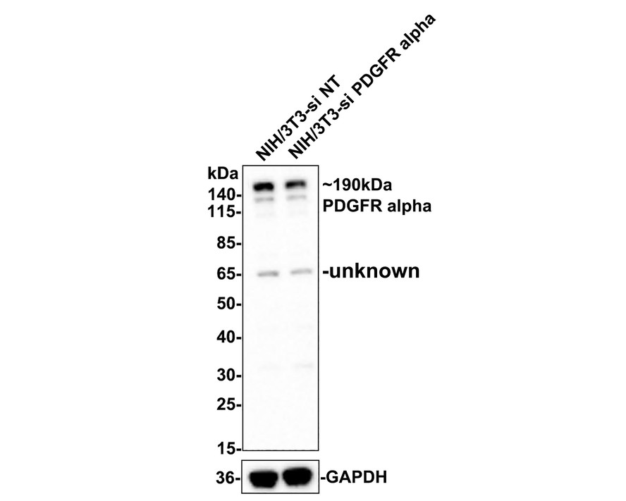
Western blot analysis of PDGFR alpha on different lysates with Rabbit anti-PDGFR alpha antibody (ET1702-49) at 1/500 dilution.
Lane 1: NIH/3T3-si NT cell lysate
Lane 2: NIH/3T3-si PDGFR alpha cell lysate
Lysates/proteins at 10 µg/Lane.
Predicted band size: 123 kDa
Observed band size: 190 kDa
Exposure time: 2 minutes;
4-20% SDS-PAGE gel.
ET1702-49 was shown to specifically react with PDGFR alpha in Hela-si NT cells. Weakened band was observed when Hela-si PDGFR alpha sample was tested. Hela-si NT and Hela-si PDGFR alpha samples were subjected to SDS-PAGE. Proteins were transferred to a PVDF membrane and blocked with 5% NFDM in TBST for 1 hour at room temperature. The primary antibody (ET1702-49, 1/500) and Loading control antibody (Rabbit anti-GAPDH, ET1601-4, 1/10,000) were used in 5% BSA at room temperature for 2 hours. Goat Anti-rabbit IgG-HRP Secondary Antibody (HA1001) at 1:300,000 dilution was used for 1 hour at room temperature. -
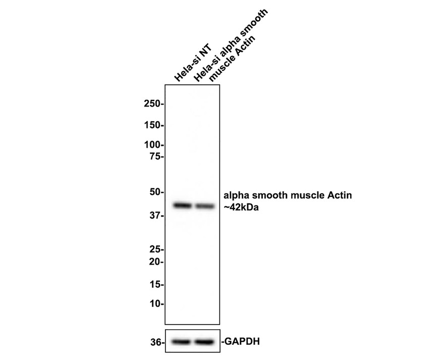
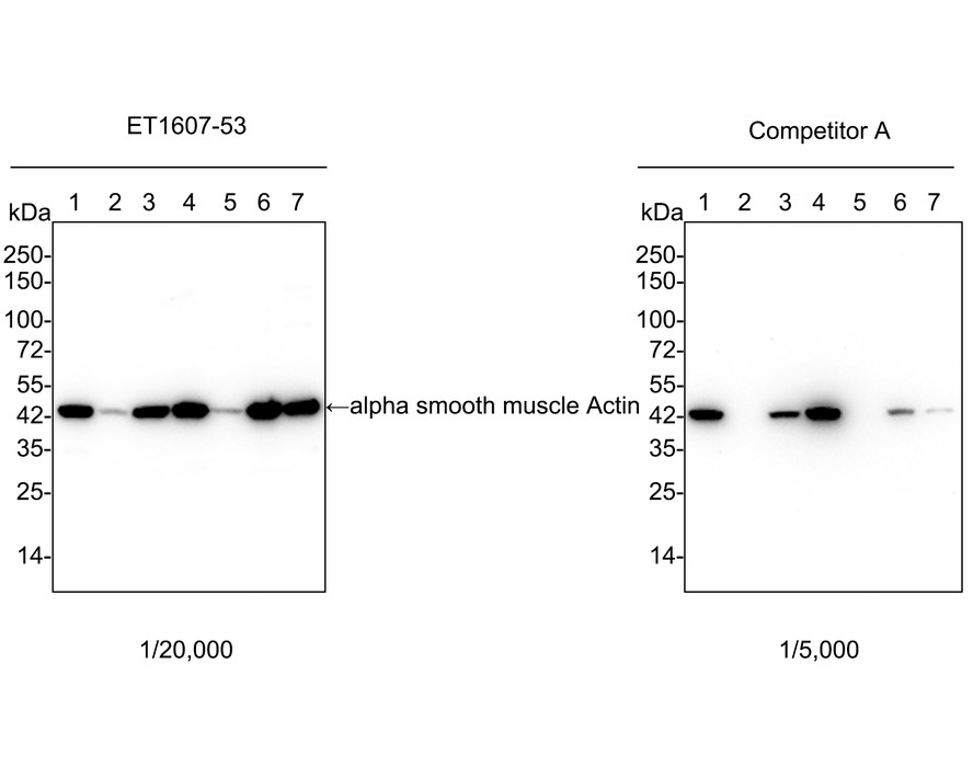
Western blot analysis of alpha smooth muscle Actin on different lysates with Rabbit anti-alpha smooth muscle Actin antibody (ET1607-53) at 1/1,000 dilution.
Lane 1: Hela-si NT cell lysate
Lane 2: Hela-si alpha smooth muscle Actin cell lysate
Lysates/proteins at 10 µg/Lane.
Predicted band size: 42 kDa
Observed band size: 42 kDa
Exposure time: 5 seconds;
4-20% SDS-PAGE gel.
ET1607-53 was shown to specifically react with alpha smooth muscle Actin in Hela-si NT cells. Weakened band was observed when Hela-si alpha smooth muscle Actin sample was tested. Hela-si NT and Hela-si alpha smooth muscle Actin samples were subjected to SDS-PAGE. Proteins were transferred to a PVDF membrane and blocked with 5% NFDM in TBST for 1 hour at room temperature. The primary antibody (ET1607-53, 1/1,000) and Loading control antibody (Rabbit anti-GAPDH, ET1601-4, 1/10,000) were used in 5% BSA at room temperature for 2 hours. Goat Anti-rabbit IgG-HRP Secondary Antibody (HA1001) at 1:100,000 dilution was used for 1 hour at room temperature. -
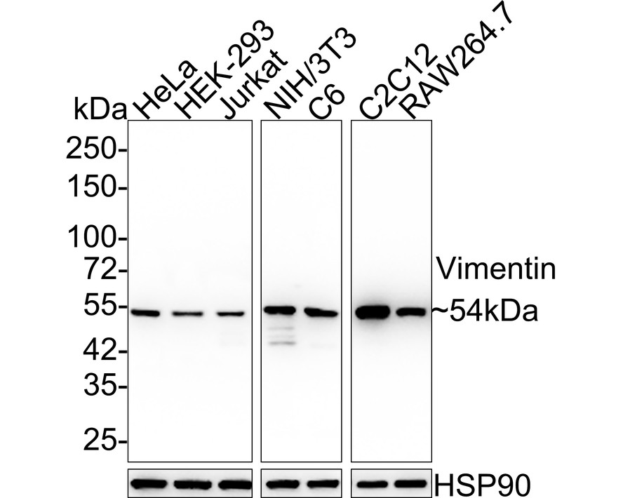
Western blot analysis of Vimentin on different lysates with Rabbit anti-Vimentin antibody (ET1610-39) at 1/20,000 dilution.
Lane 1: HeLa cell lysate (10 µg/Lane)
Lane 2: HEK-293 cell lysate (10 µg/Lane)
Lane 3: Jurkat cell lysate (10 µg/Lane)
Lane 4: NIH/3T3 cell lysate (10 µg/Lane)
Lane 5: C6 cell lysate (10 µg/Lane)
Lane 6: C2C12 cell lysate (10 µg/Lane)
Lane 7: RAW264.7 cell lysate (10 µg/Lane)
Predicted band size: 54 kDa
Observed band size: 54 kDa
Exposure time: Lane 1-5: 3 seconds; Lane 6-7: 14 seconds;
4-20% SDS-PAGE gel.
Proteins were transferred to a PVDF membrane and blocked with 5% NFDM/TBST for 1 hour at room temperature. The primary antibody (ET1610-39) at 1/20,000 dilution was used in 5% NFDM/TBST at room temperature for 2 hours. Goat Anti-Rabbit IgG - HRP Secondary Antibody (HA1001) at 1:50,000 dilution was used for 1 hour at room temperature. -
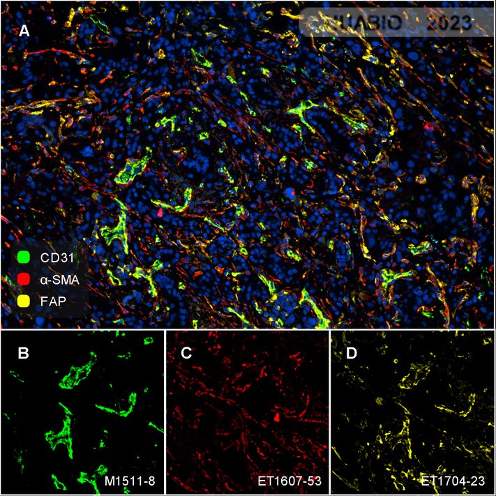
Fluorescence multiplex immunohistochemical analysis of the human pancreatic carcinoma (Formalin/PFA-fixed paraffin-embedded sections). Panel A: the merged image of anti-CD31 (M1511-8, green), anti-α-SMA (ET1607-53, red) and anti-FAP (ET1704-23, yellow) on human pancreatic carcinoma. Panel B: anti- CD31 stained on the endothelial cells. Panel C: anti-α-SMA stained on cancer-associated fibroblasts and smooth muscle cells. Panel D: anti-FAP stained on the cancer-associated fibroblasts. HRP Conjugated UltraPolymer Goat Polyclonal Antibody HA1119/HA1120 was used as a secondary antibody. The immunostaining was performed with the Sequential Immuno-staining Kit (IRISKit™MH010101, www.luminiris.cn). The section was incubated in three rounds of staining: in the order of M1511-8 (1/5000 dilution), ET1704-23 (1/1000 dilution), and ET1607-53 (1/3000 dilution) for 20 mins at room temperature. Heat mediated antigen retrieval with Tris-EDTA buffer (pH 9.0) for 30 mins at 95℃. DAPI (blue) was used as a nuclear counter stain. Image acquisition was performed with Nikon ECLIPSE Ni-E microscope.
Related Products

alpha smooth muscle Actin Recombinant Rabbit Monoclonal Antibody [SY25-03]
Application: WB,IHC-P,FC,IP
Reactivity: Human,Mouse,Rat
Conjugate: unconjugated
alpha smooth muscle Actin Recombinant Rabbit Monoclonal Antibody [SY02-64]
Application: WB,IF-Cell,IF-Tissue,IHC-P,FC,mIHC
Reactivity: Human,Mouse,Rat
Conjugate: unconjugated
