CTCF Recombinant Rabbit Monoclonal Antibody [JM10-61]


Catalog# ET1703-90
CTCF Recombinant Rabbit Monoclonal Antibody [JM10-61]
-
WB
-
IF-Cell
-
IF-Tissue
-
IHC-P
-
IP
-
FC
-
CUT&Tag-seq
-
ChIP
-
Human
-
Mouse
-
Zebrafish
-
Rat
概述
产品名称
CTCF Recombinant Rabbit Monoclonal Antibody [JM10-61]
抗体类型
Recombinant Rabbit monoclonal Antibody
免疫原
Synthetic peptide within Human CTCF aa 685-727 / 727.
种属反应性
Human, Mouse, Zebrafish, Rat
验证应用
WB, IF-Cell, IF-Tissue, IHC-P, IP, FC, CUT&Tag-seq, ChIP
分子量
Predicted band size: 83 kDa
阳性对照
MCF-7 cell lysates, 293T cell lysates, zebrafish tissue lysates, hybrid fish (crucian-carp) brain tissue lysates, HeLa, MCF-7, NIH/3T3, human breast carcinoma tissue, human endometrium tissue, mouse stomach tissue, human liver tissue, human kidney tissue, mouse colon tissue.
偶联
unconjugated
克隆号
JM10-61
RRID
产品特性
形态
Liquid
浓度
1ug/ul
存放说明
Store at +4℃ after thawing. Aliquot store at -20℃ or -80℃. Avoid repeated freeze / thaw cycles.
存储缓冲液
1*TBS (pH7.4), 0.05% BSA, 40% Glycerol. Preservative: 0.05% Sodium Azide.
亚型
IgG
纯化方式
Protein A affinity purified.
应用稀释度
-
WB
-
1:500-1:2,000
-
IF-Cell
-
1:100-1:500
-
IF-Tissue
-
1:100-1:500
-
IHC-P
-
1:50-1:2,000
-
FC
-
1:500-1:1,000
-
IP
-
Use at an assay dependent concentration.
靶点
功能
CTCF belongs to the zinc finger transcription factor family, and it recognizes unusually long and remarkably divergent DNA target sequences to influence expression of many various genes. The DNA-binding domain of CTCF is composed of 11 Zn fingers including 10 that are of C2H2 class, and 1 that is of C2HC class, and they are highly conserved between vertebrate species. CTCF functions as a repressor of the c-myc gene and as a regulator of lysozyme gene expression. In addition, CTCF associates with the essential activator domain in the promotor region of the amyloid beta-protein precursor (APP) gene to activate transcription of APP. Expression of CTCF up-regulates APP expression and thereby, enhances synapse formations between primary neurons during development. CTCF is ubiquitously expressed and localized to the nucleus. During terminal differentiation, CTCF is negatively regulated by differential phosphorylation and also by decreases in CTCF mRNA and protein expression.
背景文献
1. Harr JC et al. Directed targeting of chromatin to the nuclear lamina is mediated by chromatin state and A-type lamins. J Cell Biol 208:33-52 (2015).
2. Shi F et al. The expression of Pax6 variants is subject to posttranscriptional regulation in the developing mouse eyelid. PLoS One 8:e53919 (2013).
序列相似性
Belongs to the CTCF zinc-finger protein family.
组织特异性
Ubiquitous. Absent in primary spermatocytes.
翻译后修饰
Sumoylated on Lys-74 and Lys-689; sumoylation of CTCF contributes to the repressive function of CTCF on the MYC P2 promoter.
亚细胞定位
Nucleoplasm, Chromosome, centromere.
UNIPROT #
别名
11 zinc finger protein antibody
11 zinc finger transcriptional repressor antibody
11-zinc finger protein antibody
CCCTC binding factor (zinc finger protein) antibody
CCCTC binding factor antibody
CCCTC-binding factor antibody
Ctcf antibody
CTCF_HUMAN antibody
CTCFL paralog antibody
MRD21 antibody
展开图片
-
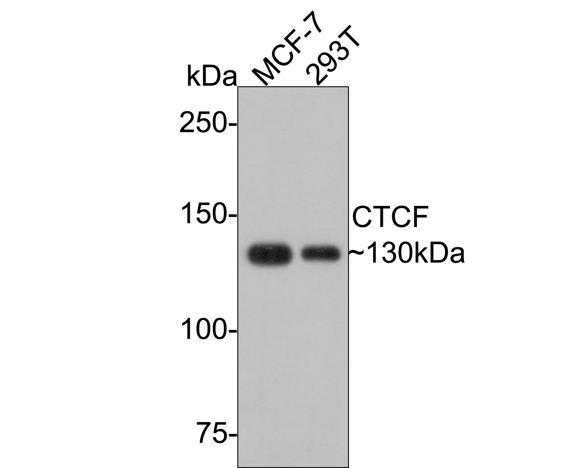
Western blot analysis of CTCF on different lysates with Rabbit anti-CTCF antibody (ET1703-90) at 1/500 dilution.
Lane 1: MCF-7 cell lysate
Lane 2: 293T cell lysate
Lysates/proteins at 10 µg/Lane.
Predicted band size: 83 kDa
Observed band size: 130 kDa
Exposure time: 30 seconds;
6% SDS-PAGE gel.
Proteins were transferred to a PVDF membrane and blocked with 5% NFDM/TBST for 1 hour at room temperature. The primary antibody (ET1703-90) at 1/500 dilution was used in 5% NFDM/TBST at room temperature for 2 hours. Goat Anti-Rabbit IgG - HRP Secondary Antibody (HA1001) at 1:200,000 dilution was used for 1 hour at room temperature. -
Western blot analysis of CTCF on zebrafish tissue lysates. Proteins were transferred to a PVDF membrane and blocked with 5% BSA in PBS for 1 hour at room temperature. The primary antibody (ET1703-90, 1/500) was used in 5% BSA at room temperature for 2 hours. Goat Anti-Rabbit IgG - HRP Secondary Antibody (HA1001) at 1:5,000 dilution was used for 1 hour at room temperature.
-
Western blot analysis of CTCF on hybrid fish (crucian-carp) brain tissue lysates. Proteins were transferred to a PVDF membrane and blocked with 5% BSA in PBS for 1 hour at room temperature. The primary antibody (ET1703-90, 1/500) was used in 5% BSA at room temperature for 2 hours. Goat Anti-Rabbit IgG - HRP Secondary Antibody (HA1001) at 1:5,000 dilution was used for 1 hour at room temperature.
-
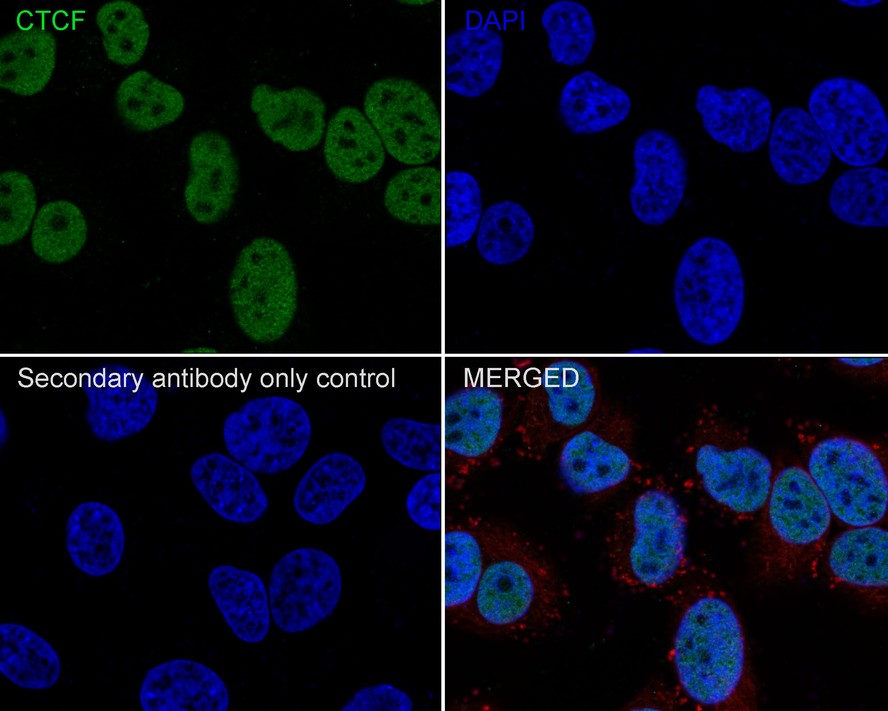
Immunocytochemistry analysis of HeLa cells labeling CTCF with Rabbit anti-CTCF antibody (ET1703-90) at 1/200 dilution.
Cells were fixed in 4% paraformaldehyde for 10 minutes at 37 ℃, permeabilized with 0.05% Triton X-100 in PBS for 20 minutes, and then blocked with 2% negative goat serum for 30 minutes at room temperature. Cells were then incubated with Rabbit anti-CTCF antibody (ET1703-90) at 1/200 dilution in 2% negative goat serum overnight at 4 ℃. Goat Anti-Rabbit IgG H&L (iFluor™ 488, HA1121) was used as the secondary antibody at 1/1,000 dilution. Nuclear DNA was labelled in blue with DAPI.
Beta tubulin (M1305-2, red) was stained at 1/200 dilution overnight at +4℃. Goat Anti-Mouse IgG H&L (iFluor™ 594, HA1126) was used as the secondary antibody at 1/1,000 dilution. -
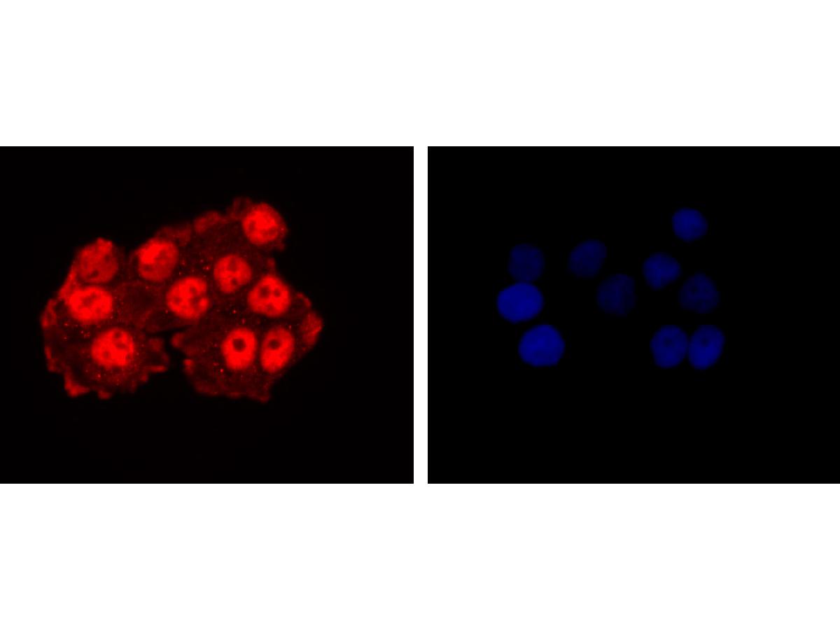
ICC staining of CTCF in HeLa cells (red). Formalin fixed cells were permeabilized with 0.1% Triton X-100 in TBS for 10 minutes at room temperature and blocked with 1% Blocker BSA for 15 minutes at room temperature. Cells were probed with the primary antibody (ET1703-90, 1/50) for 1 hour at room temperature, washed with PBS. Alexa Fluor®594 Goat anti-Rabbit IgG was used as the secondary antibody at 1/1,000 dilution. The nuclear counter stain is DAPI (blue).
-
ICC staining of CTCF in MCF-7 cells (red). Formalin fixed cells were permeabilized with 0.1% Triton X-100 in TBS for 10 minutes at room temperature and blocked with 1% Blocker BSA for 15 minutes at room temperature. Cells were probed with the primary antibody (ET1703-90, 1/50) for 1 hour at room temperature, washed with PBS. Alexa Fluor®594 Goat anti-Rabbit IgG was used as the secondary antibody at 1/1,000 dilution. The nuclear counter stain is DAPI (blue).
-
ICC staining of CTCF in NIH/3T3 cells (red). Formalin fixed cells were permeabilized with 0.1% Triton X-100 in TBS for 10 minutes at room temperature and blocked with 1% Blocker BSA for 15 minutes at room temperature. Cells were probed with the primary antibody (ET1703-90, 1/50) for 1 hour at room temperature, washed with PBS. Alexa Fluor®594 Goat anti-Rabbit IgG was used as the secondary antibody at 1/1,000 dilution. The nuclear counter stain is DAPI (blue).
-
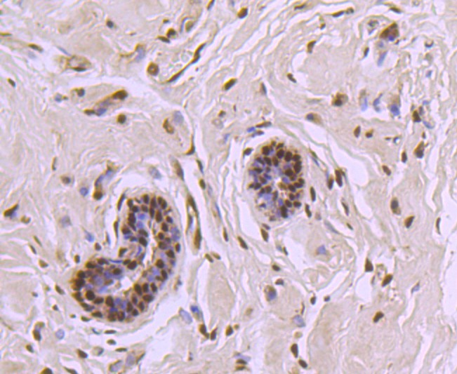
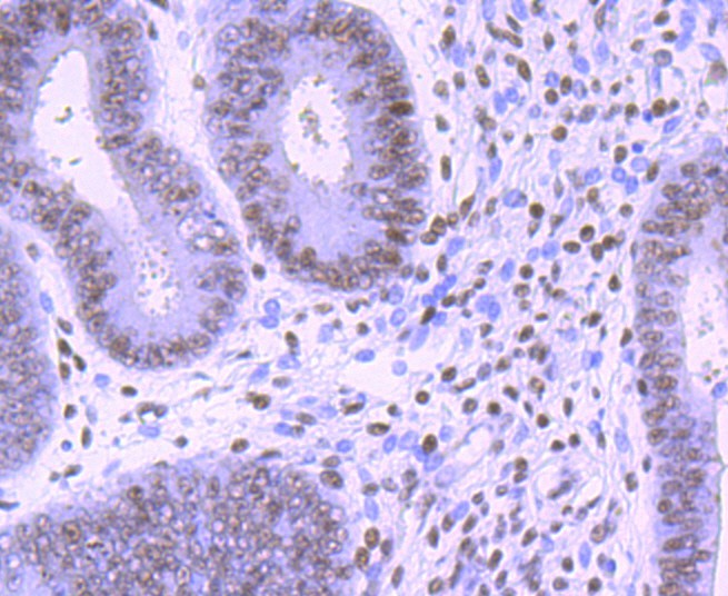
Immunohistochemical analysis of paraffin-embedded human breast carcinoma tissue using anti-CTCF antibody. The section was pre-treated using heat mediated antigen retrieval with Tris-EDTA buffer (pH 8.0-8.4) for 20 minutes.The tissues were blocked in 5% BSA for 30 minutes at room temperature, washed with ddH2O and PBS, and then probed with the primary antibody (ET1703-90, 1/50) for 30 minutes at room temperature. The detection was performed using an HRP conjugated compact polymer system. DAB was used as the chromogen. Tissues were counterstained with hematoxylin and mounted with DPX.
-
Immunohistochemical analysis of paraffin-embedded human endometrium tissue using anti-CTCF antibody. The section was pre-treated using heat mediated antigen retrieval with Tris-EDTA buffer (pH 8.0-8.4) for 20 minutes.The tissues were blocked in 5% BSA for 30 minutes at room temperature, washed with ddH2O and PBS, and then probed with the primary antibody (ET1703-90, 1/50) for 30 minutes at room temperature. The detection was performed using an HRP conjugated compact polymer system. DAB was used as the chromogen. Tissues were counterstained with hematoxylin and mounted with DPX.
-
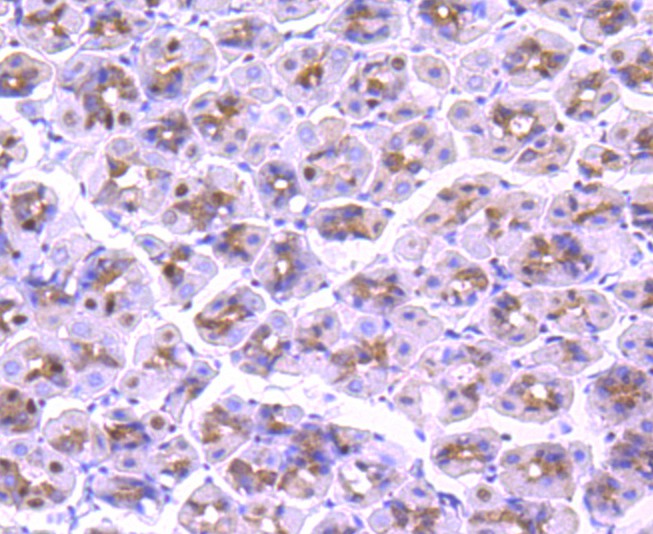
Immunohistochemical analysis of paraffin-embedded mouse stomach tissue using anti-CTCF antibody. The section was pre-treated using heat mediated antigen retrieval with Tris-EDTA buffer (pH 8.0-8.4) for 20 minutes.The tissues were blocked in 5% BSA for 30 minutes at room temperature, washed with ddH2O and PBS, and then probed with the primary antibody (ET1703-90, 1/50) for 30 minutes at room temperature. The detection was performed using an HRP conjugated compact polymer system. DAB was used as the chromogen. Tissues were counterstained with hematoxylin and mounted with DPX.
-
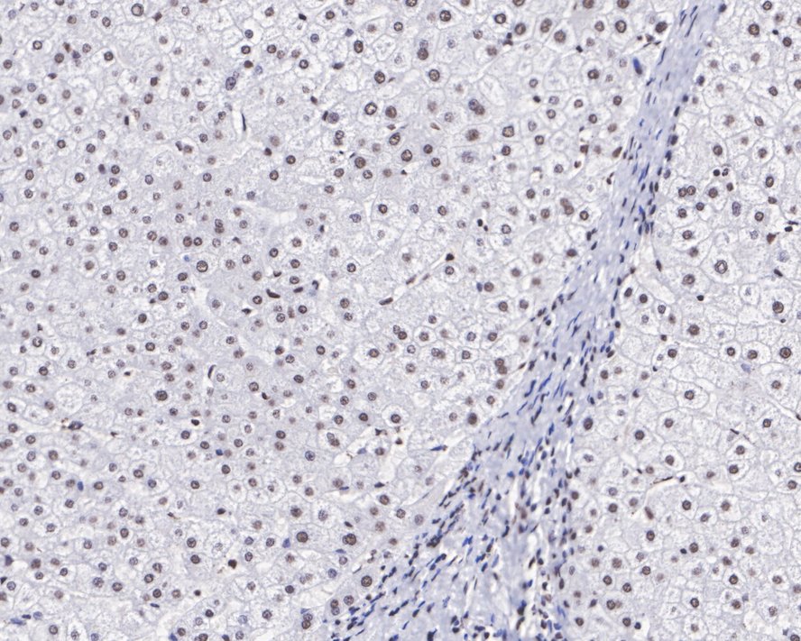
Immunohistochemical analysis of paraffin-embedded human liver tissue with Rabbit anti-CTCF antibody (ET1703-90) at 1/2,000 dilution.
The section was pre-treated using heat mediated antigen retrieval with sodium citrate buffer (pH 6.0) for 2 minutes. The tissues were blocked in 1% BSA for 20 minutes at room temperature, washed with ddH2O and PBS, and then probed with the primary antibody (ET1703-90) at 1/2,000 dilution for 1 hour at room temperature. The detection was performed using an HRP conjugated compact polymer system. DAB was used as the chromogen. Tissues were counterstained with hematoxylin and mounted with DPX. -
Immunohistochemical analysis of paraffin-embedded human kidney tissue with Rabbit anti-CTCF antibody (ET1703-90) at 1/1,000 dilution.
The section was pre-treated using heat mediated antigen retrieval with sodium citrate buffer (pH 6.0) for 2 minutes. The tissues were blocked in 1% BSA for 20 minutes at room temperature, washed with ddH2O and PBS, and then probed with the primary antibody (ET1703-90) at 1/1,000 dilution for 1 hour at room temperature. The detection was performed using an HRP conjugated compact polymer system. DAB was used as the chromogen. Tissues were counterstained with hematoxylin and mounted with DPX. -
Immunohistochemical analysis of paraffin-embedded mouse colon tissue using anti-CTCF antibody. The section was pre-treated using heat mediated antigen retrieval with Tris-EDTA buffer (pH 8.0-8.4) for 20 minutes.The tissues were blocked in 5% BSA for 30 minutes at room temperature, washed with ddH2O and PBS, and then probed with the primary antibody (ET1703-90, 1/50) for 30 minutes at room temperature. The detection was performed using an HRP conjugated compact polymer system. DAB was used as the chromogen. Tissues were counterstained with hematoxylin and mounted with DPX.
-
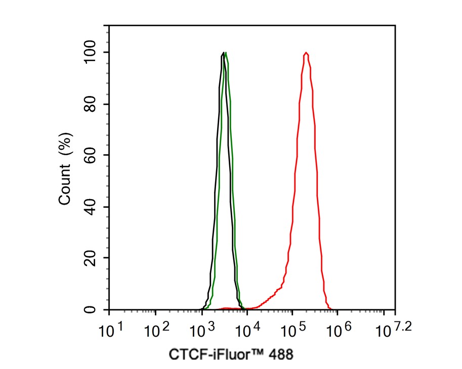
Flow cytometric analysis of HeLa cells labeling CTCF.
Cells were fixed and permeabilized. Then stained with the primary antibody (ET1703-90, 1ug/ml) (red) compared with Rabbit IgG Isotype Control (green). After incubation of the primary antibody at +4℃ for an hour, the cells were stained with a iFluor™ 488 conjugate-Goat anti-Rabbit IgG Secondary antibody (HA1121) at 1/1,000 dilution for 30 minutes at +4℃. Unlabelled sample was used as a control (cells without incubation with primary antibody; black).
Please note: All products are "FOR RESEARCH USE ONLY AND ARE NOT INTENDED FOR DIAGNOSTIC OR THERAPEUTIC USE"