SAE1 Recombinant Rabbit Monoclonal Antibody [JG35-88]

Catalog# ET7108-22
SAE1 Recombinant Rabbit Monoclonal Antibody [JG35-88]
-
WB
-
IHC-P
-
FC
-
Human
概述
产品名称
SAE1 Recombinant Rabbit Monoclonal Antibody [JG35-88]
抗体类型
Recombinant Rabbit monoclonal Antibody
免疫原
Synthetic peptide within Human SAE1 aa 201-249 / 346.
种属反应性
Human
验证应用
WB, IHC-P, FC
分子量
Predicted band size: 38 kDa
阳性对照
Jurkat cell lysate, HeLa cell lysate, 293T cell lysate, human colon cancer tissue, human skin tissue, A549.
偶联
unconjugated
克隆号
JG35-88
RRID
产品特性
形态
Liquid
浓度
1ug/ul
存放说明
Store at +4℃ after thawing. Aliquot store at -20℃. Avoid repeated freeze / thaw cycles.
存储缓冲液
1*TBS (pH7.4), 0.05% BSA, 40% Glycerol. Preservative: 0.05% Sodium Azide.
亚型
IgG
纯化方式
Protein A affinity purified.
应用稀释度
-
WB
-
1:1,000
-
IHC-P
-
1:200
-
FC
-
1:50-1:100
靶点
功能
Proteolytic degradation by the ubiquitin (Ub) system is essential for normal cell cycle progression, cellular differentiation and stress responses. Proteins conjugated to Ub are marked for progressive degradation by the 26S Proteasome. AOS-1, also designated SUMO-1-activating enzyme or ubiquitin-like 1-activating enzyme E1A, belongs to the ubiquitin-activating E1 family of proteins and plays an important role in the first step of the UBL1 conjugation pathway. AOS-1, which is a dimeric enzyme, functions as a UBLI E1 ligase, mediating the ATP-dependent activation of UBL1. AOS-1 can bind with UBLE1A and UBLE1B to form a heterodimer which can bind UBL1.
背景文献
1. Okuma T et al. In vitro SUMO-1 modification requires two enzymatic steps, E1 and E2. Biochem Biophys Res Commun 254:693-698 (1999).
2. Gong L et al. Molecular cloning and characterization of human AOS1 and UBA2, components of the sentrin-activating enzyme complex. FEBS Lett 448:185-189 (1999).
序列相似性
Belongs to the ubiquitin-activating E1 family.
组织特异性
Expression level increases during S phase and drops in G2 phase (at protein level).
亚细胞定位
Nucleus.
UNIPROT #
别名
Activator of SUMO1 antibody
AOS1 antibody
HSPC140 antibody
Sae1 antibody
SAE1_HUMAN antibody
Sentrin/SUMO activating protein AOS1 antibody
SUA1 antibody
SUMO 1 activating enzyme E1 N subunit antibody
SUMO 1 activating enzyme subunit 1 antibody
SUMO-activating enzyme subunit 1 antibody
展开图片
-
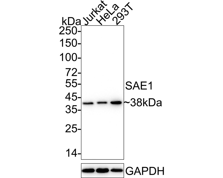
Western blot analysis of SAE1 on different lysates with Rabbit anti-SAE1 antibody (ET7108-22) at 1/1,000 dilution.
Lane 1: Jurkat cell lysate
Lane 2: HeLa cell lysate
Lane 3: 293T cell lysate
Lysates/proteins at 20 µg/Lane.
Predicted band size: 38 kDa
Observed band size: 38 kDa
Exposure time: 14 seconds; ECL: K1801;
4-20% SDS-PAGE gel.
Proteins were transferred to a PVDF membrane and blocked with 5% NFDM/TBST for 1 hour at room temperature. The primary antibody (ET7108-22) at 1/1,000 dilution was used in 5% NFDM/TBST at 4℃ overnight. Goat Anti-Rabbit IgG - HRP Secondary Antibody (HA1001) at 1/50,000 dilution was used for 1 hour at room temperature. -
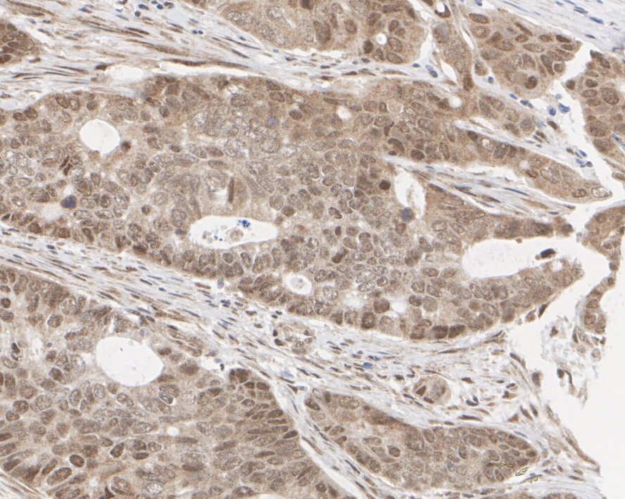
Immunohistochemical analysis of paraffin-embedded human colon cancer tissue with Rabbit anti-SAE1 antibody (ET7108-22) at 1/200 dilution.
The section was pre-treated using heat mediated antigen retrieval with sodium citrate buffer (pH 6.0) for 2 minutes. The tissues were blocked in 1% BSA for 20 minutes at room temperature, washed with ddH2O and PBS, and then probed with the primary antibody (ET7108-22) at 1/200 dilution for 1 hour at room temperature. The detection was performed using an HRP conjugated compact polymer system. DAB was used as the chromogen. Tissues were counterstained with hematoxylin and mounted with DPX. -
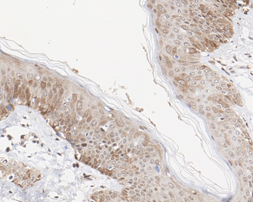
Immunohistochemical analysis of paraffin-embedded human skin tissue with Rabbit anti-SAE1 antibody (ET7108-22) at 1/200 dilution.
The section was pre-treated using heat mediated antigen retrieval with sodium citrate buffer (pH 6.0) for 2 minutes. The tissues were blocked in 1% BSA for 20 minutes at room temperature, washed with ddH2O and PBS, and then probed with the primary antibody (ET7108-22) at 1/200 dilution for 1 hour at room temperature. The detection was performed using an HRP conjugated compact polymer system. DAB was used as the chromogen. Tissues were counterstained with hematoxylin and mounted with DPX. -
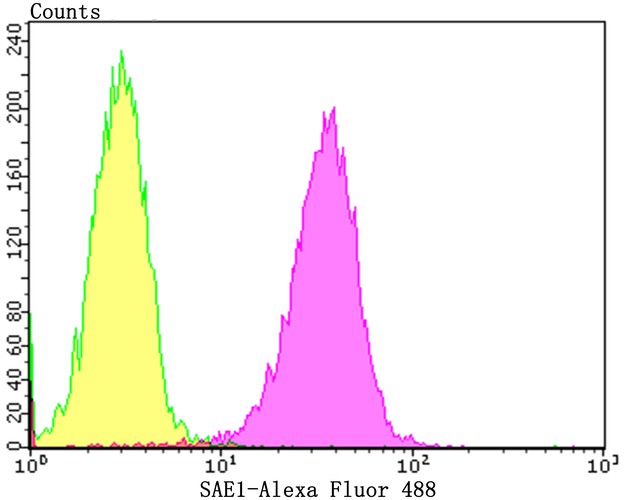
Flow cytometric analysis of A549 cells with SAE1 antibody at 1/100 dilution (yellow) compared with an unlabelled control (cells without incubation with primary antibody; purple).Alexa Fluor 488-conjugated goat anti-rabbit IgG was used as the secondary antibody.
Please note: All products are "FOR RESEARCH USE ONLY AND ARE NOT INTENDED FOR DIAGNOSTIC OR THERAPEUTIC USE"