hnRNP Q Recombinant Rabbit Monoclonal Antibody [JG35-72]


Catalog# ET7108-17
hnRNP Q Recombinant Rabbit Monoclonal Antibody [JG35-72]
-
WB
-
IHC-P
-
IF-Cell
-
IF-Tissue
-
Human
-
Rat
-
Mouse
概述
产品名称
hnRNP Q Recombinant Rabbit Monoclonal Antibody [JG35-72]
抗体类型
Recombinant Rabbit monoclonal Antibody
免疫原
Synthetic peptide within Human hnRNP Q aa 574-623 / 623.
种属反应性
Human, Rat, Mouse
验证应用
WB, IHC-P, IF-Cell, IF-Tissue
分子量
Predicted band size: 70 kDa
阳性对照
K562 cell lysates, rat brain tissue lysates, LOVO, SiHa, rat testis tissue, human thyroid tissue, human breast tissue, human kidney tissue.
偶联
unconjugated
克隆号
JG35-72
RRID
产品特性
形态
Liquid
浓度
1ug/ul
存放说明
Store at +4℃ after thawing. Aliquot store at -20℃. Avoid repeated freeze / thaw cycles.
存储缓冲液
1*TBS (pH7.4), 0.05% BSA, 40% Glycerol. Preservative: 0.05% Sodium Azide.
亚型
IgG
纯化方式
Protein A affinity purified.
应用稀释度
-
WB
-
1:500-1:2,000
-
IF-Cell
-
1:50-1:200
-
IF-Tissue
-
1:50-1:200
-
IHC-P
-
1:50-1:200
靶点
功能
Pre-mRNA splicing is a critical step in the posttranscriptional regulation of gene expression. Heterogeneous nuclear ribonucleoprotein Q (hnRNP Q) is involved in RNA processing and is necessary for efficient pre-mRNA splicing. hnRNP is widely expressed and developmentally regulated. hnRNP Q interacts with survival motor neuron protein (SMN). Loss of function of SMN results in spinal muscular atrophy, a common neurodegenerative disease. The most common deletion in SMN genes disrupts the interaction between SMN and hnRNP Q. hnRNP Q is upregulated after midnight, and this upregulation correlates with an abrupt decline in AANAT, the key enzyme in melatonin synthesis. Rhythmic AANAT mRNA degradation mediated in part by hnRNP Q implicates this enzyme in the regulation of circadian oscillation.
背景文献
1. Mourelatos Z et al. SMN interacts with a novel family of hnRNP and spliceosomal proteins. EMBO J 20:5443-5452 (2001).
2. Grosset C et al. A mechanism for translationally coupled mRNA turnover: interaction between the poly(A) tail and a c-fos RNA coding determinant via a protein complex. Cell 103:29-40 (2000).
组织特异性
Ubiquitously expressed. Detected in heart, brain, pancreas, placenta, spleen, lung, liver, skeletal muscle, kidney, thymus, prostate, uterus, small intestine, colon, peripheral blood and testis.
翻译后修饰
Phosphorylated on tyrosine. The membrane-bound form found in microsomes is phosphorylated in vitro by insulin receptor tyrosine kinase (INSR). Phosphorylation is inhibited upon binding to RNA, whereas the cytoplasmic form is poorly phosphorylated (By similarity).
亚细胞定位
Cytoplasm, Endoplasmic reticulum, Microsome, Nucleus, Spliceosome.
UNIPROT #
别名
cytoplasmic RNA-interacting protein antibody
dJ3J17.2 antibody
Glycine and tyrosine rich RNA binding protein antibody
Glycine- and tyrosine-rich RNA-binding protein antibody
GRY RBP antibody
GRY-RBP antibody
GRYRBP antibody
Heterogeneous nuclear ribonucleoprotein Q antibody
hnRNP Q antibody
HNRPQ antibody
展开图片
-
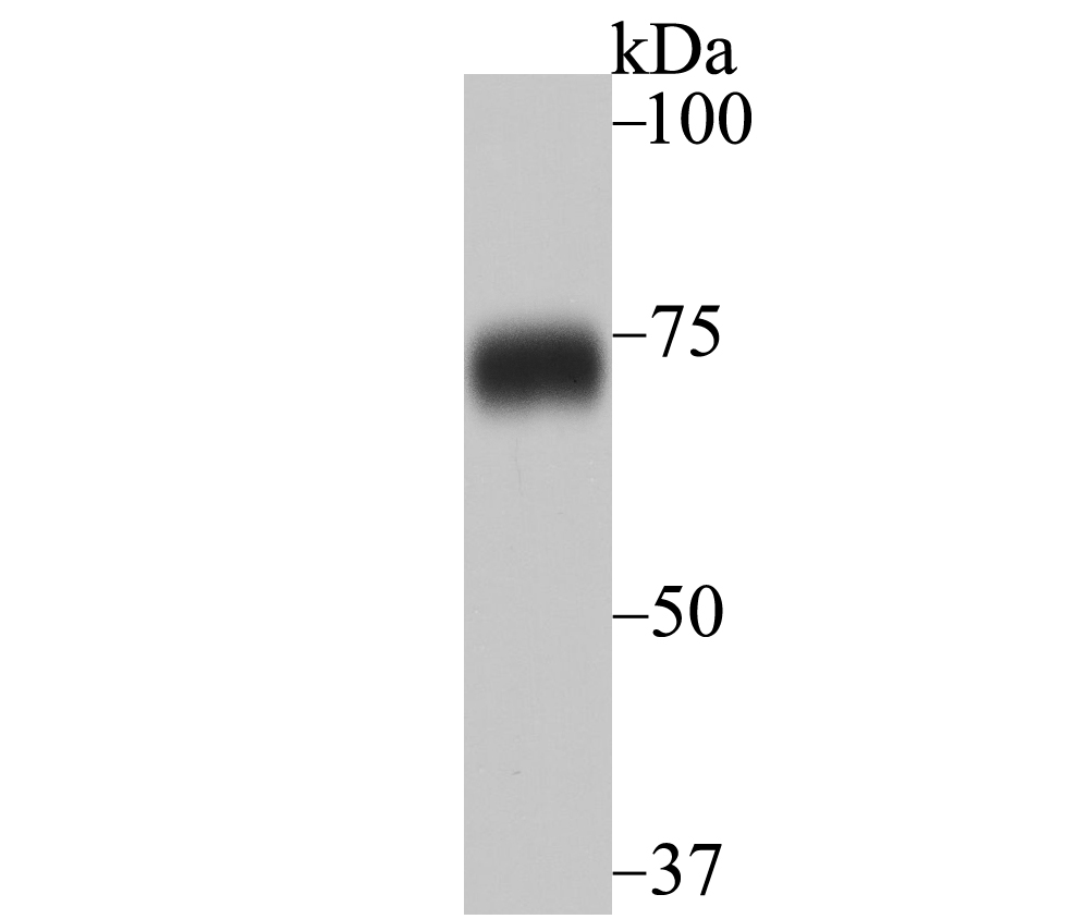
Western blot analysis of hnRNP Q on K562 cell lysates. Proteins were transferred to a PVDF membrane and blocked with 5% BSA in PBS for 1 hour at room temperature. The primary antibody (ET7108-17, 1/500) was used in 5% BSA at room temperature for 2 hours. Goat Anti-Rabbit IgG - HRP Secondary Antibody (HA1001) at 1:200,000 dilution was used for 1 hour at room temperature.
-
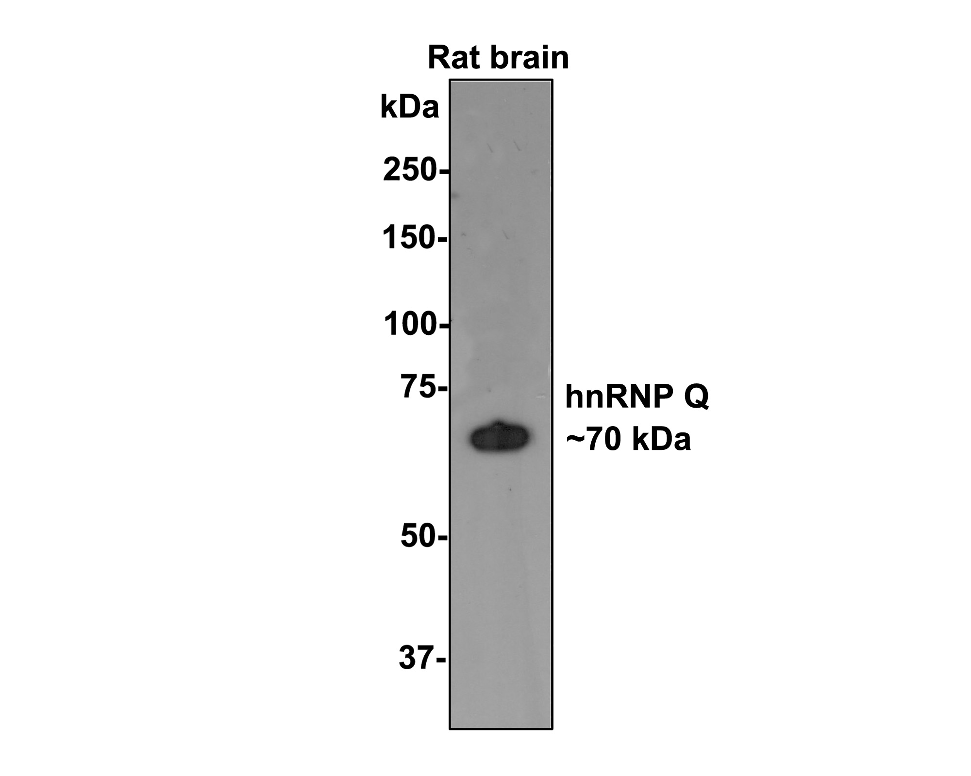
Western blot analysis of hnRNP Q on rat brain tissue lysates with Rabbit anti-hnRNP Q antibody (ET7108-17) at 1/500 dilution.
Lysates/proteins at 20 µg/Lane.
Predicted band size: 70 kDa
Observed band size: 70 kDa
Exposure time: 2 minutes;
8% SDS-PAGE gel.
Proteins were transferred to a PVDF membrane and blocked with 5% NFDM/TBST for 1 hour at room temperature. The primary antibody (ET7108-17) at 1/500 dilution was used in 5% NFDM/TBST at room temperature for 2 hours. Goat Anti-Rabbit IgG - HRP Secondary Antibody (HA1001) at 1:200,000 dilution was used for 1 hour at room temperature. -
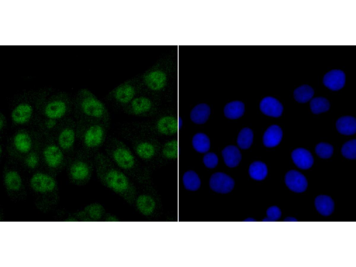
ICC staining of hnRNP Q in LOVO cells (green). Formalin fixed cells were permeabilized with 0.1% Triton X-100 in TBS for 10 minutes at room temperature and blocked with 10% negative goat serum for 15 minutes at room temperature. Cells were probed with the primary antibody (ET7108-17, 1/50) for 1 hour at room temperature, washed with PBS. Alexa Fluor®488 conjugate-Goat anti-Rabbit IgG was used as the secondary antibody at 1/1,000 dilution. The nuclear counter stain is DAPI (blue).
-
ICC staining of hnRNP Q in SiHa cells (green). Formalin fixed cells were permeabilized with 0.1% Triton X-100 in TBS for 10 minutes at room temperature and blocked with 10% negative goat serum for 15 minutes at room temperature. Cells were probed with the primary antibody (ET7108-17, 1/50) for 1 hour at room temperature, washed with PBS. Alexa Fluor®488 conjugate-Goat anti-Rabbit IgG was used as the secondary antibody at 1/1,000 dilution. The nuclear counter stain is DAPI (blue).
-
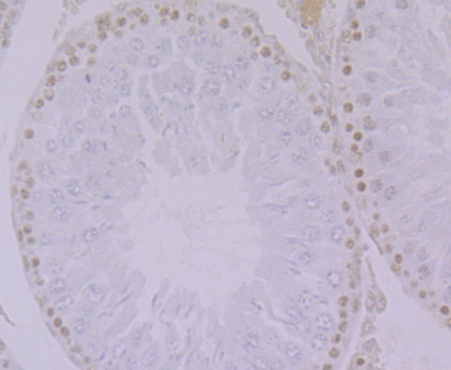
Immunohistochemical analysis of paraffin-embedded rat testis tissue using anti-hnRNP Q antibody. The section was pre-treated using heat mediated antigen retrieval with Tris-EDTA buffer (pH 9.0) for 20 minutes.The tissues were blocked in 1% BSA for 30 minutes at room temperature, washed with ddH2O and PBS, and then probed with the primary antibody (ET7108-17, 1/50) for 30 minutes at room temperature. The detection was performed using an HRP conjugated compact polymer system. DAB was used as the chromogen. Tissues were counterstained with hematoxylin and mounted with DPX.
-
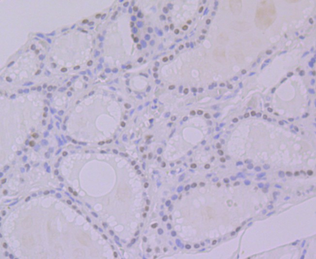
Immunohistochemical analysis of paraffin-embedded human thyroid tissue using anti-hnRNP Q antibody. The section was pre-treated using heat mediated antigen retrieval with Tris-EDTA buffer (pH 9.0) for 20 minutes.The tissues were blocked in 1% BSA for 30 minutes at room temperature, washed with ddH2O and PBS, and then probed with the primary antibody (ET7108-17, 1/50) for 30 minutes at room temperature. The detection was performed using an HRP conjugated compact polymer system. DAB was used as the chromogen. Tissues were counterstained with hematoxylin and mounted with DPX.
-
Immunohistochemical analysis of paraffin-embedded human kidney tissue using anti-hnRNP Q antibody. The section was pre-treated using heat mediated antigen retrieval with Tris-EDTA buffer (pH 9.0) for 20 minutes.The tissues were blocked in 1% BSA for 30 minutes at room temperature, washed with ddH2O and PBS, and then probed with the primary antibody (ET7108-17, 1/50) for 30 minutes at room temperature. The detection was performed using an HRP conjugated compact polymer system. DAB was used as the chromogen. Tissues were counterstained with hematoxylin and mounted with DPX.
-
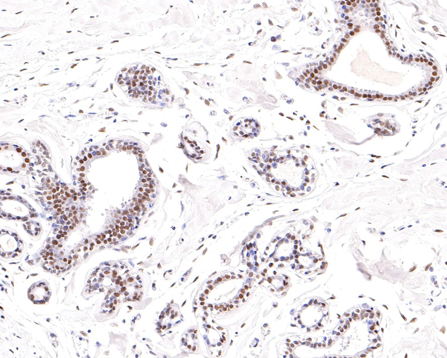
Immunohistochemical analysis of paraffin-embedded human breast tissue with Rabbit anti-hnRNP Q antibody (ET7108-17) at 1/200 dilution.
The section was pre-treated using heat mediated antigen retrieval with sodium citrate buffer (pH 6.0) for 2 minutes. The tissues were blocked in 1% BSA for 20 minutes at room temperature, washed with ddH2O and PBS, and then probed with the primary antibody (ET7108-17) at 1/200 dilution for 1 hour at room temperature. The detection was performed using an HRP conjugated compact polymer system. DAB was used as the chromogen. Tissues were counterstained with hematoxylin and mounted with DPX.
Please note: All products are "FOR RESEARCH USE ONLY AND ARE NOT INTENDED FOR DIAGNOSTIC OR THERAPEUTIC USE"