Kir2.1 Recombinant Rabbit Monoclonal Antibody [JE54-56]


Catalog# HA721227
Kir2.1 Recombinant Rabbit Monoclonal Antibody [JE54-56]
-
WB
-
IF-Cell
-
IHC-P
-
Human
-
Mouse
-
Rat
概述
产品名称
Kir2.1 Recombinant Rabbit Monoclonal Antibody [JE54-56]
抗体类型
Recombinant Rabbit monoclonal Antibody
免疫原
Synthetic peptide within Human Kir2.1 aa 91-140 / 427.
种属反应性
Human, Mouse, Rat
验证应用
WB, IF-Cell, IHC-P
分子量
Predicted band size: 48 kDa
阳性对照
N2A, SW620, rat uterus tissue, human placenta tissue, mouse brain tissue.
偶联
unconjugated
克隆号
JE54-56
RRID
产品特性
形态
Liquid
浓度
1ug/ul
存放说明
Store at +4℃ after thawing. Aliquot store at -20℃. Avoid repeated freeze / thaw cycles.
存储缓冲液
1*TBS (pH7.4), 1%BSA, 50%Glycerol. Preservative: 0.05% Sodium Azide.
亚型
IgG
纯化方式
Protein A affinity purified.
应用稀释度
-
WB
-
1:1,000
-
IF-Cell
-
1:50-1:100
-
IHC-P
-
1:200-1:1,000
靶点
功能
Potassium channels are present in most mammalian cells, where they participate in a wide range of physiologic responses. The protein encoded by this gene is an integral membrane protein and inward-rectifier type potassium channel. The encoded protein, which has a greater tendency to allow potassium to flow into a cell rather than out of a cell, probably participates in establishing action potential waveform and excitability of neuronal and muscle tissues. Mutations in this gene have been associated with Andersen syndrome, which is characterized by periodic paralysis, cardiac arrhythmias, and dysmorphic features.
背景文献
1. Despang A. et. al. Functional dissection of the Sox9-Kcnj2 locus identifies nonessential and instructive roles of TAD architecture. Nat Genet. 2019 Aug
2. Fukumura S. et. al. Functional analysis of a double-point mutation in the KCNJ2 gene identified in a family with Andersen-Tawil syndrome. J Neurol Sci. 2019 Dec
亚细胞定位
Membrane.
别名
Cardiac inward rectifier potassium channel antibody
HHBIRK 1 antibody
HHBIRK1 antibody
HHIRK 1 antibody
HHIRK1 antibody
HIRK 1 antibody
hIRK1 antibody
Inward rectifier K antibody
Inward rectifier K(+) channel Kir2.1 antibody
Inward rectifier potassium channel 2 antibody
展开图片
-
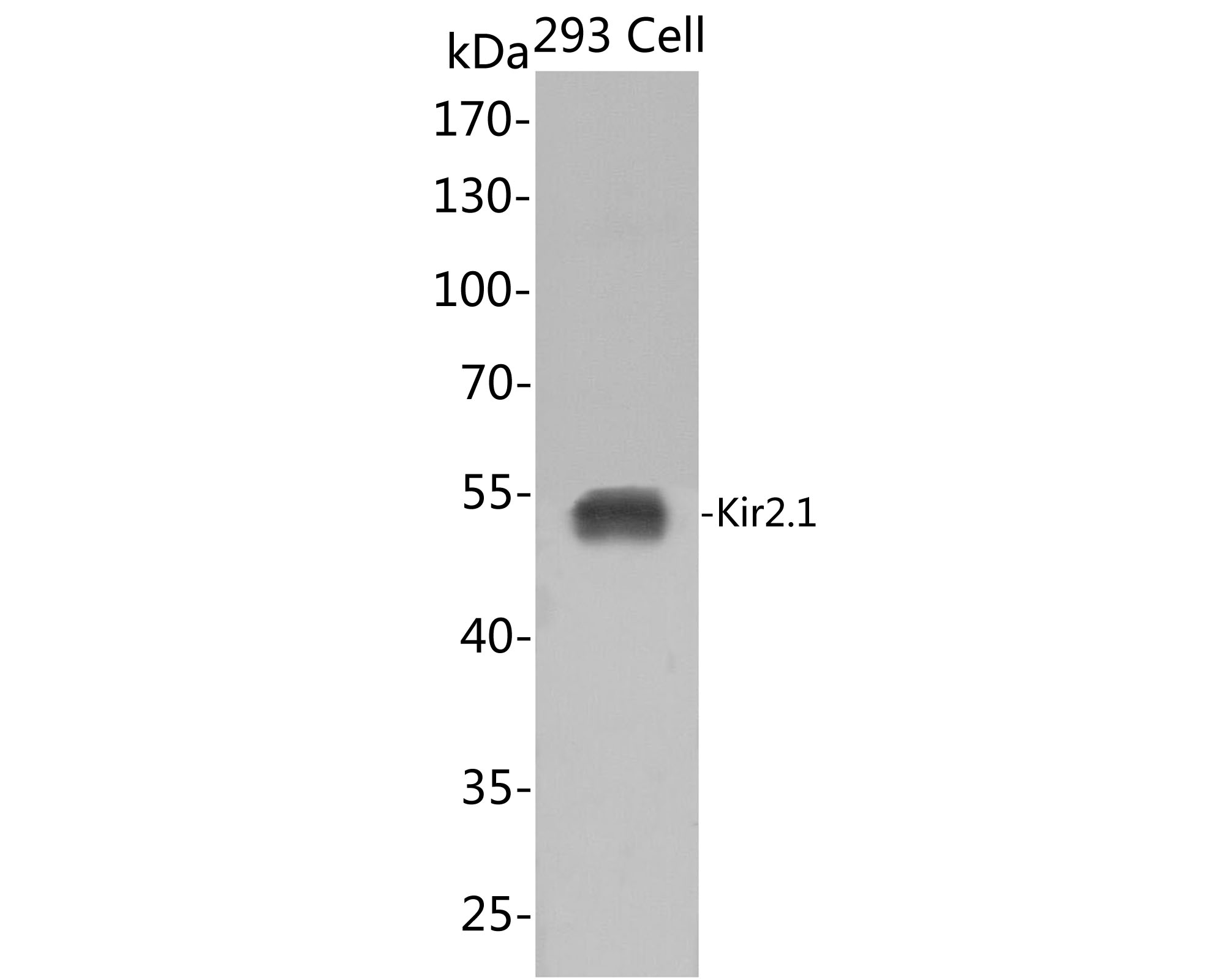
Western blot analysis of Kir2.1 on 293 cell lysates with Rabbit anti-Kir2.1 antibody (HA721227) at 1/1,000 dilution.
Predicted band size: 48 kDa
Observed band size: 48 kDa
Exposure time: 30s;
10% SDS-PAGE gel.
Proteins were transferred to a PVDF membrane and blocked with 5% NFDM/TBST for 1 hour at room temperature. The primary antibody (HA721227) at 1/1,000 dilution was used in 5% NFDM/TBST at room temperature for 2 hours. Goat Anti-Rabbit IgG - HRP Secondary Antibody (HA1001) at 1:300,000 dilution was used for 1 hour at room temperature. -
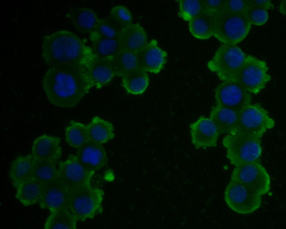
ICC staining of Kir2.1 in N2A cells (green). Formalin fixed cells were permeabilized with 0.1% Triton X-100 in TBS for 10 minutes at room temperature and blocked with 1% Blocker BSA for 15 minutes at room temperature. Cells were probed with the primary antibody (HA721227, 1/100) for 1 hour at room temperature, washed with PBS. Alexa Fluor®488 Goat anti-Rabbit IgG was used as the secondary antibody at 1/1,000 dilution. The nuclear counter stain is DAPI (blue).
-
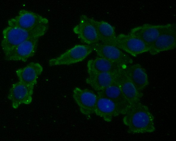
ICC staining of Kir2.1 in SW620 cells (green). Formalin fixed cells were permeabilized with 0.1% Triton X-100 in TBS for 10 minutes at room temperature and blocked with 1% Blocker BSA for 15 minutes at room temperature. Cells were probed with the primary antibody (HA721227, 1/100) for 1 hour at room temperature, washed with PBS. Alexa Fluor®488 Goat anti-Rabbit IgG was used as the secondary antibody at 1/1,000 dilution. The nuclear counter stain is DAPI (blue).
-
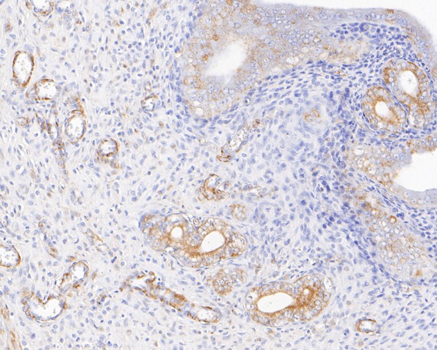
Immunohistochemical analysis of paraffin-embedded rat uterus tissue using anti-Kir2.1 antibody. The section was pre-treated using heat mediated antigen retrieval with sodium citrate buffer (pH 6.0) for 20 minutes. The tissues were blocked in 5% BSA for 30 minutes at room temperature, washed with ddH2O and PBS, and then probed with the primary antibody (HA721227, 1/400) for 30 minutes at room temperature. The detection was performed using an HRP conjugated compact polymer system. DAB was used as the chromogen. Tissues were counterstained with hematoxylin and mounted with DPX.
-
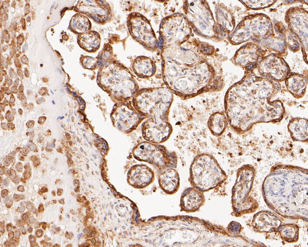
Immunohistochemical analysis of paraffin-embedded human placenta tissue using anti-Kir2.1 antibody. The section was pre-treated using heat mediated antigen retrieval with sodium citrate buffer (pH 6.0) for 20 minutes. The tissues were blocked in 5% BSA for 30 minutes at room temperature, washed with ddH2O and PBS, and then probed with the primary antibody (HA721227, 1/400) for 30 minutes at room temperature. The detection was performed using an HRP conjugated compact polymer system. DAB was used as the chromogen. Tissues were counterstained with hematoxylin and mounted with DPX.
-
Immunohistochemical analysis of paraffin-embedded mouse brain tissue using anti-Kir2.1 antibody. The section was pre-treated using heat mediated antigen retrieval with sodium citrate buffer (pH 6.0) for 20 minutes. The tissues were blocked in 5% BSA for 30 minutes at room temperature, washed with ddH2O and PBS, and then probed with the primary antibody (HA721227, 1/800) for 30 minutes at room temperature. The detection was performed using an HRP conjugated compact polymer system. DAB was used as the chromogen. Tissues were counterstained with hematoxylin and mounted with DPX.
Please note: All products are "FOR RESEARCH USE ONLY AND ARE NOT INTENDED FOR DIAGNOSTIC OR THERAPEUTIC USE"