TRP1 Recombinant Rabbit Monoclonal Antibody [JU36-48]


Catalog# ET7106-66
TRP1 Recombinant Rabbit Monoclonal Antibody [JU36-48]
-
WB
-
IF-Cell
-
IHC-P
-
IP
-
Human
概述
产品名称
TRP1 Recombinant Rabbit Monoclonal Antibody [JU36-48]
抗体类型
Recombinant Rabbit monoclonal Antibody
免疫原
Recombinant protein within Human TRP1 aa 411-537 / 537.
种属反应性
Human
验证应用
WB, IF-Cell, IHC-P, IP
分子量
72 kDa
阳性对照
Melanoma tissue lysates, HUVEC, A431, human skin tissue, human malignant melanoma tissue.
偶联
unconjugated
克隆号
JU36-48
RRID
产品特性
形态
Liquid
浓度
1ug/ul
存放说明
Store at +4℃ after thawing. Aliquot store at -20℃ or -80℃. Avoid repeated freeze / thaw cycles.
存储缓冲液
1*TBS (pH7.4), 0.05% BSA, 40% Glycerol. Preservative: 0.05% Sodium Azide.
亚型
IgG
纯化方式
Protein A affinity purified.
应用稀释度
-
WB
-
1:500-1:2,000
-
IF-Cell
-
1:50-1:100
-
IHC-P
-
1:50-1:500
-
IP
-
1:10-1:50
靶点
功能
Tyrosinase (TYR), a type I membrane protein and copper-containing enzyme, is involved in the production of melanin, the primary pigment found in vertebrates. Melanin biogenesis requires the enzymatic activity of TYR, which catalyzes the critical and rate-limiting step of tyrosine hydroxylation in the biosynthesis of melanin. Defects effecting TYR activity result in various forms of albinism. The TYR-related proteins, TRP1 and TRP2, are also specifically expressed in melanocytes, and they likewise contribute to the synthesis of melanin within the melanosomes. The TRPs, including TYR, all share a similar transmembrane region, contain two metal-binding regions and a cysteine-rich epidermal growth factor motif, and are localized in the melanosomal membrane. These proteins, however, have distinct catalytic activity, and they individually contribute to the biosynthesis of melanin biopolymers. The TRPs are believed to exists as a multi-enzyme complex, as these proteins form aggregates together, and the expression of TRP1 also helps stabilize TYR in melanocytes.
背景文献
1. Kenny E E et al. Melanesian blond hair is caused by an amino acid change in TYRP1. Science 336:554-554 (2012).
2. Rooryck C et al. Oculocutaneous albinism with TYRP1 gene mutations in a Caucasian patient. Pigment Cell Res 19:239-242 (2006).
序列相似性
Belongs to the tyrosinase family.
组织特异性
Pigment cells.
翻译后修饰
Glycosylated.
亚细胞定位
Membrane.
UNIPROT #
别名
5 antibody
5,6 dihydroxyindole 2 carboxylic acid oxidase antibody
6-dihydroxyindole-2-carboxylic acid oxidase antibody
b-PROTEIN antibody
CAS2 antibody
Catalase B antibody
CATB antibody
DHICA oxidase antibody
Glycoprotein 75 antibody
GP75 antibody
展开图片
-
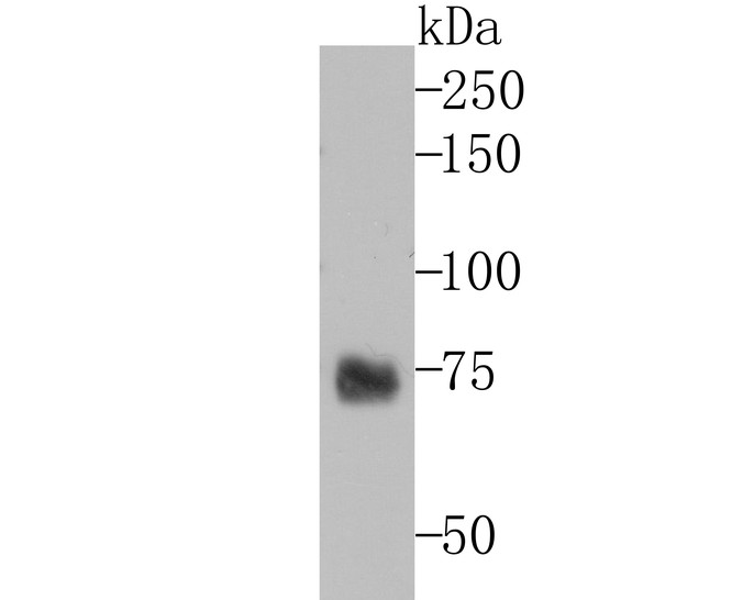
Western blot analysis of TRP1 on melanoma tissue lysates. Proteins were transferred to a PVDF membrane and blocked with 5% BSA in PBS for 1 hour at room temperature. The primary antibody (ET7106-66, 1/500) was used in 5% BSA at room temperature for 2 hours. Goat Anti-Rabbit IgG - HRP Secondary Antibody (HA1001) at 1:200,000 dilution was used for 1 hour at room temperature.
-
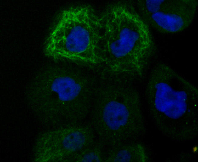
ICC staining of TRP1 in HUVEC cells (green). Formalin fixed cells were permeabilized with 0.1% Triton X-100 in TBS for 10 minutes at room temperature and blocked with 10% negative goat serum for 15 minutes at room temperature. Cells were probed with the primary antibody (ET7106-66, 1/50) for 1 hour at room temperature, washed with PBS. Alexa Fluor®488 conjugate-Goat anti-Rabbit IgG was used as the secondary antibody at 1/1,000 dilution. The nuclear counter stain is DAPI (blue).
-
ICC staining of TRP1 in A431 cells (green). Formalin fixed cells were permeabilized with 0.1% Triton X-100 in TBS for 10 minutes at room temperature and blocked with 10% negative goat serum for 15 minutes at room temperature. Cells were probed with the primary antibody (ET7106-66, 1/50) for 1 hour at room temperature, washed with PBS. Alexa Fluor®488 conjugate-Goat anti-Rabbit IgG was used as the secondary antibody at 1/1,000 dilution. The nuclear counter stain is DAPI (blue).
-
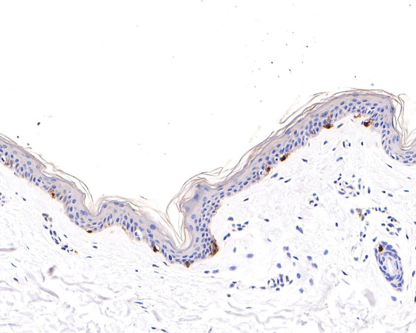
Immunohistochemical analysis of paraffin-embedded human skin tissue using anti-TRP1 antibody. The section was pre-treated using heat mediated antigen retrieval with Tris-EDTA buffer (pH 9.0) for 20 minutes.The tissues were blocked in 1% BSA for 30 minutes at room temperature, washed with ddH2O and PBS, and then probed with the primary antibody (ET7106-66, 1/100) for 30 minutes at room temperature. The detection was performed using an HRP conjugated compact polymer system. DAB was used as the chromogen. Tissues were counterstained with hematoxylin and mounted with DPX.
-
Immunohistochemical analysis of paraffin-embedded human malignant melanoma tissue using anti-TRP1 antibody. The section was pre-treated using heat mediated antigen retrieval with Tris-EDTA buffer (pH 9.0) for 20 minutes.The tissues were blocked in 1% BSA for 30 minutes at room temperature, washed with ddH2O and PBS, and then probed with the primary antibody (ET7106-66, 1/400) for 30 minutes at room temperature. The detection was performed using an HRP conjugated compact polymer system. DAB was used as the chromogen. Tissues were counterstained with hematoxylin and mounted with DPX.
Please note: All products are "FOR RESEARCH USE ONLY AND ARE NOT INTENDED FOR DIAGNOSTIC OR THERAPEUTIC USE"